Movie
Movie Controller


+ Open data

- Basic information
| Entry | Database: EMDB / ID: EMD-3848 | |||||||||
|---|---|---|---|---|---|---|---|---|---|---|
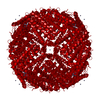
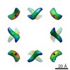
| Title | Single particle cryo-STEM of ferritin with 48 Zn atoms | |||||||||
 Map data Map data | Ferritin with 48 Zn atoms from single particle cryo-STEM data | |||||||||
 Sample Sample |
| |||||||||
| Biological species |   Homo sapiens (human) Homo sapiens (human) | |||||||||
| Method | single particle reconstruction / cryo EM / Resolution: 21.0 Å | |||||||||
 Authors Authors | Elad N / Bellapadrona G / Houben L / Sagi I / Elbaum M | |||||||||
 Citation Citation | Journal: Proc Natl Acad Sci U S A / Year: 2017 Title: Detection of isolated protein-bound metal ions by single-particle cryo-STEM. Authors: Nadav Elad / Giuliano Bellapadrona / Lothar Houben / Irit Sagi / Michael Elbaum /  Abstract: Metal ions play essential roles in many aspects of biological chemistry. Detecting their presence and location in proteins and cells is important for understanding biological function. Conventional ...Metal ions play essential roles in many aspects of biological chemistry. Detecting their presence and location in proteins and cells is important for understanding biological function. Conventional structural methods such as X-ray crystallography and cryo-transmission electron microscopy can identify metal atoms on protein only if the protein structure is solved to atomic resolution. We demonstrate here the detection of isolated atoms of Zn and Fe on ferritin, using cryogenic annular dark-field scanning transmission electron microscopy (cryo-STEM) coupled with single-particle 3D reconstructions. Zn atoms are found in a pattern that matches precisely their location at the ferroxidase sites determined earlier by X-ray crystallography. By contrast, the Fe distribution is smeared along an arc corresponding to the proposed path from the ferroxidase sites to the mineral nucleation sites along the twofold axes. In this case the single-particle reconstruction is interpreted as a probability distribution function based on the average of individual locations. These results establish conditions for detection of isolated metal atoms in the broader context of electron cryo-microscopy and tomography. | |||||||||
| History |
|
- Structure visualization
| Movie |
 Movie viewer Movie viewer |
|---|---|
| Structure viewer | EM map: SurfViewMolmilJmol/JSmol |
| Supplemental images |

- Downloads & links
-EMDB archive
| Map data | emd_3848.map.gz | 926.3 KB | EMDB map data format | |
|---|---|---|---|---|
| Header (meta data) | emd-3848-v30.xmlemd-3848.xml | 12.7 KB 12.7 KB | Display Display | EMDB header |
| Images | 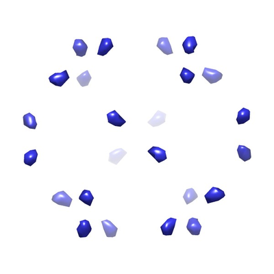 emd_3848.png emd_3848.png | 49.9 KB | ||
| Archive directory |  http://ftp.pdbj.org/pub/emdb/structures/EMD-3848ftp://ftp.pdbj.org/pub/emdb/structures/EMD-3848 http://ftp.pdbj.org/pub/emdb/structures/EMD-3848ftp://ftp.pdbj.org/pub/emdb/structures/EMD-3848 | HTTPS FTP |
-Related structure data
-Links
| EMDB pages | EMDB (EBI/PDBe) / EMDataResource |
|---|
-Map
| File | Download / File: emd_3848.map.gz / Format: CCP4 / Size: 2 MB / Type: IMAGE STORED AS FLOATING POINT NUMBER (4 BYTES) | ||||||||||||||||||||||||||||||||||||||||||||||||||||||||||||
|---|---|---|---|---|---|---|---|---|---|---|---|---|---|---|---|---|---|---|---|---|---|---|---|---|---|---|---|---|---|---|---|---|---|---|---|---|---|---|---|---|---|---|---|---|---|---|---|---|---|---|---|---|---|---|---|---|---|---|---|---|---|
| Annotation | Ferritin with 48 Zn atoms from single particle cryo-STEM data | ||||||||||||||||||||||||||||||||||||||||||||||||||||||||||||
| Voxel size | X=Y=Z: 3.3 Å | ||||||||||||||||||||||||||||||||||||||||||||||||||||||||||||
| Density |
| ||||||||||||||||||||||||||||||||||||||||||||||||||||||||||||
| Symmetry | Space group: 1 | ||||||||||||||||||||||||||||||||||||||||||||||||||||||||||||
| Details | EMDB XML:
CCP4 map header:
| ||||||||||||||||||||||||||||||||||||||||||||||||||||||||||||
-Supplemental data
- Sample components
-Entire : Human heavy chain ferritin with Zn
| Entire | Name: Human heavy chain ferritin with Zn |
|---|---|
| Components |
|
-Supramolecule #1: Human heavy chain ferritin with Zn
| Supramolecule | Name: Human heavy chain ferritin with Zn / type: complex / ID: 1 / Parent: 0 / Macromolecule list: #1 Details: Bacterially expressed and purified human heavy chain ferritin mixed with a 48 fold molar excess of Zn(II)SO4 |
|---|---|
| Source (natural) | Organism: Homo sapiens (human) |
| Recombinant expression | Organism:  Escherichia coli (E. coli) Escherichia coli (E. coli) |
| Molecular weight | Theoretical: 504 KDa |
-Experimental details
-Structure determination
| Method | cryo EM |
|---|---|
 Processing Processing | single particle reconstruction |
| Aggregation state | particle |
-Sample preparation
| Concentration | 0.13 mg/mL | ||||||
|---|---|---|---|---|---|---|---|
| Buffer | pH: 7.2 Component:
| ||||||
| Grid | Model: Quantifoil R2/1 / Material: COPPER / Mesh: 200 / Support film - #0 - Film type ID: 1 / Support film - #0 - Material: CARBON / Support film - #0 - topology: HOLEY / Support film - #1 - Film type ID: 2 / Support film - #1 - Material: CARBON / Support film - #1 - topology: CONTINUOUS / Pretreatment - Type: GLOW DISCHARGE / Pretreatment - Atmosphere: AIR | ||||||
| Vitrification | Cryogen name: ETHANE / Chamber humidity: 95 % / Chamber temperature: 297 K / Instrument: LEICA EM GP | ||||||
| Details | For metal loading, stock protein (2.9 mg/ml) was diluted to 0.25 mg/ml and mixed with a 48 fold molar excess of Zn(II)SO4. |
- Electron microscopy
| Microscope | FEI TECNAI 20 |
|---|---|
| Electron beam | Acceleration voltage: 200 kV / Electron source: FIELD EMISSION GUN |
| Electron optics | C2 aperture diameter: 70.0 µm / Illumination mode: OTHER / Imaging mode: DARK FIELD / Cs: 2.0 mm / Nominal magnification: 450000 |
| Sample stage | Specimen holder model: GATAN 626 SINGLE TILT LIQUID NITROGEN CRYO TRANSFER HOLDER Cooling holder cryogen: NITROGEN |
| Details | STEM mode imaging using Fischione model 3000 HAADF detector. Camera length: 520 mm. |
| Image recording | Film or detector model: OTHER / Digitization - Dimensions - Width: 2048 pixel / Digitization - Dimensions - Height: 2048 pixel / Number grids imaged: 1 / Number real images: 15 / Average electron dose: 127.0 e/Å2 Details: ADF images collected with a Fischione Model 3000 high-angle annular DF detector |
-Image processing
| Particle selection | Number selected: 647 / Details: Manually selected |
|---|---|
| Startup model | Type of model: PDB ENTRY PDB model - PDB ID: Details: The initial model was prepared from the human ferritin heavy chain crystal structure from which all heavy atoms were removed. The protein PDB coordinates were converted to structure factors ...Details: The initial model was prepared from the human ferritin heavy chain crystal structure from which all heavy atoms were removed. The protein PDB coordinates were converted to structure factors and low pass filtered to 4 nm. |
| Initial angle assignment | Type: PROJECTION MATCHING / Software - Name: RELION (ver. 1.4) |
| Final angle assignment | Type: PROJECTION MATCHING / Software - Name: RELION (ver. 1.4) |
| Final reconstruction | Number classes used: 1 / Applied symmetry - Point group: O (octahedral) / Resolution.type: BY AUTHOR / Resolution: 21.0 Å / Resolution method: FSC 0.143 CUT-OFF / Software - Name: RELION (ver. 1.4) / Number images used: 647 |
| Details | Fischione 3000 HAADF |