 Movie
Movie Controller

[English] 日本語
 Yorodumi
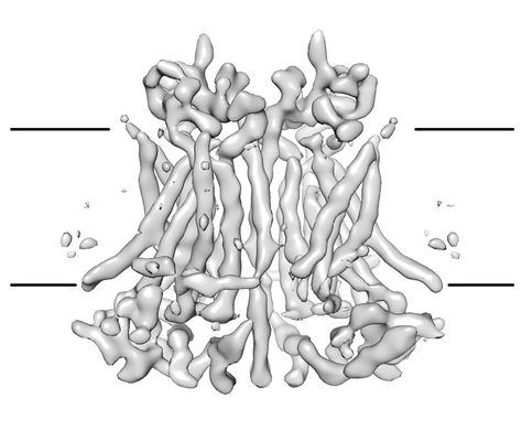
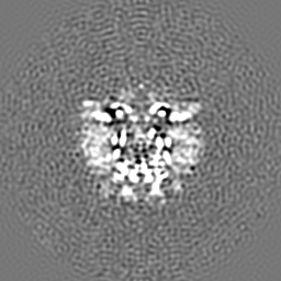
- EMDB-3658: cryo-EM structure of the mTMEM16A ion channel at 6.6 A resolution. -
+ Open data

- Basic information
| Entry | Database: EMDB / ID: EMD-3658 | |||||||||
|---|---|---|---|---|---|---|---|---|---|---|
| Title | cryo-EM structure of the mTMEM16A ion channel at 6.6 A resolution. | |||||||||
 Map data Map data | None | |||||||||
 Sample Sample |
| |||||||||
| Function / homology |  Function and homology information Function and homology informationglial cell projection elongation / trachea development / iodide transmembrane transporter activity / iodide transport / intracellularly calcium-gated chloride channel activity / mucus secretion / cellular response to peptide / Stimuli-sensing channels / voltage-gated chloride channel activity / calcium-activated cation channel activity ...glial cell projection elongation / trachea development / iodide transmembrane transporter activity / iodide transport / intracellularly calcium-gated chloride channel activity / mucus secretion / cellular response to peptide / Stimuli-sensing channels / voltage-gated chloride channel activity / calcium-activated cation channel activity / protein localization to membrane / chloride transport /  chloride channel activity / positive regulation of insulin secretion involved in cellular response to glucose stimulus / detection of temperature stimulus involved in sensory perception of pain / chloride channel complex / monoatomic cation transport / chloride transmembrane transport / regulation of membrane potential / cell projection / establishment of localization in cell / presynaptic membrane / cellular response to heat / phospholipase C-activating G protein-coupled receptor signaling pathway / apical plasma membrane / external side of plasma membrane / signaling receptor binding / glutamatergic synapse / protein homodimerization activity / nucleoplasm / identical protein binding / metal ion binding / plasma membrane chloride channel activity / positive regulation of insulin secretion involved in cellular response to glucose stimulus / detection of temperature stimulus involved in sensory perception of pain / chloride channel complex / monoatomic cation transport / chloride transmembrane transport / regulation of membrane potential / cell projection / establishment of localization in cell / presynaptic membrane / cellular response to heat / phospholipase C-activating G protein-coupled receptor signaling pathway / apical plasma membrane / external side of plasma membrane / signaling receptor binding / glutamatergic synapse / protein homodimerization activity / nucleoplasm / identical protein binding / metal ion binding / plasma membraneSimilarity search - Function | |||||||||
| Biological species |  Mus musculus (house mouse) Mus musculus (house mouse) | |||||||||
| Method | single particle reconstruction / cryo EM / Resolution: 6.6 Å | |||||||||
 Authors Authors | Paulino C / Neldner Y / Lam KM / Kalienkova V / Brunner JD / Schenck S / Dutzler R | |||||||||
 Citation Citation | Journal: Elife / Year: 2017 Title: Structural basis for anion conduction in the calcium-activated chloride channel TMEM16A. Authors: Cristina Paulino / Yvonne Neldner / Andy Km Lam / Valeria Kalienkova / Janine Denise Brunner / Stephan Schenck / Raimund Dutzler /  Abstract: The calcium-activated chloride channel TMEM16A is a member of a conserved protein family that comprises ion channels and lipid scramblases. Although the structure of the scramblase nhTMEM16 has ...The calcium-activated chloride channel TMEM16A is a member of a conserved protein family that comprises ion channels and lipid scramblases. Although the structure of the scramblase nhTMEM16 has defined the architecture of the family, it was unknown how a channel has adapted to cope with its distinct functional properties. Here we have addressed this question by the structure determination of mouse TMEM16A by cryo-electron microscopy and a complementary functional characterization. The protein shows a similar organization to nhTMEM16, except for changes at the site of catalysis. There, the conformation of transmembrane helices constituting a membrane-spanning furrow that provides a path for lipids in scramblases has changed to form an enclosed aqueous pore that is largely shielded from the membrane. Our study thus reveals the structural basis of anion conduction in a TMEM16 channel and it defines the foundation for the diverse functional behavior in the TMEM16 family. | |||||||||
| History |
|
- Structure visualization
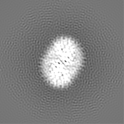
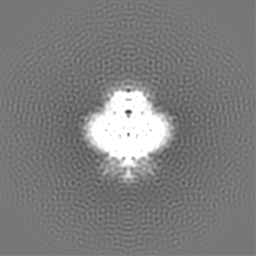
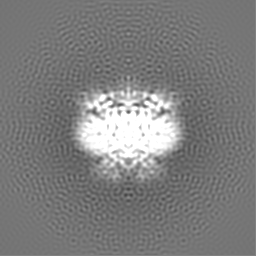
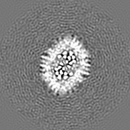
| Movie |
Movie viewer |
|---|---|
| Structure viewer | EM map: SurfViewMolmilJmol/JSmol |
| Supplemental images |
- Downloads & links
-EMDB archive
| Map data | emd_3658.map.gz | 57.3 MB | EMDB map data format | |
|---|---|---|---|---|
| Header (meta data) | emd-3658-v30.xmlemd-3658.xml | 19.1 KB 19.1 KB | Display Display | EMDB header |
| FSC (resolution estimation) | emd_3658_fsc.xml | 8.9 KB | Display | FSC data file |
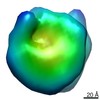
| Images |  emd_3658.png emd_3658.png | 62.8 KB | ||
| Others | emd_3658_additional.map.gz | 59.4 MB | ||
| Archive directory |  http://ftp.pdbj.org/pub/emdb/structures/EMD-3658ftp://ftp.pdbj.org/pub/emdb/structures/EMD-3658 http://ftp.pdbj.org/pub/emdb/structures/EMD-3658ftp://ftp.pdbj.org/pub/emdb/structures/EMD-3658 | HTTPS FTP |
-Related structure data
| Related structure data |  5nl2MC M: atomic model generated by this map C: citing same article ( |
|---|---|
| Similar structure data |


-Links
| EMDB pages | EMDB (EBI/PDBe) / EMDataResource |
|---|
-Map
| File | Download / File: emd_3658.map.gz / Format: CCP4 / Size: 64 MB / Type: IMAGE STORED AS FLOATING POINT NUMBER (4 BYTES) | ||||||||||||||||||||||||||||||||||||||||||||||||||||||||||||
|---|---|---|---|---|---|---|---|---|---|---|---|---|---|---|---|---|---|---|---|---|---|---|---|---|---|---|---|---|---|---|---|---|---|---|---|---|---|---|---|---|---|---|---|---|---|---|---|---|---|---|---|---|---|---|---|---|---|---|---|---|---|
| Annotation | None | ||||||||||||||||||||||||||||||||||||||||||||||||||||||||||||
| Voxel size | X=Y=Z: 1.35 Å | ||||||||||||||||||||||||||||||||||||||||||||||||||||||||||||
| Density |
| ||||||||||||||||||||||||||||||||||||||||||||||||||||||||||||
| Symmetry | Space group: 1 | ||||||||||||||||||||||||||||||||||||||||||||||||||||||||||||
| Details | EMDB XML:
CCP4 map header:
| ||||||||||||||||||||||||||||||||||||||||||||||||||||||||||||
-Supplemental data
-Additional map: None
| File | emd_3658_additional.map | ||||||||||||
|---|---|---|---|---|---|---|---|---|---|---|---|---|---|
| Annotation | None | ||||||||||||
| Projections & Slices |
| ||||||||||||
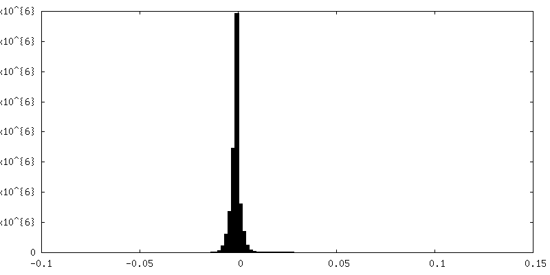
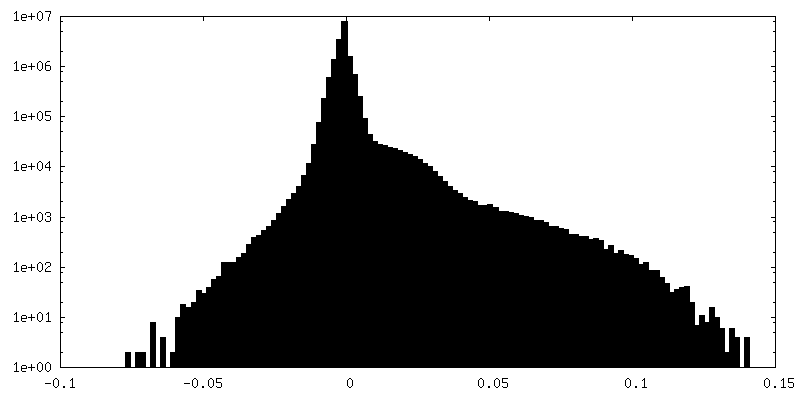
| Density Histograms |
 Z
Z Y
Y X
X


- Sample components
-Entire : mTMEM16A
| Entire | Name: mTMEM16A |
|---|---|
| Components |
|
-Supramolecule #1: mTMEM16A
| Supramolecule | Name: mTMEM16A / type: complex / ID: 1 / Parent: 0 / Macromolecule list: all |
|---|---|
| Source (natural) | Organism: Mus musculus (house mouse) |
| Recombinant expression | Organism:  Homo sapiens (human) / Recombinant strain: HEK293 / Recombinant cell: stabel mTMEM16A cell line (Flp-In System) Homo sapiens (human) / Recombinant strain: HEK293 / Recombinant cell: stabel mTMEM16A cell line (Flp-In System) |
| Molecular weight | Experimental: 110.916 KDa |
-Macromolecule #1: Anoctamin-1
| Macromolecule | Name: Anoctamin-1 / type: protein_or_peptide / ID: 1 / Number of copies: 2 / Enantiomer: LEVO |
|---|---|
| Source (natural) | Organism: Mus musculus (house mouse) |
| Molecular weight | Theoretical: 111.058992 KDa |
| Recombinant expression | Organism: Homo sapiens (human) |
| Sequence | String: MRVPEKYSTL PAEDRSVHIV NICAIEDLGY LPSEGTLLNS LSVDPDAECK YGLYFRDGKR KVDYILVYHH KRASGSRTLA RRGLQNDMV LGTRSVRQDQ PLPGKGSPVD AGSPEVPMDY HEDDKRFRRE EYEGNLLEAG LELENDEDTK IHGVGFVKIH A PWHVLCRE ...String: MRVPEKYSTL PAEDRSVHIV NICAIEDLGY LPSEGTLLNS LSVDPDAECK YGLYFRDGKR KVDYILVYHH KRASGSRTLA RRGLQNDMV LGTRSVRQDQ PLPGKGSPVD AGSPEVPMDY HEDDKRFRRE EYEGNLLEAG LELENDEDTK IHGVGFVKIH A PWHVLCRE AEFLKLKMPT KKVYHISETR GLLKTINSVL QKITDPIQPK VAEHRPQTTK RLSYPFSREK QHLFDLTDRD SF FDSKTRS TIVYEILKRT TCTKAKYSMG ITSLLANGVY SAAYPLHDGD YEGDNVEFND RKLLYEEWAS YGVFYKYQPI DLV RKYFGE KVGLYFAWLG AYTQMLIPAS IVGVIVFLYG CATVDENIPS MEMCDQRYNI TMCPLCDKTC SYWKMSSACA TARA SHLFD NPATVFFSVF MALWAATFME HWKRKQMRLN YRWDLTGFEE EEEAVKDHPR AEYEARVLEK SLRKESRNKE TDKVK LTWR DRFPAYFTNL VSIIFMIAVT FAIVLGVIIY RISTAAALAM NSSPSVRSNI RVTVTATAVI INLVVIILLD EVYGCI ARW LTKIEVPKTE KSFEERLTFK AFLLKFVNSY TPIFYVAFFK GRFVGRPGDY VYIFRSFRME ECAPGGCLME LCIQLSI IM LGKQLIQNNL FEIGIPKMKK FIRYLKLRRQ SPSDREEYVK RKQRYEVDFN LEPFAGLTPE YMEMIIQFGF VTLFVASF P LAPLFALLNN IIEIRLDAKK FVTELRRPVA IRAKDIGIWY NILRGVGKLA VIINAFVISF TSDFIPRLVY LYMYSQNGT MHGFVNHTLS SFNVSDFQNG TAPNDPLDLG YEVQICRYKD YREPPWSEHK YDISKDFWAV LAARLAFVIV FQNLVMFMSD FVDWVIPDI PKDISQQIHK EKVLMVELFM REEQGKQQLL DTWMEKEKPR DVPCNNHSPT THPEAGDGSP VPSYEYHGDA L |
-Experimental details
-Structure determination
| Method | cryo EM |
|---|---|
 Processing Processing | single particle reconstruction |
| Aggregation state | particle |
-Sample preparation
| Concentration | 3.08 mg/mL |
|---|---|
| Buffer | pH: 7.5 / Component - Concentration: 20.0 mM / Component - Name: Hepes / Details: 20 mM Hepes 150 mM NaCl 0.5 mM CaCl2 <0.12% digitonin |
| Grid | Model: Quantifoil R1.2/1.3 / Material: GOLD / Mesh: 200 / Support film - Material: CARBON / Support film - topology: HOLEY / Pretreatment - Type: GLOW DISCHARGE |
| Vitrification | Cryogen name: ETHANE / Chamber humidity: 100 % / Chamber temperature: 293 K / Instrument: FEI VITROBOT MARK IV / Details: 2.5 ul sample volume 1-5 sec blotting time. |
| Details | full-length (wild-type isoform ac) deglycosylated mTMEM16A |
- Electron microscopy
| Microscope | FEI TITAN KRIOS |
|---|---|
| Electron beam | Acceleration voltage: 300 kV / Electron source: FIELD EMISSION GUN |
| Electron optics | C2 aperture diameter: 100.0 µm / Calibrated defocus max: 3.8 µm / Calibrated defocus min: 0.5 µm / Calibrated magnification: 37037 / Illumination mode: FLOOD BEAM / Imaging mode: BRIGHT FIELDBright-field microscopy / Cs: 2.7 mm / Nominal defocus max: 3.8 µm / Nominal defocus min: 0.5 µm / Nominal magnification: 37037 |
| Specialist optics | Energy filter - Name: GIF / Energy filter - Lower energy threshold: -10 eV / Energy filter - Upper energy threshold: +10 eV |
| Sample stage | Specimen holder model: FEI TITAN KRIOS AUTOGRID HOLDER / Cooling holder cryogen: NITROGEN |
| Temperature | Min: 80.0 K / Max: 100.0 K |
| Image recording | Film or detector model: GATAN K2 SUMMIT (4k x 4k) / Detector mode: SUPER-RESOLUTION / Digitization - Dimensions - Width: 7420 pixel / Digitization - Dimensions - Height: 7676 pixel / Digitization - Sampling interval: 5.0 µm / Digitization - Frames/image: 1-50 / Number grids imaged: 5 / Number real images: 5503 / Average exposure time: 15.0 sec. / Average electron dose: 80.0 e/Å2 Details: Data were collected in an automated fashion on a K2 Summit detector (Gatan) set to super-resolution mode with a pixel size of 0.675 A and a defocus range of -0.5 to -3.8 um using SerialEM. ...Details: Data were collected in an automated fashion on a K2 Summit detector (Gatan) set to super-resolution mode with a pixel size of 0.675 A and a defocus range of -0.5 to -3.8 um using SerialEM. Images were recorded for 15 sec with an initial sub-frame exposure time of 300 ms (50 frames total) with a dose of 1.5 e-/A^2/frame, and later with a sub-frame exposure time of 150 ms (100 frames total) with a dose of 0.8 e-/A^2/frame, resulting in a total accumulated dose on the specimen level of approximately 80 e-/A^2. |
| Experimental equipment |  Model: Titan Krios / Image courtesy: FEI Company |
-Image processing
| Particle selection | Number selected: 755348 Details: Images showing a strong drift, higher defocus than -3.8 um or a bad CTF estimation were discarded, resulting in 4,178 images used for further analysis. Image processing was performed using ...Details: Images showing a strong drift, higher defocus than -3.8 um or a bad CTF estimation were discarded, resulting in 4,178 images used for further analysis. Image processing was performed using the software package RELION1.4 and at a later stage RELION2.0. Approximately 4,000 particles were manually picked to generate templates for automated particle selection. Following automated picking in RELION, false positives were eliminated manually or through a first round of 2D classification resulting in 755,348 particles. |
|---|---|
| CTF correction | Software - Name: CTFFIND (ver. 4.1.5) Details: The contrast transfer function (CTF) parameters were estimated on the movie frames by ctffind4.1 |
| Startup model | Type of model: PDB ENTRY PDB model - PDB ID: |
| Initial angle assignment | Type: ANGULAR RECONSTITUTION / Software - Name: RELION (ver. 2) |
| Final 3D classification | Software - Name: RELION (ver. 2) |
| Final angle assignment | Type: ANGULAR RECONSTITUTION / Software - Name: RELION (ver. 2) |
| Final reconstruction | Applied symmetry - Point group: C2 (2 fold cyclic) / Resolution.type: BY AUTHOR / Resolution: 6.6 Å / Resolution method: FSC 0.143 CUT-OFF / Software - Name: RELION (ver. 2) / Number images used: 213243 |
| Details | Fourier cropping (final pixel size 1.35 A), motion correction and dose-weighting of frames were performed by MotionCor2 |
| FSC plot (resolution estimation) | 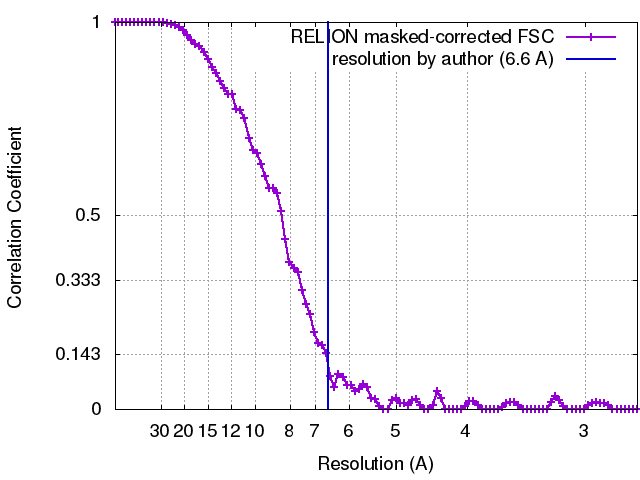 |

