 Movie
Movie Controller

[English] 日本語
 Yorodumi
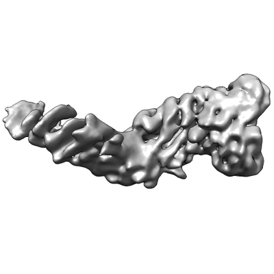
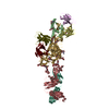
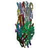
- EMDB-3584: Cryo-EM structure of the human Separase-Securin complex at medium... -
+ Open data

- Basic information
| Entry | Database: EMDB / ID: EMD-3584 | |||||||||
|---|---|---|---|---|---|---|---|---|---|---|
| Title | Cryo-EM structure of the human Separase-Securin complex at medium resolution | |||||||||
 Map data Map data | Cryo-EM structure of the human Separase-Securin complex | |||||||||
 Sample Sample |
| |||||||||
| Biological species |   Homo sapiens (human) Homo sapiens (human) | |||||||||
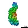
| Method | single particle reconstruction / cryo EM / Resolution: 7.0 Å | |||||||||
 Authors Authors | Boland A / Martin TG / Zhang Z / Yang J / Bai XC / Chang L / Scheres SHW / Barford D | |||||||||
 Citation Citation | Journal: Nat Struct Mol Biol / Year: 2017 Title: Cryo-EM structure of a metazoan separase-securin complex at near-atomic resolution. Authors: Andreas Boland / Thomas G Martin / Ziguo Zhang / Jing Yang / Xiao-Chen Bai / Leifu Chang / Sjors H W Scheres / David Barford /  Abstract: Separase is a caspase-family protease that initiates chromatid segregation by cleaving the kleisin subunits (Scc1 and Rec8) of cohesin, and regulates centrosome duplication and mitotic spindle ...Separase is a caspase-family protease that initiates chromatid segregation by cleaving the kleisin subunits (Scc1 and Rec8) of cohesin, and regulates centrosome duplication and mitotic spindle function through cleavage of kendrin and Slk19. To understand the mechanisms of securin regulation of separase, we used single-particle cryo-electron microscopy (cryo-EM) to determine a near-atomic-resolution structure of the Caenorhabditis elegans separase-securin complex. Separase adopts a triangular-shaped bilobal architecture comprising an N-terminal tetratricopeptide repeat (TPR)-like α-solenoid domain docked onto the conserved C-terminal protease domain. Securin engages separase in an extended antiparallel conformation, interacting with both lobes. It inhibits separase by interacting with the catalytic site through a pseudosubstrate mechanism, thus revealing that in the inhibited separase-securin complex, the catalytic site adopts a conformation compatible with substrate binding. Securin is protected from cleavage because an aliphatic side chain at the P1 position represses protease activity by disrupting the organization of catalytic site residues. | |||||||||
| History |
|
- Structure visualization
| Movie |
 Movie viewer Movie viewer |
|---|---|
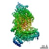
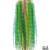
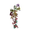
| Structure viewer | EM map: SurfViewMolmilJmol/JSmol |
| Supplemental images |
- Downloads & links
-EMDB archive
| Map data | emd_3584.map.gz | 40.8 MB | EMDB map data format | |
|---|---|---|---|---|
| Header (meta data) | emd-3584-v30.xmlemd-3584.xml | 14.9 KB 14.9 KB | Display Display | EMDB header |
| Images |  emd_3584.png emd_3584.png | 36.6 KB | ||
| Archive directory |  http://ftp.pdbj.org/pub/emdb/structures/EMD-3584ftp://ftp.pdbj.org/pub/emdb/structures/EMD-3584 http://ftp.pdbj.org/pub/emdb/structures/EMD-3584ftp://ftp.pdbj.org/pub/emdb/structures/EMD-3584 | HTTPS FTP |
-Related structure data


-Links
| EMDB pages | EMDB (EBI/PDBe) / EMDataResource |
|---|
-Map
| File | Download / File: emd_3584.map.gz / Format: CCP4 / Size: 52.7 MB / Type: IMAGE STORED AS FLOATING POINT NUMBER (4 BYTES) | ||||||||||||||||||||||||||||||||||||||||||||||||||||||||||||
|---|---|---|---|---|---|---|---|---|---|---|---|---|---|---|---|---|---|---|---|---|---|---|---|---|---|---|---|---|---|---|---|---|---|---|---|---|---|---|---|---|---|---|---|---|---|---|---|---|---|---|---|---|---|---|---|---|---|---|---|---|---|
| Annotation | Cryo-EM structure of the human Separase-Securin complex | ||||||||||||||||||||||||||||||||||||||||||||||||||||||||||||
| Voxel size | X=Y=Z: 1.05 Å | ||||||||||||||||||||||||||||||||||||||||||||||||||||||||||||
| Density |
| ||||||||||||||||||||||||||||||||||||||||||||||||||||||||||||
| Symmetry | Space group: 1 | ||||||||||||||||||||||||||||||||||||||||||||||||||||||||||||
| Details | EMDB XML:
CCP4 map header:
| ||||||||||||||||||||||||||||||||||||||||||||||||||||||||||||
-Supplemental data
- Sample components
-Entire : Homo sapiens Separase-Securin complex
| Entire | Name: Homo sapiens Separase-Securin complex |
|---|---|
| Components |
|
-Supramolecule #1: Homo sapiens Separase-Securin complex
| Supramolecule | Name: Homo sapiens Separase-Securin complex / type: complex / ID: 1 / Parent: 0 / Macromolecule list: all Details: H. sapiens Separase-Securin complex at medium resolution refined with Relion. Sample exhibits strong preferred orientation of particles. |
|---|---|
| Source (natural) | Organism: Homo sapiens (human) |
| Recombinant expression | Organism:  Trichoplusia ni (cabbage looper) Trichoplusia ni (cabbage looper) |
| Molecular weight | Theoretical: 255 KDa |
-Macromolecule #1: Separase-Securin complex
| Macromolecule | Name: Separase-Securin complex / type: protein_or_peptide / ID: 1 / Enantiomer: LEVO |
|---|---|
| Source (natural) | Organism: Homo sapiens (human) |
| Recombinant expression | Organism: Trichoplusia ni (cabbage looper) |
| Sequence | String: MRSFKRVNFG TLLSSQKEAE ELLPALKEFL SNPPAGFPSS RSDAERRQAC DAILRACNQQ LTAKLACPR HLGSLLELAE LACDGYLVST PQRPPLYLER ILFVLLRNAA AQGSPEATLR L AQPLHACL VQCSREAAPQ DYEAVARGSF SLLWKGAEAL LERRAAFAAR ...String: MRSFKRVNFG TLLSSQKEAE ELLPALKEFL SNPPAGFPSS RSDAERRQAC DAILRACNQQ LTAKLACPR HLGSLLELAE LACDGYLVST PQRPPLYLER ILFVLLRNAA AQGSPEATLR L AQPLHACL VQCSREAAPQ DYEAVARGSF SLLWKGAEAL LERRAAFAAR LKALSFLVLL ED ESTPCEV PHFASPTACR AVAAHQLFDA SGHGLNEADA DFLDDLLSRH VIRALVGERG SSS GLLSPQ RALCLLELTL EHCRRFCWSR HHDKAISAVE KAHSYLRNTN LAPSLQLCQL GVKL LQVGE EGPQAVAKLL IKASAVLSKS MEAPSPPLRA LYESCQFFLS GLERGTKRRY RLDAI LSLF AFLGGYCSLL QQLRDDGVYG GSSKQQQSFL QMYFQGLHLY TVVVYDFAQG CQIVDL ADL TQLVDSCKST VVWMLEALEG LSGQELTDHM GMTASYTSNL AYSFYSHKLY AEACAIS EP LCQHLGLVKP GTYPEVPPEK LHRCFRLQVE SLKKLGKQAQ GCKMVILWLA ALQPCSPE H MAEPVTFWVR VKMDAARAGD KELQLKTLRD SLSGWDPETL ALLLREELQA YKAVRADTG QERFNIICDL LELSPEETPA GAWARATHLV ELAQVLCYHD FTQQTNCSAL DAIREALQLL DSVRPEAQA RDQLLDDKAQ ALLWLYICTL EAKMQEGIER DRRAQAPGNL EEFEVNDLNY E DKLQEDRF LYSNIAFNLA ADAAQSKCLD QALALWKELL TKGQAPAVRC LQQTAASLQI LA ALYQLVA KPMQALEVLL LLRIVSERLK DHSKAAGSSC HITQLLLTLG CPSYAQLHLE EAA SSLKHL DQTTDTYLLL SLTCDLLRSQ LYWTHQKVTK GVSLLLSVLR DPALQKSSKA WYLL RVQVL QLVAAYLSLP SNNLSHSLWE QLCAQGWQTP EIALIDSHKL LRSIILLLMG SDILS TQKA AVETSFLDYG ENLVQKWQVL SEVLSCSEKL VCHLGRLGSV SEAKAFCLEA LKLTTK LQI PRQCALFLVL KGELELARND IDLCQSDLQQ VLFLLESCTE FGGVTQHLDS VKKVHLQ KG KQQAQVPCPP QLPEEELFLR GPALELVATV AKEPGPIAPS TNSSPVLKTK PQPIPNFL S HSPTCDCSLC ASPVLTAVCL RWVLVTAGVR LAMGHQAQGL DLLQVVLKGC PEAAERLTQ ALQASLNHKT PPSLVPSLLD EILAQAYTLL ALEGLNQPSN ESLQKVLQSG LKFVAARIPH LEPWRASLL LIWALTKLGG LSCCTTQLFA SSWGWQPPLI KSVPGSEPSK TQGQKRSGRG R QKLASAPL RLNNTSQKGL EGRGLPCTPK PPDRIRQAGP HVPFTVFEEV CPTESKPEVP QA PRVQQRV QTRLKVNFSD DSDLEDPVSA EAWLAEEPKR RGTASRGRGR ARKGLSLKTD AVV APGSAP GNPGLNGRSR RAKKVASRHC EERRPQRASD QARPGPEIMR TIPEEELTDN WRKM SFEIL RGSDGEDSAS GGKTPAPGPE AASGEWELLR LDSSKKKLPS PCPDKESDKD LGPRL RLPS APVATGLSTL DSICDSLSVA FRGISHCPPS GLYAHLCRFL ALCLGHRDPY ATAFLV TES VSITCRHQLL THLHRQLSKA QKHRGSLEIA DQLQGLSLQE MPGDVPLARI QRLFSFR AL ESGHFPQPEK ESFQERLALI PSGVTVCVLA LATLQPGTVG NTLLLTRLEK DSPPVSVQ I PTGQNKLHLR SVLNEFDAIQ KAQKENSSCT DKREWWTGRL ALDHRMEVLI ASLEKSVLG CWKGLLLPSS EEPGPAQEAS RLQELLQDCG WKYPDRTLLK IMLSGAGALT PQDIQALAYG LCPTQPERA QELLNEAVGR LQGLTVPSNS HLVLVLDKDL QKLPWESMPS LQALPVTRLP S FRFLLSYS IIKEYGASPV LSQGVDPRST FYVLNPHNNL SSTEEQFRAN FSSEAGWRGV VG EVPRPEQ VQEALTKHDL YIYAGHGAGA RFLDGQAVLR LSCRAVALLF GCSSAALAVR GNL EGAGIV LKYIMAGCPL FLGNLWDVTD RDIDRYTEAL LQGWLGAGPG APLLYYVNQA RQAP RLKYL IGAAPIAYGL PVSLR |
-Experimental details
-Structure determination
| Method | cryo EM |
|---|---|
 Processing Processing | single particle reconstruction |
| Aggregation state | particle |
-Sample preparation
| Concentration | 0.02 mg/mL |
|---|---|
| Buffer | pH: 7.8 |
| Grid | Model: Quantifoil R1.2/1.3 / Material: GOLD / Mesh: 300 / Support film - Material: GRAPHENE OXIDE / Support film - topology: HOLEY ARRAY / Pretreatment - Type: GLOW DISCHARGE / Pretreatment - Atmosphere: AIR Details: Glow discharging was performed before applying graphene oxide solution onto the grid. Grid was washed three times, dried and sample was applied. For detailed information see: https: ...Details: Glow discharging was performed before applying graphene oxide solution onto the grid. Grid was washed three times, dried and sample was applied. For detailed information see: https://figshare.com/articles/Graphene_Oxide_Grid_Preparation/3178669 |
| Vitrification | Cryogen name: ETHANE / Chamber humidity: 100 % / Chamber temperature: 277 K / Details: Custom Manual Plunger. |
| Details | Monodisperse sample |
- Electron microscopy
| Microscope | FEI TITAN KRIOS |
|---|---|
| Electron beam | Acceleration voltage: 300 kV / Electron source: FIELD EMISSION GUN |
| Electron optics | C2 aperture diameter: 70.0 µm / Illumination mode: FLOOD BEAM / Imaging mode: BRIGHT FIELDBright-field microscopy / Cs: 2.7 mm |
| Specialist optics | Energy filter - Name: GIF / Energy filter - Lower energy threshold: -20 eV / Energy filter - Upper energy threshold: 20 eV |
| Sample stage | Specimen holder model: FEI TITAN KRIOS AUTOGRID HOLDER / Cooling holder cryogen: NITROGEN |
| Image recording | Film or detector model: GATAN K2 SUMMIT (4k x 4k) / Detector mode: SUPER-RESOLUTION / Digitization - Dimensions - Width: 3710 pixel / Digitization - Dimensions - Height: 3710 pixel / Average electron dose: 1.25 e/Å2 |
| Experimental equipment |  Model: Titan Krios / Image courtesy: FEI Company |
-Image processing
| CTF correction | Software - Name: Gctf (ver. 1.06) |
|---|---|
| Startup model | Type of model: NONE / Details: Initial model was created using SIMPLE PRIME |
| Initial angle assignment | Type: ANGULAR RECONSTITUTION / Software - Name: RELION (ver. 2.0) |
| Final angle assignment | Type: ANGULAR RECONSTITUTION / Software - Name: RELION (ver. 2.0) |
| Final reconstruction | Applied symmetry - Point group: C1 (asymmetric) / Resolution.type: BY AUTHOR / Resolution: 7.0 Å / Resolution method: FSC 0.143 CUT-OFF / Software - Name: RELION (ver. 2.0) / Number images used: 103696 |
-Atomic model buiding 1
| Refinement | Space: REAL / Protocol: AB INITIO MODEL |
|---|
