Movie
Movie Controller


+ Open data

- Basic information
| Entry | Database: EMDB / ID: EMD-3460 | |||||||||
|---|---|---|---|---|---|---|---|---|---|---|
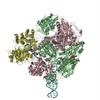
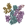
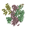
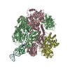
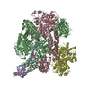
| Title | Cryo-EM structure of Lambda Phage protein GamS bound to RecBCD. | |||||||||
 Map data Map data | ||||||||||
 Sample Sample |
| |||||||||
| Function / homology |  Function and homology information Function and homology informationsymbiont-mediated evasion of DNA end degradation by host / deoxyribonuclease inhibitor activity /  exodeoxyribonuclease V / exodeoxyribonuclease V activity / exodeoxyribonuclease V complex / DNA 5'-3' helicase / DNA 3'-5' helicase / clearance of foreign intracellular DNA / DNA translocase activity / single-stranded DNA helicase activity ...symbiont-mediated evasion of DNA end degradation by host / deoxyribonuclease inhibitor activity / exodeoxyribonuclease V / exodeoxyribonuclease V activity / exodeoxyribonuclease V complex / DNA 5'-3' helicase / DNA 3'-5' helicase / clearance of foreign intracellular DNA / DNA translocase activity / single-stranded DNA helicase activity / recombinational repair / 3'-5' DNA helicase activity / ATP-dependent activity, acting on DNA / DNA helicase activity / DNA endonuclease activity / helicase activity / double-strand break repair via homologous recombination / response to radiation / DNA recombination / DNA damage response / magnesium ion binding / ATP hydrolysis activity / DNA binding / ATP binding / cytosol exodeoxyribonuclease V / exodeoxyribonuclease V activity / exodeoxyribonuclease V complex / DNA 5'-3' helicase / DNA 3'-5' helicase / clearance of foreign intracellular DNA / DNA translocase activity / single-stranded DNA helicase activity ...symbiont-mediated evasion of DNA end degradation by host / deoxyribonuclease inhibitor activity / exodeoxyribonuclease V / exodeoxyribonuclease V activity / exodeoxyribonuclease V complex / DNA 5'-3' helicase / DNA 3'-5' helicase / clearance of foreign intracellular DNA / DNA translocase activity / single-stranded DNA helicase activity / recombinational repair / 3'-5' DNA helicase activity / ATP-dependent activity, acting on DNA / DNA helicase activity / DNA endonuclease activity / helicase activity / double-strand break repair via homologous recombination / response to radiation / DNA recombination / DNA damage response / magnesium ion binding / ATP hydrolysis activity / DNA binding / ATP binding / cytosolSimilarity search - Function | |||||||||
| Biological species |  Escherichia coli (E. coli) / Escherichia coli (E. coli) /  Enterobacteria phage lambda (virus) Enterobacteria phage lambda (virus) | |||||||||
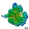
| Method | single particle reconstruction / cryo EM / Resolution: 3.8 Å | |||||||||
 Authors Authors | Wilkinson M / Chaban Y / Wigley DB | |||||||||
 Citation Citation | Journal: Elife / Year: 2016 Title: Structural basis for the inhibition of RecBCD by Gam and its synergistic antibacterial effect with quinolones. Authors: Martin Wilkinson / Luca Troman / Wan Ak Wan Nur Ismah / Yuriy Chaban / Matthew B Avison / Mark S Dillingham / Dale B Wigley /  Abstract: Our previous paper (Wilkinson , 2016) used high-resolution cryo-electron microscopy to solve the structure of the RecBCD complex, which acts in both the repair of double-stranded DNA breaks and the ...Our previous paper (Wilkinson , 2016) used high-resolution cryo-electron microscopy to solve the structure of the RecBCD complex, which acts in both the repair of double-stranded DNA breaks and the degradation of bacteriophage DNA. To counteract the latter activity, bacteriophage λ encodes a small protein inhibitor called Gam that binds to RecBCD and inactivates the complex. Here, we show that Gam inhibits RecBCD by competing at the DNA-binding site. The interaction surface is extensive and involves molecular mimicry of the DNA substrate. We also show that expression of Gam in or increases sensitivity to fluoroquinolones; antibacterials that kill cells by inhibiting topoisomerases and inducing double-stranded DNA breaks. Furthermore, fluoroquinolone-resistance in clinical isolates is reversed by expression of Gam. Together, our data explain the synthetic lethality observed between topoisomerase-induced DNA breaks and the RecBCD gene products, suggesting a new co-antibacterial strategy. | |||||||||
| History |
|
- Structure visualization
| Movie |
Movie viewer |
|---|---|
| Structure viewer | EM map: SurfViewMolmilJmol/JSmol |
| Supplemental images |

- Downloads & links
-EMDB archive
| Map data | emd_3460.map.gz | 20 MB | EMDB map data format | |
|---|---|---|---|---|
| Header (meta data) | emd-3460-v30.xmlemd-3460.xml | 23.5 KB 23.5 KB | Display Display | EMDB header |
| Images | 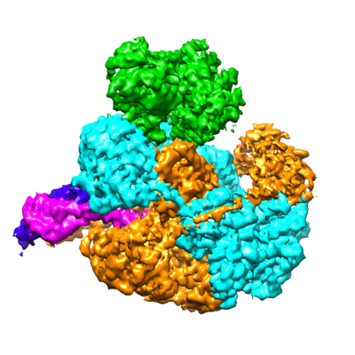 emd_3460.png emd_3460.png | 143.8 KB | ||
| Archive directory |  http://ftp.pdbj.org/pub/emdb/structures/EMD-3460ftp://ftp.pdbj.org/pub/emdb/structures/EMD-3460 http://ftp.pdbj.org/pub/emdb/structures/EMD-3460ftp://ftp.pdbj.org/pub/emdb/structures/EMD-3460 | HTTPS FTP |
-Related structure data
| Related structure data |  5mbvMC M: atomic model generated by this map C: citing same article ( |
|---|---|
| Similar structure data |


-Links
| EMDB pages | EMDB (EBI/PDBe) / EMDataResource |
|---|---|
| Related items in Molecule of the Month |


-Map
| File | Download / File: emd_3460.map.gz / Format: CCP4 / Size: 22.2 MB / Type: IMAGE STORED AS FLOATING POINT NUMBER (4 BYTES) | ||||||||||||||||||||||||||||||||||||||||||||||||||||||||||||
|---|---|---|---|---|---|---|---|---|---|---|---|---|---|---|---|---|---|---|---|---|---|---|---|---|---|---|---|---|---|---|---|---|---|---|---|---|---|---|---|---|---|---|---|---|---|---|---|---|---|---|---|---|---|---|---|---|---|---|---|---|---|
| Voxel size | X=Y=Z: 1.34 Å | ||||||||||||||||||||||||||||||||||||||||||||||||||||||||||||
| Density |
| ||||||||||||||||||||||||||||||||||||||||||||||||||||||||||||
| Symmetry | Space group: 1 | ||||||||||||||||||||||||||||||||||||||||||||||||||||||||||||
| Details | EMDB XML:
CCP4 map header:
| ||||||||||||||||||||||||||||||||||||||||||||||||||||||||||||
-Supplemental data
- Sample components
-Entire : RecBCD complex bound to inhibitory protein lambda GamS
| Entire | Name: RecBCD complex bound to inhibitory protein lambda GamS |
|---|---|
| Components |
|
-Supramolecule #1: RecBCD complex bound to inhibitory protein lambda GamS
| Supramolecule | Name: RecBCD complex bound to inhibitory protein lambda GamS type: complex / ID: 1 / Parent: 0 / Macromolecule list: #1-#5 Details: RecBCD and GamS were expressed and purified separately. They were mixed with an excess of GamS then passed through a size exclusion column to separate out free GamS. |
|---|---|
| Molecular weight | Theoretical: 350 KDa |
-Supramolecule #2: RecBCD helicase/nuclease complex
| Supramolecule | Name: RecBCD helicase/nuclease complex / type: complex / ID: 2 / Parent: 1 / Macromolecule list: #1-#3 |
|---|---|
| Source (natural) | Organism: Escherichia coli (E. coli) |
| Recombinant expression | Organism: Escherichia coli (E. coli) / Recombinant strain: BL21(DE3) / Recombinant plasmid: Multiple |
| Molecular weight | Theoretical: 330 KDa |
-Supramolecule #3: Lambda GamS
| Supramolecule | Name: Lambda GamS / type: complex / ID: 3 / Parent: 1 / Macromolecule list: #4-#5 |
|---|---|
| Source (natural) | Organism: Enterobacteria phage lambda (virus) |
| Recombinant expression | Organism: Escherichia coli (E. coli) / Recombinant strain: BL21(DE3) / Recombinant plasmid: pET22b |
| Molecular weight | Theoretical: 20 KDa |
-Macromolecule #1: RecBCD enzyme subunit RecB
| Macromolecule | Name: RecBCD enzyme subunit RecB / type: protein_or_peptide / ID: 1 / Number of copies: 1 / Enantiomer: LEVO / EC number: exodeoxyribonuclease V |
|---|---|
| Source (natural) | Organism: Escherichia coli (E. coli) |
| Molecular weight | Theoretical: 134.167703 KDa |
| Recombinant expression | Organism: Escherichia coli BL21(DE3) (bacteria) |
| Sequence | String: GMSDVAETLD PLRLPLQGER LIEASAGTGK TFTIAALYLR LLLGLGGSAA FPRPLTVEEL LVVTFTEAAT AELRGRIRSN IHELRIACL RETTDNPLYE RLLEEIDDKA QAAQWLLLAE RQMDEAAVFT IHGFCQRMLN LNAFESGMLF EQQLIEDESL L RYQACADF ...String: GMSDVAETLD PLRLPLQGER LIEASAGTGK TFTIAALYLR LLLGLGGSAA FPRPLTVEEL LVVTFTEAAT AELRGRIRSN IHELRIACL RETTDNPLYE RLLEEIDDKA QAAQWLLLAE RQMDEAAVFT IHGFCQRMLN LNAFESGMLF EQQLIEDESL L RYQACADF WRRHCYPLPR EIAQVVFETW KGPQALLRDI NRYLQGEAPV IKAPPPDDET LASRHAQIVA RIDTVKQQWR DA VGELDAL IESSGIDRRK FNRSNQAKWI DKISAWAEEE TNSYQLPESL EKFSQRFLED RTKAGGETPR HPLFEAIDQL LAE PLSIRD LVITRALAEI RETVAREKRR RGELGFDDML SRLDSALRSE SGEVLAAAIR TRFPVAMIDE FQDTDPQQYR IFRR IWHHQ PETALLLIGD PKQAIYAFRG ADIFTYMKAR SEVHAHYTLD TNWRSAPGMV NSVNKLFSQT DDAFMFREIP FIPVK SAGK NQALRFVFKG ETQPAMKMWL MEGESCGVGD YQSTMAQVCA AQIRDWLQAG QRGEALLMNG DDARPVRASD ISVLVR SRQ EAAQVRDALT LLEIPSVYLS NRDSVFETLE AQEMLWLLQA VMTPERENTL RSALATSMMG LNALDIETLN NDEHAWD VV VEEFDGYRQI WRKRGVMPML RALMSARNIA ENLLATAGGE RRLTDILHIS ELLQEAGTQL ESEHALVRWL SQHILEPD S NASSQQMRLE SDKHLVQIVT IHKSKGLEYP LVWLPFITNF RVQEQAFYHD RHSFEAVLDL NAAPESVDLA EAERLAEDL RLLYVALTRS VWHCSLGVAP LVRRRGDKKG DTDVHQSALG RLLQKGEPQD AAGLRTCIEA LCDDDIAWQT AQTGDNQPWQ VNDVSTAEL NAKTLQRLPG DNWRVTSYSG LQQRGHGIAQ DLMPRLDVDA AGVASVVEEP TLTPHQFPRG ASPGTFLHSL F EDLDFTQP VDPNWVREKL ELGGFESQWE PVLTEWITAV LQAPLNETGV SLSQLSARNK QVEMEFYLPI SEPLIASQLD TL IRQFDPL SAGCPPLEFM QVRGMLKGFI DLVFRHEGRY YLLDYKSNWL GEDSSAYTQQ AMAAAMQAHR YDLQYQLYTL ALH RYLRHR IADYDYEHHF GGVIYLFLRG VDKEHPQQGI YTTRPNAGLI ALMDEMFAGM TLEEA |
-Macromolecule #2: RecBCD enzyme subunit RecC
| Macromolecule | Name: RecBCD enzyme subunit RecC / type: protein_or_peptide / ID: 2 / Number of copies: 1 / Enantiomer: LEVO / EC number: exodeoxyribonuclease V |
|---|---|
| Source (natural) | Organism: Escherichia coli (E. coli) |
| Molecular weight | Theoretical: 128.974102 KDa |
| Recombinant expression | Organism: Escherichia coli BL21(DE3) (bacteria) |
| Sequence | String: MLRVYHSNRL DVLEALMEFI VERERLDDPF EPEMILVQST GMAQWLQMTL SQKFGIAANI DFPLPASFIW DMFVRVLPEI PKESAFNKQ SMSWKLMTLL PQLLEREDFT LLRHYLTDDS DKRKLFQLSS KAADLFDQYL VYRPDWLAQW ETGHLVEGLG E AQAWQAPL ...String: MLRVYHSNRL DVLEALMEFI VERERLDDPF EPEMILVQST GMAQWLQMTL SQKFGIAANI DFPLPASFIW DMFVRVLPEI PKESAFNKQ SMSWKLMTLL PQLLEREDFT LLRHYLTDDS DKRKLFQLSS KAADLFDQYL VYRPDWLAQW ETGHLVEGLG E AQAWQAPL WKALVEYTHQ LGQPRWHRAN LYQRFIETLE SATTCPPGLP SRVFICGISA LPPVYLQALQ ALGKHIEIHL LF TNPCRYY WGDIKDPAYL AKLLTRQRRH SFEDRELPLF RDSENAGQLF NSDGEQDVGN PLLASWGKLG RDYIYLLSDL ESS QELDAF VDVTPDNLLH NIQSDILELE NRAVAGVNIE EFSRSDNKRP LDPLDSSITF HVCHSPQREV EVLHDRLLAM LEED PTLTP RDIIVMVADI DSYSPFIQAV FGSAPADRYL PYAISDRRAR QSHPVLEAFI SLLSLPDSRF VSEDVLALLD VPVLA ARFD ITEEGLRYLR QWVNESGIRW GIDDDNVREL ELPATGQHTW RFGLTRMLLG YAMESAQGEW QSVLPYDESS GLIAEL VGH LASLLMQLNI WRRGLAQERP LEEWLPVCRD MLNAFFLPDA ETEAAMTLIE QQWQAIIAEG LGAQYGDAVP LSLLRDE LA QRLDQERISQ RFLAGPVNIC TLMPMRSIPF KVVCLLGMND GVYPRQLAPL GFDLMSQKPK RGDRSRRDDD RYLFLEAL I SAQQKLYISY IGRSIQDNSE RFPSVLVQEL IDYIGQSHYL PGDEALNCDE SEARVKAHLT CLHTRMPFDP QNYQPGERQ SYAREWLPAA SQAGKAHSEF VQPLPFTLPE TVPLETLQRF WAHPVRAFFQ MRLQVNFRTE DSEIPDTEPF ILEGLSRYQI NQQLLNALV EQDDAERLFR RFRAAGDLPY GAFGEIFWET QCQEMQQLAD RVIACRQPGQ SMEIDLACNG VQITGWLPQV Q PDGLLRWR PSLLSVAQGM QLWLEHLVYC ASGGNGESRL FLRKDGEWRF PPLAAEQALH YLSQLIEGYR EGMSAPLLVL PE SGGAWLK TCYDAQNDAM LDDDSTLQKA RTKFLQAYEG NMMVRGEGDD IWYQRLWRQL TPETMEAIVE QSQRFLLPLF RFN QS |
-Macromolecule #3: RecBCD enzyme subunit RecD
| Macromolecule | Name: RecBCD enzyme subunit RecD / type: protein_or_peptide / ID: 3 / Number of copies: 1 / Enantiomer: LEVO / EC number: exodeoxyribonuclease V |
|---|---|
| Source (natural) | Organism: Escherichia coli (E. coli) |
| Molecular weight | Theoretical: 67.047422 KDa |
| Recombinant expression | Organism: Escherichia coli BL21(DE3) (bacteria) |
| Sequence | String: MGKLQKQLLE AVEHKQLRPL DVQFALTVAG DEHPAVTLAA ALLSHDAGEG HVCLPLSRLE NNEASHPLLA TCVSEIGELQ NWEECLLAS QAVSRGDEPT PMILCGDRLY LNRMWCNERT VARFFNEVNH AIEVDEALLA QTLDKLFPVS DEINWQKVAA A VALTRRIS ...String: MGKLQKQLLE AVEHKQLRPL DVQFALTVAG DEHPAVTLAA ALLSHDAGEG HVCLPLSRLE NNEASHPLLA TCVSEIGELQ NWEECLLAS QAVSRGDEPT PMILCGDRLY LNRMWCNERT VARFFNEVNH AIEVDEALLA QTLDKLFPVS DEINWQKVAA A VALTRRIS VISGGPGTGK TTTVAKLLAA LIQMADGERC RIRLAAPTGK AAARLTESLG KALRQLPLTD EQKKRIPEDA ST LHRLLGA QPGSQRLRHH AGNPLHLDVL VVDEASMIDL PMMSRLIDAL PDHARVIFLG DRDQLASVEA GAVLGDICAY ANA GFTAER ARQLSRLTGT HVPAGTGTEA ASLRDSLCLL QKSYRFGSDS GIGQLAAAIN RGDKTAVKTV FQQDFTDIEK RLLQ SGEDY IAMLEEALAG YGRYLDLLQA RAEPDLIIQA FNEYQLLCAL REGPFGVAGL NERIEQFMQQ KRKIHRHPHS RWYEG RPVM IARNDSALGL FNGDIGIALD RGQGTRVWFA MPDGNIKSVQ PSRLPEHETT WAMTVHKSQG SEFDHAALIL PSQRTP VVT RELVYTAVTR ARRRLSLYAD ERILSAAIAT RTERRSGLAA LFSSRE |
-Macromolecule #4: Host-nuclease inhibitor protein gam
| Macromolecule | Name: Host-nuclease inhibitor protein gam / type: protein_or_peptide / ID: 4 Details: GamL is cleaved at position Y44 when expressed in E.coli into GamS (see paper - pubmed id 17544443). We had the GamS gene synthesised to start at M41 for this project. It purifies as a dimer. Number of copies: 2 / Enantiomer: LEVO |
|---|---|
| Source (natural) | Organism: Enterobacteria phage lambda (virus) |
| Molecular weight | Theoretical: 11.661924 KDa |
| Recombinant expression | Organism: Escherichia coli BL21(DE3) (bacteria) |
| Sequence | String: MNAYYIQDRL EAQSWARHYQ QLAREEKEAE LADDMEKGIP QHLFESLCID HLQRHGASKK SITRAFDDDV EFQERMAEHI RYMVETIAH HQVDIDSEV |
-Experimental details
-Structure determination
| Method | cryo EM |
|---|---|
 Processing Processing | single particle reconstruction |
| Aggregation state | particle |
-Sample preparation
| Concentration | 1.4 mg/mL | ||||||||
|---|---|---|---|---|---|---|---|---|---|
| Buffer | pH: 7.5 Component:
| ||||||||
| Grid | Model: C-flat-1/1 / Mesh: 400 / Support film - Material: CARBON / Support film - topology: HOLEY / Pretreatment - Type: GLOW DISCHARGE Details: Grids were thinned by glow discharge in 30s steps with 1 minute wait in between treatments. Then left for 1-2 weeks prior to overnight treatment with 1 mM Ampiphol A8-35 to render surface ...Details: Grids were thinned by glow discharge in 30s steps with 1 minute wait in between treatments. Then left for 1-2 weeks prior to overnight treatment with 1 mM Ampiphol A8-35 to render surface hydrophilic. Grids were washed with 5 drops of water prior to use. EMS | ||||||||
| Vitrification | Cryogen name: ETHANE / Chamber humidity: 90 % / Chamber temperature: 277 K / Instrument: FEI VITROBOT MARK IV / Details: 3 ul sample applied Blot force of -4 for 1 s. |
- Electron microscopy
| Microscope | FEI TITAN KRIOS |
|---|---|
| Electron beam | Acceleration voltage: 300 kV / Electron source: FIELD EMISSION GUN |
| Electron optics | C2 aperture diameter: 70.0 µm / Calibrated defocus max: 2.9 µm / Calibrated defocus min: 0.6 µm / Calibrated magnification: 37313 / Illumination mode: FLOOD BEAM / Imaging mode: BRIGHT FIELDBright-field microscopy / Cs: 2.7 mm / Nominal defocus max: 2.4 µm / Nominal defocus min: 1.2 µm / Nominal magnification: 105000 |
| Sample stage | Specimen holder model: FEI TITAN KRIOS AUTOGRID HOLDER / Cooling holder cryogen: NITROGEN |
| Image recording | Film or detector model: GATAN K2 SUMMIT (4k x 4k) / Detector mode: COUNTING / Digitization - Sampling interval: 5.0 µm / Digitization - Frames/image: 1-25 / Number grids imaged: 1 / Number real images: 334 / Average exposure time: 0.4 sec. / Average electron dose: 1.4 e/Å2 |
| Experimental equipment |  Model: Titan Krios / Image courtesy: FEI Company |
-Image processing
| Particle selection | Number selected: 134124 |
|---|---|
| CTF correction | Software - Name: Gctf (ver. 0.5) / Details: CTF correction done at start of processing |
| Startup model | Type of model: PDB ENTRY PDB model - PDB ID: Details: The DNA was removed from the model so there was nothing to bias density where the inhibitor protein Gam was thought to bind. |
| Initial angle assignment | Type: ANGULAR RECONSTITUTION / Software - Name: RELION (ver. 1.4) |
| Final 3D classification | Number classes: 10 / Software - Name: RELION (ver. 1.4) Details: 3D classification without alignment (after running 3D refinement) |
| Final angle assignment | Type: ANGULAR RECONSTITUTION / Software - Name: RELION (ver. 1.4) |
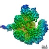
| Final reconstruction | Applied symmetry - Point group: C1 (asymmetric) / Resolution.type: BY AUTHOR / Resolution: 3.8 Å / Resolution method: FSC 0.143 CUT-OFF / Software - Name: RELION (ver. 1.4) / Number images used: 122796 |
| Details | Frames were aligned using motioncorr prior to processing. |

-Atomic model buiding 1
| Initial model | PDB ID: |
|---|---|
| Refinement | Protocol: RIGID BODY FIT / Target criteria: Cross-correlation coefficient |
| Output model | PDB-5mbv: |