 Movie
Movie Controller

[English] 日本語
 Yorodumi
- EMDB-3137: Multiple capsid-stabilizing protein-RNA and protein-protein inter... -
+ Open data

- Basic information
| Entry | Database: EMDB / ID: EMD-3137 | |||||||||
|---|---|---|---|---|---|---|---|---|---|---|
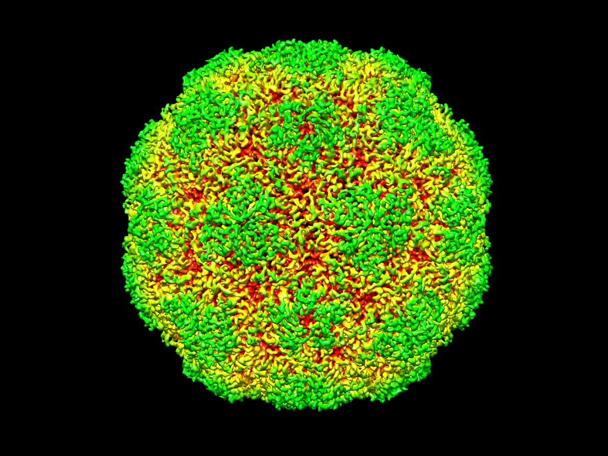
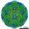
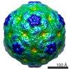
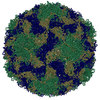
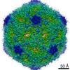
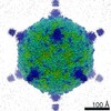
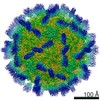
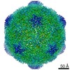
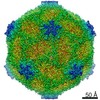
| Title | Multiple capsid-stabilizing protein-RNA and protein-protein interactions revealed in a high-resolution structure of an emerging picornavirus causing neonatal sepsis | |||||||||
 Map data Map data | Reconstruction of human parechovirus 3 | |||||||||
 Sample Sample |
| |||||||||
 Keywords Keywords |  picornavirus / parechovirus / human parechovirus 3 / HPeV3 / neonatal sepsis / cryoEM / image processing / single particle anaylsis picornavirus / parechovirus / human parechovirus 3 / HPeV3 / neonatal sepsis / cryoEM / image processing / single particle anaylsis | |||||||||
| Function / homology |  Function and homology information Function and homology informationRNA-protein covalent cross-linking / : / host cell cytoplasmic vesicle membrane / cytoplasmic vesicle membrane / : / viral capsid / protein complex oligomerization / monoatomic ion channel activity / RNA helicase activity / symbiont entry into host cell ...RNA-protein covalent cross-linking / : / host cell cytoplasmic vesicle membrane / cytoplasmic vesicle membrane / : / viral capsid / protein complex oligomerization / monoatomic ion channel activity / RNA helicase activity / symbiont entry into host cell / viral RNA genome replication / cysteine-type endopeptidase activity / RNA-dependent RNA polymerase activity / DNA-templated transcription / structural molecule activity / virion attachment to host cell / RNA binding / ATP bindingSimilarity search - Function | |||||||||
| Biological species |  Human parechovirus 3 Human parechovirus 3 | |||||||||
| Method | single particle reconstruction / cryo EM / negative staining / Resolution: 4.3 Å | |||||||||
 Authors Authors | Shakeel S / Westerhuis BM / Domanska A / Koning RI / Matadeen R / Koster AJ / Bakker AQ / Beaumont T / Wolthers KC / Butcher SJ | |||||||||
 Citation Citation | Journal: Nat Commun / Year: 2016 Title: Multiple capsid-stabilizing interactions revealed in a high-resolution structure of an emerging picornavirus causing neonatal sepsis. Authors: Shabih Shakeel / Brenda M Westerhuis / Ausra Domanska / Roman I Koning / Rishi Matadeen / Abraham J Koster / Arjen Q Bakker / Tim Beaumont / Katja C Wolthers / Sarah J Butcher /   Abstract: The poorly studied picornavirus, human parechovirus 3 (HPeV3) causes neonatal sepsis with no therapies available. Our 4.3-Å resolution structure of HPeV3 on its own and at 15 Å resolution in ...The poorly studied picornavirus, human parechovirus 3 (HPeV3) causes neonatal sepsis with no therapies available. Our 4.3-Å resolution structure of HPeV3 on its own and at 15 Å resolution in complex with human monoclonal antibody Fabs demonstrates the expected picornavirus capsid structure with three distinct features. First, 25% of the HPeV3 RNA genome in 60 sites is highly ordered as confirmed by asymmetric reconstruction, and interacts with conserved regions of the capsid proteins VP1 and VP3. Second, the VP0 N terminus stabilizes the capsid inner surface, in contrast to other picornaviruses where on expulsion as VP4, it forms an RNA translocation channel. Last, VP1's hydrophobic pocket, the binding site for the antipicornaviral drug, pleconaril, is blocked and thus inappropriate for antiviral development. Together, these results suggest a direction for development of neutralizing antibodies, antiviral drugs based on targeting the RNA-protein interactions and dissection of virus assembly on the basis of RNA nucleation. | |||||||||
| History |
|
- Structure visualization
| Movie |
Movie viewer |
|---|---|
| Structure viewer | EM map: SurfViewMolmilJmol/JSmol |
| Supplemental images |
- Downloads & links
-EMDB archive
| Map data | emd_3137.map.gz | 227.5 MB | EMDB map data format | |
|---|---|---|---|---|
| Header (meta data) | emd-3137-v30.xmlemd-3137.xml | 11.4 KB 11.4 KB | Display Display | EMDB header |
| FSC (resolution estimation) | emd_3137_fsc.xml | 13.7 KB | Display | FSC data file |
| Images | emd_3137.tif | 1.2 MB | ||
| Archive directory |  http://ftp.pdbj.org/pub/emdb/structures/EMD-3137ftp://ftp.pdbj.org/pub/emdb/structures/EMD-3137 http://ftp.pdbj.org/pub/emdb/structures/EMD-3137ftp://ftp.pdbj.org/pub/emdb/structures/EMD-3137 | HTTPS FTP |
-Related structure data
| Related structure data |  5apmMC  3138C  3322C M: atomic model generated by this map C: citing same article ( |
|---|---|
| Similar structure data | |
| EM raw data | EMPIAR-10033 (Title: Multiple capsid-stabilizing protein-RNA and protein-protein interactions revealed in a high-resolution structure of an emerging picornavirus causing neonatal sepsis Data size: 3.3 TB Data #1: Selected aligned movie frames, aligned-averaged micrographs and particle coordinates of human parechovirus 3 (HPeV3) virions [Aligned movie frames (.mrcs extension), aligned-averaged ...Data #1: Selected aligned movie frames, aligned-averaged micrographs and particle coordinates of human parechovirus 3 (HPeV3) virions [Aligned movie frames (.mrcs extension), aligned-averaged micrographs (.mrc extension) and particle coordinates (.box extension)] Data #2: Unaligned movie frames of human parechovirus 3 (HPeV3) virions [micrographs - multiframe]) |

-Links
| EMDB pages | EMDB (EBI/PDBe) / EMDataResource |
|---|---|
| Related items in Molecule of the Month |

-Map
| File | Download / File: emd_3137.map.gz / Format: CCP4 / Size: 238.4 MB / Type: IMAGE STORED AS FLOATING POINT NUMBER (4 BYTES) | ||||||||||||||||||||||||||||||||||||||||||||||||||||||||||||
|---|---|---|---|---|---|---|---|---|---|---|---|---|---|---|---|---|---|---|---|---|---|---|---|---|---|---|---|---|---|---|---|---|---|---|---|---|---|---|---|---|---|---|---|---|---|---|---|---|---|---|---|---|---|---|---|---|---|---|---|---|---|
| Annotation | Reconstruction of human parechovirus 3 | ||||||||||||||||||||||||||||||||||||||||||||||||||||||||||||
| Voxel size | X=Y=Z: 1.14 Å | ||||||||||||||||||||||||||||||||||||||||||||||||||||||||||||
| Density |
| ||||||||||||||||||||||||||||||||||||||||||||||||||||||||||||
| Symmetry | Space group: 1 | ||||||||||||||||||||||||||||||||||||||||||||||||||||||||||||
| Details | EMDB XML:
CCP4 map header:
| ||||||||||||||||||||||||||||||||||||||||||||||||||||||||||||
-Supplemental data
- Sample components
-Entire : Human parechovirus 3 capsid structure
| Entire | Name: Human parechovirus 3 capsid structure |
|---|---|
| Components |
|
-Supramolecule #1000: Human parechovirus 3 capsid structure
| Supramolecule | Name: Human parechovirus 3 capsid structure / type: sample / ID: 1000 Details: The purified human parechovirus 3 virions were monodisperse. They were formaldehyde fixed. Oligomeric state: icosahedrally-symmetric virus with 60 copies each of VP0, VP3 and VP1 Number unique components: 3 |
|---|---|
| Molecular weight | Experimental: 5 MDa / Theoretical: 5 MDa Method: theoretical weight determination from protein sequences, excluding viral genome. |
-Supramolecule #1: Human parechovirus 3
| Supramolecule | Name: Human parechovirus 3 / type: virus / ID: 1 / Name.synonym: HPeV3 / NCBI-ID: 195055 / Sci species name: Human parechovirus 3 / Virus type: VIRION / Virus isolate: OTHER / Virus enveloped: No / Virus empty: No / Syn species name: HPeV3 |
|---|---|
| Host (natural) | Organism:  Homo sapiens (human) / synonym: VERTEBRATES Homo sapiens (human) / synonym: VERTEBRATES |
| Host system | Organism:  Chlorocebus aethiops (grivet) / Recombinant cell: Vero Chlorocebus aethiops (grivet) / Recombinant cell: Vero |
| Molecular weight | Experimental: 5 MDa / Theoretical: 5 MDa |
| Virus shell | Shell ID: 1 / Diameter: 280 Å / T number (triangulation number): 1 |
-Experimental details
-Structure determination
| Method | negative staining, cryo EM |
|---|---|
 Processing Processing | single particle reconstruction |
| Aggregation state | particle |
-Sample preparation
| Concentration | 1 mg/mL |
|---|---|
| Buffer | pH: 7.5 / Details: 10 mM Tris-HCL, 150 mM NaCl, 1 mM MgCl2 |
| Staining | Type: NEGATIVE / Details: Unstained |
| Grid | Details: Quantifoil holey carbon on copper grids |
| Vitrification | Cryogen name: ETHANE / Chamber humidity: 92 % / Chamber temperature: 92 K / Instrument: LEICA EM GP / Method: Blot for 2 seconds on one side before plunging. |
- Electron microscopy
| Microscope | FEI TITAN KRIOS |
|---|---|
| Electron beam | Acceleration voltage: 300 kV / Electron source: FIELD EMISSION GUN |
| Electron optics | Illumination mode: FLOOD BEAM / Imaging mode: BRIGHT FIELDBright-field microscopy / Cs: 0.01 mm / Nominal defocus max: 2.34 µm / Nominal defocus min: 0.42 µm / Nominal magnification: 59000 |
| Sample stage | Specimen holder: Liquid nitrogen cooled. / Specimen holder model: FEI TITAN KRIOS AUTOGRID HOLDER |
| Temperature | Min: 80 K / Max: 95 K / Average: 87 K |
| Alignment procedure | Legacy - Astigmatism: Done as part of Cs corrector routine. |
| Details | Cs corrector was used during imaging. |
| Date | Jan 21, 2014 |
| Image recording | Category: CCD / Film or detector model: FEI FALCON II (4k x 4k) / Number real images: 6604 / Average electron dose: 36 e/Å2 Details: Total number of images collected were 6604. Total number of images used in the reconstruction were 1028. Every image is the average of seven aligned frames recorded by the direct electron detector. |
| Experimental equipment |  Model: Titan Krios / Image courtesy: FEI Company |
-Image processing
| CTF correction | Details: Each micrograph |
|---|---|
| Final two d classification | Number classes: 4 |
| Final reconstruction | Applied symmetry - Point group: I (icosahedral) / Algorithm: OTHER / Resolution.type: BY AUTHOR / Resolution: 4.3 Å / Resolution method: OTHER Software - Name: Ethan, CTFFIND3, EMAN1, EMAN2, AUTO3DEM, RELION, ResMap Number images used: 8889 |
| Details | Movie alignment done using motioncorr software. CTF estimated using CTFFIND3. Particles were selected using Ethan. Bad particles discarded by eye in Eman1 BOXER. Random model generated using Auto3dem. 2d and 3d class averaging done in Relion. Final reconstruction done in Relion. |
| FSC plot (resolution estimation) | 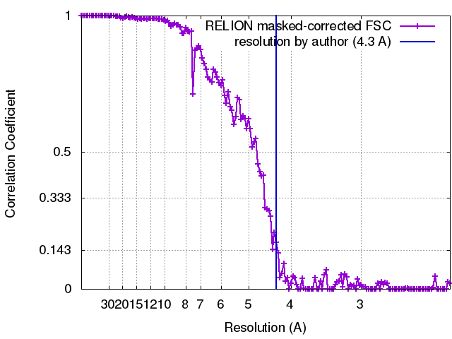 |
