Movie
Movie Controller


+ Open data

- Basic information
| Entry | Database: EMDB / ID: EMD-3019 | |||||||||
|---|---|---|---|---|---|---|---|---|---|---|
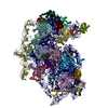
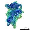
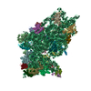
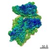
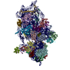
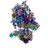
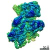
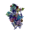
| Title | Structure of HCV IRES bound to the human ribosome | |||||||||
 Map data Map data | Reconstruction of hepatitis C virus IRES bound to human ribosome | |||||||||
 Sample Sample |
| |||||||||
 Keywords Keywords |  Human ribosome / IRES / Hepatitis C virus / translation initiation Human ribosome / IRES / Hepatitis C virus / translation initiation | |||||||||
| Function / homology |  Function and homology information Function and homology informationpositive regulation of cysteine-type endopeptidase activity involved in execution phase of apoptosis / negative regulation of endoplasmic reticulum unfolded protein response / oxidized pyrimidine DNA binding / response to TNF agonist / positive regulation of base-excision repair / protein tyrosine kinase inhibitor activity / positive regulation of intrinsic apoptotic signaling pathway in response to DNA damage / positive regulation of respiratory burst involved in inflammatory response / positive regulation of gastrulation / IRE1-RACK1-PP2A complex ...positive regulation of cysteine-type endopeptidase activity involved in execution phase of apoptosis / negative regulation of endoplasmic reticulum unfolded protein response / oxidized pyrimidine DNA binding / response to TNF agonist / positive regulation of base-excision repair / protein tyrosine kinase inhibitor activity / positive regulation of intrinsic apoptotic signaling pathway in response to DNA damage / positive regulation of respiratory burst involved in inflammatory response / positive regulation of gastrulation / IRE1-RACK1-PP2A complex / nucleolus organization / response to extracellular stimulus / positive regulation of endodeoxyribonuclease activity / positive regulation of Golgi to plasma membrane protein transport / exit from mitosis / TNFR1-mediated ceramide production / negative regulation of DNA repair / negative regulation of RNA splicing / laminin receptor activity / optic nerve development / oxidized purine DNA binding / negative regulation of intrinsic apoptotic signaling pathway in response to hydrogen peroxide / supercoiled DNA binding / neural crest cell differentiation / retinal ganglion cell axon guidance / NF-kappaB complex / rRNA modification in the nucleus and cytosol / negative regulation of phagocytosis / ubiquitin-like protein conjugating enzyme binding / regulation of establishment of cell polarity / positive regulation of ubiquitin-protein transferase activity / Formation of the ternary complex, and subsequently, the 43S complex / erythrocyte homeostasis / cytoplasmic side of rough endoplasmic reticulum membrane / positive regulation of signal transduction by p53 class mediator / ubiquitin ligase inhibitor activity / pigmentation / protein kinase A binding / negative regulation of ubiquitin protein ligase activity / Ribosomal scanning and start codon recognition / ion channel inhibitor activity / Translation initiation complex formation / phagocytic cup / positive regulation of mitochondrial depolarization / negative regulation of Wnt signaling pathway / positive regulation of T cell receptor signaling pathway / positive regulation of activated T cell proliferation / fibroblast growth factor binding / regulation of cell division / SARS-CoV-1 modulates host translation machinery / iron-sulfur cluster binding / Protein hydroxylation / TOR signaling / BH3 domain binding / mTORC1-mediated signalling / endonucleolytic cleavage to generate mature 3'-end of SSU-rRNA from (SSU-rRNA, 5.8S rRNA, LSU-rRNA) / Peptide chain elongation / Selenocysteine synthesis / monocyte chemotaxis / cysteine-type endopeptidase activator activity involved in apoptotic process / Formation of a pool of free 40S subunits / ribosomal small subunit export from nucleus / positive regulation of cyclic-nucleotide phosphodiesterase activity / Eukaryotic Translation Termination / Response of EIF2AK4 (GCN2) to amino acid deficiency / translation regulator activity / SRP-dependent cotranslational protein targeting to membrane / positive regulation of intrinsic apoptotic signaling pathway by p53 class mediator / Viral mRNA Translation / Nonsense Mediated Decay (NMD) independent of the Exon Junction Complex (EJC) / GTP hydrolysis and joining of the 60S ribosomal subunit / negative regulation of respiratory burst involved in inflammatory response / negative regulation of phosphatidylinositol 3-kinase/protein kinase B signal transduction / L13a-mediated translational silencing of Ceruloplasmin expression / Major pathway of rRNA processing in the nucleolus and cytosol / gastrulation / endonucleolytic cleavage in ITS1 to separate SSU-rRNA from 5.8S rRNA and LSU-rRNA from tricistronic rRNA transcript (SSU-rRNA, 5.8S rRNA, LSU-rRNA) / spindle assembly / regulation of translational fidelity / MDM2/MDM4 family protein binding / laminin binding / Protein methylation / Nonsense Mediated Decay (NMD) enhanced by the Exon Junction Complex (EJC) / rough endoplasmic reticulum / Amplification of signal from unattached kinetochores via a MAD2 inhibitory signal / Nuclear events stimulated by ALK signaling in cancer / negative regulation of smoothened signaling pathway / rescue of stalled ribosome / signaling adaptor activity / positive regulation of cell cycle / negative regulation of peptidyl-serine phosphorylation / stress granule assembly / translation initiation factor binding / maturation of SSU-rRNA / positive regulation of intrinsic apoptotic signaling pathway / Mitotic Prometaphase / maturation of SSU-rRNA from tricistronic rRNA transcript (SSU-rRNA, 5.8S rRNA, LSU-rRNA) / class I DNA-(apurinic or apyrimidinic site) endonuclease activity / EML4 and NUDC in mitotic spindle formation / positive regulation of apoptotic signaling pathwaySimilarity search - Function | |||||||||
| Biological species |  Homo sapiens (human) / Homo sapiens (human) /  Hepatitis C virus Hepatitis C virus | |||||||||
| Method | single particle reconstruction / cryo EM / Resolution: 3.9 Å | |||||||||
 Authors Authors | Quade N / Leibundgut M / Boehringer D / van den Heuvel J / Ban N | |||||||||
 Citation Citation | Journal: Nat Commun / Year: 2015 Title: Cryo-EM structure of Hepatitis C virus IRES bound to the human ribosome at 3.9-Å resolution. Authors: Nick Quade / Daniel Boehringer / Marc Leibundgut / Joop van den Heuvel / Nenad Ban /   Abstract: Hepatitis C virus (HCV), a widespread human pathogen, is dependent on a highly structured 5'-untranslated region of its mRNA, referred to as internal ribosome entry site (IRES), for the translation ...Hepatitis C virus (HCV), a widespread human pathogen, is dependent on a highly structured 5'-untranslated region of its mRNA, referred to as internal ribosome entry site (IRES), for the translation of all of its proteins. The HCV IRES initiates translation by directly binding to the small ribosomal subunit (40S), circumventing the need for many eukaryotic translation initiation factors required for mRNA scanning. Here we present the cryo-EM structure of the human 40S ribosomal subunit in complex with the HCV IRES at 3.9 Å resolution, determined by focused refinement of an 80S ribosome-HCV IRES complex. The structure reveals the molecular details of the interactions between the IRES and the 40S, showing that expansion segment 7 (ES7) of the 18S rRNA acts as a central anchor point for the HCV IRES. The structural data rationalizes previous biochemical and genetic evidence regarding the initiation mechanism of the HCV and other related IRESs. | |||||||||
| History |
|
- Structure visualization
| Movie |
Movie viewer |
|---|---|
| Structure viewer | EM map: SurfViewMolmilJmol/JSmol |
| Supplemental images |

- Downloads & links
-EMDB archive
| Map data | emd_3019.map.gz | 7.1 MB | EMDB map data format | |
|---|---|---|---|---|
| Header (meta data) | emd-3019-v30.xmlemd-3019.xml | 14.7 KB 14.7 KB | Display Display | EMDB header |
| Images | 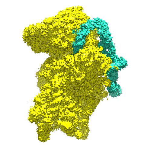 emd_3019.png emd_3019.png | 201.9 KB | ||
| Archive directory |  http://ftp.pdbj.org/pub/emdb/structures/EMD-3019ftp://ftp.pdbj.org/pub/emdb/structures/EMD-3019 http://ftp.pdbj.org/pub/emdb/structures/EMD-3019ftp://ftp.pdbj.org/pub/emdb/structures/EMD-3019 | HTTPS FTP |
-Related structure data
| Related structure data | 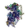 5a2qMC M: atomic model generated by this map C: citing same article ( |
|---|---|
| Similar structure data |


-Links
| EMDB pages | EMDB (EBI/PDBe) / EMDataResource |
|---|---|
| Related items in Molecule of the Month |


-Map
| File | Download / File: emd_3019.map.gz / Format: CCP4 / Size: 37.5 MB / Type: IMAGE STORED AS FLOATING POINT NUMBER (4 BYTES) | ||||||||||||||||||||||||||||||||||||||||||||||||||||||||||||||||||||
|---|---|---|---|---|---|---|---|---|---|---|---|---|---|---|---|---|---|---|---|---|---|---|---|---|---|---|---|---|---|---|---|---|---|---|---|---|---|---|---|---|---|---|---|---|---|---|---|---|---|---|---|---|---|---|---|---|---|---|---|---|---|---|---|---|---|---|---|---|---|
| Annotation | Reconstruction of hepatitis C virus IRES bound to human ribosome | ||||||||||||||||||||||||||||||||||||||||||||||||||||||||||||||||||||
| Voxel size | X=Y=Z: 1.39 Å | ||||||||||||||||||||||||||||||||||||||||||||||||||||||||||||||||||||
| Density |
| ||||||||||||||||||||||||||||||||||||||||||||||||||||||||||||||||||||
| Symmetry | Space group: 1 | ||||||||||||||||||||||||||||||||||||||||||||||||||||||||||||||||||||
| Details | EMDB XML:
CCP4 map header:
| ||||||||||||||||||||||||||||||||||||||||||||||||||||||||||||||||||||
-Supplemental data
- Sample components
-Entire : Hepatitis C virus IRES bound to human ribosome
| Entire | Name: Hepatitis C virus IRES bound to human ribosome |
|---|---|
| Components |
|
-Supramolecule #1000: Hepatitis C virus IRES bound to human ribosome
| Supramolecule | Name: Hepatitis C virus IRES bound to human ribosome / type: sample / ID: 1000 / Oligomeric state: IRES bound to ribosome / Number unique components: 2 |
|---|---|
| Molecular weight | Theoretical: 1.45 MDa |
-Supramolecule #1: 40S ribosome
| Supramolecule | Name: 40S ribosome / type: complex / ID: 1 / Recombinant expression: No / Ribosome-details: ribosome-eukaryote: SSU 40S, SSU RNA 18S |
|---|---|
| Ref GO | divclassse qspanoncli ckpopupspa nclassgree n(this)spandata popltspanc lassquotlo adingbarqu otgtltimgs rcquotimgl oadinggifq uotdecodin gquotasync quotgtltsp angtdataur lajaxphp?m odetaxoamp ... divclassse qspanoncli ckpopupspa nclassgree n(this)spandata popltspanc lassquotlo adingbarqu otgtltimgs rcquotimgl oadinggifq uotdecodin gquotasync quotgtltsp angtdataur lajaxphp?m odetaxoamp kGO3A00058 40ampajax1 classpoptr giGO000584 0ispandiv |
| Source (natural) | Organism: Homo sapiens (human) / synonym: Human / Cell: HEK293-6E / Location in cell: Cytosole |
| Molecular weight | Theoretical: 1.35 MDa |
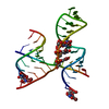
-Macromolecule #1: Hepatitis-C virus IRES
| Macromolecule | Name: Hepatitis-C virus IRES / type: rna / ID: 1 / Name.synonym: HCV IRES / Classification: OTHER / Structure: DOUBLE HELIX / Synthetic?: No |
|---|---|
| Source (natural) | Organism: Hepatitis C virus |
| Molecular weight | Theoretical: 100 KDa |
| Sequence | String: CTCCCCTGTG AGGAACTACT GTCTTCACGC AGAAAGCGTC TAGCCATGGC GTTAGTATGA GTGTCGTGCA GCCTCCAGGA CCCCCCCTCC CGGGAGAGCC ATAGTGGTCT GCGGAACCGG TGAGTACACC GGAATTGCCA GGACGACCGG GTCCTTTCTT GGATAAACCC ...String: CTCCCCTGTG AGGAACTACT GTCTTCACGC AGAAAGCGTC TAGCCATGGC GTTAGTATGA GTGTCGTGCA GCCTCCAGGA CCCCCCCTCC CGGGAGAGCC ATAGTGGTCT GCGGAACCGG TGAGTACACC GGAATTGCCA GGACGACCGG GTCCTTTCTT GGATAAACCC GCTCAATGCC TGGAGATTTG GGCGTGCCCC CGCAAGACTG CTAGCCGAGT AGTGTTGGGT CGCGAAAGGC CTTGTGGTAC TGCCTGATAG GGTGCTTGCG AGTGCCCCGG GAGGTCTCGT AGACCGTGCA CCATGAGCAC AAATC |
 Processing
 Electron microscopy