 Movie
Movie Controller

[English] 日本語
 Yorodumi
- EMDB-1929: Modular architecture of eukaryotic RNase P and RNase MRP revealed... -
+ Open data

- Basic information
| Entry | Database: EMDB / ID: EMD-1929 | |||||||||
|---|---|---|---|---|---|---|---|---|---|---|
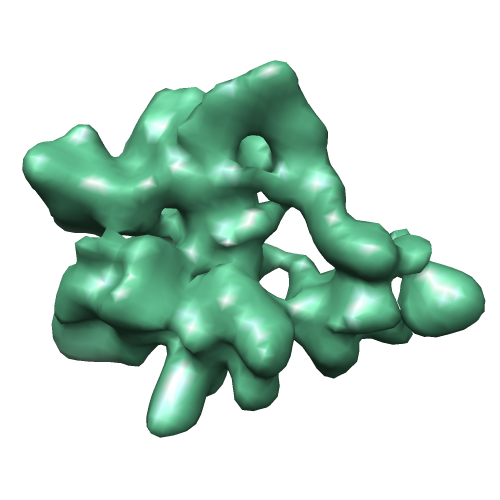
| Title | Modular architecture of eukaryotic RNase P and RNase MRP revealed by electron microscopy | |||||||||
 Map data Map data | RNase P Ribonuclease P Ribonuclease P | |||||||||
 Sample Sample |
| |||||||||
 Keywords Keywords | RNase P / RNA-processing / ribozyme | |||||||||
| Biological species |  Saccharomyces cerevisiae (brewer's yeast) Saccharomyces cerevisiae (brewer's yeast) | |||||||||
| Method | single particle reconstruction / cryo EM / negative staining / Resolution: 17.0 Å | |||||||||
 Authors Authors | Hipp K / Galani K / Batisse C / Prinz S / Bottcher B | |||||||||
 Citation Citation | Journal: Nucleic Acids Res / Year: 2012 Title: Modular architecture of eukaryotic RNase P and RNase MRP revealed by electron microscopy. Authors: Katharina Hipp / Kyriaki Galani / Claire Batisse / Simone Prinz / Bettina Böttcher /  Abstract: Ribonuclease P (RNase P) and RNase MRP are closely related ribonucleoprotein enzymes, which process RNA substrates including tRNA precursors for RNase P and 5.8 S rRNA precursors, as well as some ...Ribonuclease P (RNase P) and RNase MRP are closely related ribonucleoprotein enzymes, which process RNA substrates including tRNA precursors for RNase P and 5.8 S rRNA precursors, as well as some mRNAs, for RNase MRP. The structures of RNase P and RNase MRP have not yet been solved, so it is unclear how the proteins contribute to the structure of the complexes and how substrate specificity is determined. Using electron microscopy and image processing we show that eukaryotic RNase P and RNase MRP have a modular architecture, where proteins stabilize the RNA fold and contribute to cavities, channels and chambers between the modules. Such features are located at strategic positions for substrate recognition by shape and coordination of the cleaved-off sequence. These are also the sites of greatest difference between RNase P and RNase MRP, highlighting the importance of the adaptation of this region to the different substrates. | |||||||||
| History |
|
- Structure visualization
| Movie |
 Movie viewer Movie viewer |
|---|---|
| Structure viewer | EM map: SurfViewMolmilJmol/JSmol |
| Supplemental images |
- Downloads & links
-EMDB archive
| Map data | emd_1929.map.gz | 3.3 MB | EMDB map data format | |
|---|---|---|---|---|
| Header (meta data) | emd-1929-v30.xmlemd-1929.xml | 9.1 KB 9.1 KB | Display Display | EMDB header |
| Images |  1929.png 1929.png | 109.9 KB | ||
| Archive directory |  http://ftp.pdbj.org/pub/emdb/structures/EMD-1929ftp://ftp.pdbj.org/pub/emdb/structures/EMD-1929 http://ftp.pdbj.org/pub/emdb/structures/EMD-1929ftp://ftp.pdbj.org/pub/emdb/structures/EMD-1929 | HTTPS FTP |
-Related structure data


-Links
| EMDB pages | EMDB (EBI/PDBe) / EMDataResource |
|---|
-Map
| File | Download / File: emd_1929.map.gz / Format: CCP4 / Size: 6.4 MB / Type: IMAGE STORED AS FLOATING POINT NUMBER (4 BYTES) | ||||||||||||||||||||||||||||||||||||||||||||||||||||||||||||||||||||
|---|---|---|---|---|---|---|---|---|---|---|---|---|---|---|---|---|---|---|---|---|---|---|---|---|---|---|---|---|---|---|---|---|---|---|---|---|---|---|---|---|---|---|---|---|---|---|---|---|---|---|---|---|---|---|---|---|---|---|---|---|---|---|---|---|---|---|---|---|---|
| Annotation | RNase P | ||||||||||||||||||||||||||||||||||||||||||||||||||||||||||||||||||||
| Voxel size | X=Y=Z: 2.2 Å | ||||||||||||||||||||||||||||||||||||||||||||||||||||||||||||||||||||
| Density |
| ||||||||||||||||||||||||||||||||||||||||||||||||||||||||||||||||||||
| Symmetry | Space group: 1 | ||||||||||||||||||||||||||||||||||||||||||||||||||||||||||||||||||||
| Details | EMDB XML:
CCP4 map header:
| ||||||||||||||||||||||||||||||||||||||||||||||||||||||||||||||||||||
-Supplemental data
- Sample components
-Entire : RNase P
| Entire | Name: RNase PRibonuclease P |
|---|---|
| Components |
|
-Supramolecule #1000: RNase P
| Supramolecule | Name: RNase P / type: sample / ID: 1000 / Number unique components: 1 |
|---|---|
| Molecular weight | Theoretical: 410 KDa |
-Macromolecule #1: Ribonuclease P
| Macromolecule | Name: Ribonuclease P / type: protein_or_peptide / ID: 1 / Name.synonym: RNase P / Oligomeric state: monomer / Recombinant expression: No |
|---|---|
| Source (natural) | Organism: Saccharomyces cerevisiae (brewer's yeast) / synonym: Baker's yeast / Organelle: Nucleus |
| Molecular weight | Theoretical: 410 KDa |
-Experimental details
-Structure determination
| Method | negative staining, cryo EM |
|---|---|
 Processing Processing | single particle reconstruction |
| Aggregation state | particle |
-Sample preparation
| Buffer | pH: 7.5 Details: 50 mM Tris HCl pH 7.5, 100 mM NaCl, 10 mM MgCl2, 1 mM DTT |
|---|---|
| Staining | Type: NEGATIVE Details: sandwich, cryo negative stain with 1% uranyl acetate |
| Grid | Details: 400 mesh copper grid |
| Vitrification | Cryogen name: NITROGEN / Chamber temperature: 95 K / Instrument: OTHER Method: samples were stained and frozen after partly drying, by dipping grids into liquid nitrogen |
- Electron microscopy
| Microscope | FEI/PHILIPS CM200FEG |
|---|---|
| Electron beam | Acceleration voltage: 200 kV / Electron source: FIELD EMISSION GUN |
| Electron optics | Illumination mode: FLOOD BEAM / Imaging mode: BRIGHT FIELDBright-field microscopy / Cs: 2 mm / Nominal defocus max: 1.1 µm / Nominal defocus min: 0.68 µm / Nominal magnification: 66000 |
| Sample stage | Specimen holder: side entry, liquid nitrogen cooled / Specimen holder model: GATAN LIQUID NITROGEN |
| Temperature | Min: 95 K / Max: 95 K / Average: 95 K |
| Alignment procedure | Legacy - Astigmatism: astigmatism was corrected at 200000 times magnification on carbon film |
| Image recording | Category: CCD / Film or detector model: GENERIC TVIPS (2k x 2k) / Number real images: 1861 / Average electron dose: 20 e/Å2 / Bits/pixel: 12 |
| Tilt angle min | 0 |
| Tilt angle max | 0 |
-Image processing
| CTF correction | Details: each particle, phase flipping |
|---|---|
| Final reconstruction | Applied symmetry - Point group: C1 (asymmetric) / Algorithm: OTHER / Resolution.type: BY AUTHOR / Resolution: 17.0 Å / Resolution method: FSC 0.5 CUT-OFF / Software - Name: spider, imagic, xmipp Details: final maps were calculated from 20 defocus groups, number of particles in certain orientations were limited to counterbalance preferred orientations Number images used: 24373 |
| Details | particle were selected automatically, particle orientations were determined by projection matching |
