 Movie
Movie Controller


+ Open data

- Basic information
| Entry | Database: EMDB / ID: EMD-9566 | |||||||||||||||||||||
|---|---|---|---|---|---|---|---|---|---|---|---|---|---|---|---|---|---|---|---|---|---|---|
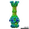
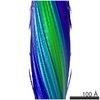
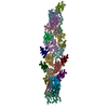
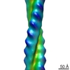
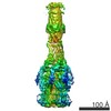
| Title | Human RAD51 presynaptic complex | |||||||||||||||||||||
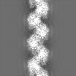
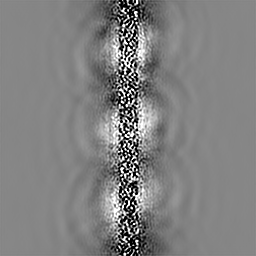
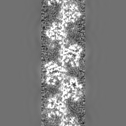
 Map data Map data | Human RAD51-ssDNA formed presynaptic complex | |||||||||||||||||||||
 Sample Sample |
| |||||||||||||||||||||
| Function / homology |  Function and homology information Function and homology informationpresynaptic intermediate filament cytoskeleton / mitotic recombination-dependent replication fork processing / chromosome organization involved in meiotic cell cycle / cellular response to camptothecin / DNA recombinase assembly / telomere maintenance via telomere lengthening / positive regulation of DNA ligation /  mitotic recombination / double-strand break repair involved in meiotic recombination / nuclear ubiquitin ligase complex ...presynaptic intermediate filament cytoskeleton / mitotic recombination-dependent replication fork processing / chromosome organization involved in meiotic cell cycle / cellular response to camptothecin / DNA recombinase assembly / telomere maintenance via telomere lengthening / positive regulation of DNA ligation / mitotic recombination / double-strand break repair involved in meiotic recombination / nuclear ubiquitin ligase complex / DNA strand invasion / replication-born double-strand break repair via sister chromatid exchange / cellular response to hydroxyurea / DNA strand exchange activity / lateral element / telomere maintenance via recombination / regulation of DNA damage checkpoint / Impaired BRCA2 binding to PALB2 / single-stranded DNA helicase activity / reciprocal meiotic recombination / Defective homologous recombination repair (HRR) due to BRCA1 loss of function / Defective HDR through Homologous Recombination Repair (HRR) due to PALB2 loss of BRCA1 binding function / Defective HDR through Homologous Recombination Repair (HRR) due to PALB2 loss of BRCA2/RAD51/RAD51C binding function / Homologous DNA Pairing and Strand Exchange / Resolution of D-loop Structures through Synthesis-Dependent Strand Annealing (SDSA) / Resolution of D-loop Structures through Holliday Junction Intermediates / HDR through Single Strand Annealing (SSA) / Impaired BRCA2 binding to RAD51 / ATP-dependent DNA damage sensor activity / regulation of double-strand break repair via homologous recombination / nuclear chromosome / replication fork processing / DNA unwinding involved in DNA replication / Transcriptional Regulation by E2F6 / Presynaptic phase of homologous DNA pairing and strand exchange / ATP-dependent activity, acting on DNA / interstrand cross-link repair / DNA polymerase binding / condensed chromosome / meiotic cell cycle / condensed nuclear chromosome / male germ cell nucleus / cellular response to ionizing radiation / double-strand break repair via homologous recombination / regulation of protein phosphorylation / HDR through Homologous Recombination (HRR) / PML body / Meiotic recombination / single-stranded DNA binding / site of double-strand break / double-stranded DNA binding / DNA recombination / chromosome, telomeric region / mitochondrial matrix / DNA repair / centrosome / DNA damage response / chromatin binding / chromatin / nucleolus / perinuclear region of cytoplasm / enzyme binding / ATP hydrolysis activity / protein-containing complex / mitochondrion / nucleoplasm / ATP binding / identical protein binding / nucleus / cytosol / cytoplasm mitotic recombination / double-strand break repair involved in meiotic recombination / nuclear ubiquitin ligase complex ...presynaptic intermediate filament cytoskeleton / mitotic recombination-dependent replication fork processing / chromosome organization involved in meiotic cell cycle / cellular response to camptothecin / DNA recombinase assembly / telomere maintenance via telomere lengthening / positive regulation of DNA ligation / mitotic recombination / double-strand break repair involved in meiotic recombination / nuclear ubiquitin ligase complex / DNA strand invasion / replication-born double-strand break repair via sister chromatid exchange / cellular response to hydroxyurea / DNA strand exchange activity / lateral element / telomere maintenance via recombination / regulation of DNA damage checkpoint / Impaired BRCA2 binding to PALB2 / single-stranded DNA helicase activity / reciprocal meiotic recombination / Defective homologous recombination repair (HRR) due to BRCA1 loss of function / Defective HDR through Homologous Recombination Repair (HRR) due to PALB2 loss of BRCA1 binding function / Defective HDR through Homologous Recombination Repair (HRR) due to PALB2 loss of BRCA2/RAD51/RAD51C binding function / Homologous DNA Pairing and Strand Exchange / Resolution of D-loop Structures through Synthesis-Dependent Strand Annealing (SDSA) / Resolution of D-loop Structures through Holliday Junction Intermediates / HDR through Single Strand Annealing (SSA) / Impaired BRCA2 binding to RAD51 / ATP-dependent DNA damage sensor activity / regulation of double-strand break repair via homologous recombination / nuclear chromosome / replication fork processing / DNA unwinding involved in DNA replication / Transcriptional Regulation by E2F6 / Presynaptic phase of homologous DNA pairing and strand exchange / ATP-dependent activity, acting on DNA / interstrand cross-link repair / DNA polymerase binding / condensed chromosome / meiotic cell cycle / condensed nuclear chromosome / male germ cell nucleus / cellular response to ionizing radiation / double-strand break repair via homologous recombination / regulation of protein phosphorylation / HDR through Homologous Recombination (HRR) / PML body / Meiotic recombination / single-stranded DNA binding / site of double-strand break / double-stranded DNA binding / DNA recombination / chromosome, telomeric region / mitochondrial matrix / DNA repair / centrosome / DNA damage response / chromatin binding / chromatin / nucleolus / perinuclear region of cytoplasm / enzyme binding / ATP hydrolysis activity / protein-containing complex / mitochondrion / nucleoplasm / ATP binding / identical protein binding / nucleus / cytosol / cytoplasmSimilarity search - Function | |||||||||||||||||||||
| Biological species |  human (human) / Homo sapiens (human) human (human) / Homo sapiens (human) | |||||||||||||||||||||
| Method | helical reconstruction / cryo EM / Resolution: 4.4 Å | |||||||||||||||||||||
 Authors Authors | Xu J / Zhao L / Xu Y / Zhao W / Sung P / Wang HW | |||||||||||||||||||||
| Funding support |  China, China,  United States, 6 items United States, 6 items
| |||||||||||||||||||||
 Citation Citation | Journal: Nat Struct Mol Biol / Year: 2017 Title: Cryo-EM structures of human RAD51 recombinase filaments during catalysis of DNA-strand exchange. Authors: Jingfei Xu / Lingyun Zhao / Yuanyuan Xu / Weixing Zhao / Patrick Sung / Hong-Wei Wang / Abstract: The central step in eukaryotic homologous recombination (HR) is ATP-dependent DNA-strand exchange mediated by the Rad51 recombinase. In this process, Rad51 assembles on single-stranded DNA (ssDNA) ...The central step in eukaryotic homologous recombination (HR) is ATP-dependent DNA-strand exchange mediated by the Rad51 recombinase. In this process, Rad51 assembles on single-stranded DNA (ssDNA) and generates a helical filament that is able to search for and invade homologous double-stranded DNA (dsDNA), thus leading to strand separation and formation of new base pairs between the initiating ssDNA and the complementary strand within the duplex. Here, we used cryo-EM to solve the structures of human RAD51 in complex with DNA molecules, in presynaptic and postsynaptic states, at near-atomic resolution. Our structures reveal both conserved and distinct structural features of the human RAD51-DNA complexes compared with their prokaryotic counterpart. Notably, we also captured the structure of an arrested synaptic complex. Our results provide new insight into the molecular mechanisms of the DNA homology search and strand-exchange processes. | |||||||||||||||||||||
| History |
|
- Structure visualization
| Movie |
Movie viewer |
|---|---|
| Structure viewer | EM map: SurfViewMolmilJmol/JSmol |
| Supplemental images |

- Downloads & links
-EMDB archive
| Map data | emd_9566.map.gz | 56.9 MB | EMDB map data format | |
|---|---|---|---|---|
| Header (meta data) | emd-9566-v30.xmlemd-9566.xml | 18.5 KB 18.5 KB | Display Display | EMDB header |
| Images | 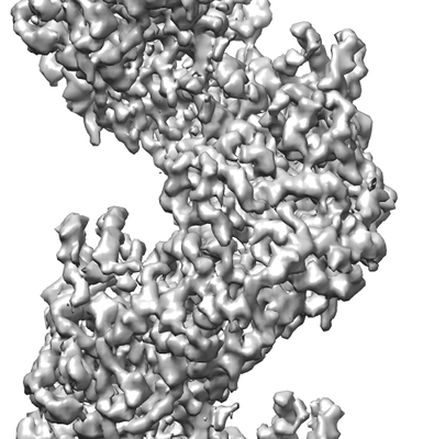 emd_9566.png emd_9566.png | 96.1 KB | ||
| Archive directory |  http://ftp.pdbj.org/pub/emdb/structures/EMD-9566ftp://ftp.pdbj.org/pub/emdb/structures/EMD-9566 http://ftp.pdbj.org/pub/emdb/structures/EMD-9566ftp://ftp.pdbj.org/pub/emdb/structures/EMD-9566 | HTTPS FTP |
-Related structure data
| Related structure data |  5h1bMC  9567C 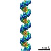 9568C  5h1cC C: citing same article ( M: atomic model generated by this map |
|---|---|
| Similar structure data |


-Links
| EMDB pages | EMDB (EBI/PDBe) / EMDataResource |
|---|---|
| Related items in Molecule of the Month |


-Map
| File | Download / File: emd_9566.map.gz / Format: CCP4 / Size: 64 MB / Type: IMAGE STORED AS FLOATING POINT NUMBER (4 BYTES) | ||||||||||||||||||||||||||||||||||||||||||||||||||||||||||||||||||||
|---|---|---|---|---|---|---|---|---|---|---|---|---|---|---|---|---|---|---|---|---|---|---|---|---|---|---|---|---|---|---|---|---|---|---|---|---|---|---|---|---|---|---|---|---|---|---|---|---|---|---|---|---|---|---|---|---|---|---|---|---|---|---|---|---|---|---|---|---|---|
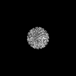
| Annotation | Human RAD51-ssDNA formed presynaptic complex | ||||||||||||||||||||||||||||||||||||||||||||||||||||||||||||||||||||
| Projections & slices | Image control
Images are generated by Spider. | ||||||||||||||||||||||||||||||||||||||||||||||||||||||||||||||||||||
| Voxel size | X=Y=Z: 1.306 Å | ||||||||||||||||||||||||||||||||||||||||||||||||||||||||||||||||||||
| Density |
| ||||||||||||||||||||||||||||||||||||||||||||||||||||||||||||||||||||
| Symmetry | Space group: 1 | ||||||||||||||||||||||||||||||||||||||||||||||||||||||||||||||||||||
| Details | EMDB XML:
CCP4 map header:
| ||||||||||||||||||||||||||||||||||||||||||||||||||||||||||||||||||||
 Z (Sec.)
 Y (Row.)
 X (Col.)


-Supplemental data
- Sample components
-Entire : Human RAD51 and ssDNA formed presynaptic complex
| Entire | Name: Human RAD51 and ssDNA formed presynaptic complex |
|---|---|
| Components |
|
-Supramolecule #1: Human RAD51 and ssDNA formed presynaptic complex
| Supramolecule | Name: Human RAD51 and ssDNA formed presynaptic complex / type: complex / ID: 1 / Parent: 0 / Macromolecule list: #1-#2 |
|---|---|
| Source (natural) | Organism: human (human) |
| Molecular weight | Theoretical: 23.56 kDa/nm |
-Supramolecule #2: Human RAD51
| Supramolecule | Name: Human RAD51 / type: complex / ID: 2 / Parent: 1 / Macromolecule list: #1 |
|---|
-Supramolecule #3: ssDNA
| Supramolecule | Name: ssDNA / type: complex / ID: 3 / Parent: 1 / Macromolecule list: #2 |
|---|
-Macromolecule #1: DNA repair protein RAD51 homolog 1
| Macromolecule | Name: DNA repair protein RAD51 homolog 1 / type: protein_or_peptide / ID: 1 / Number of copies: 3 / Enantiomer: LEVO |
|---|---|
| Source (natural) | Organism: Homo sapiens (human) |
| Molecular weight | Theoretical: 37.008074 KDa |
| Recombinant expression | Organism:  Escherichia coli 'BL21-Gold(DE3)pLysS AG' (bacteria) Escherichia coli 'BL21-Gold(DE3)pLysS AG' (bacteria) |
| Sequence | String: MAMQMQLEAN ADTSVEEESF GPQPISRLEQ CGINANDVKK LEEAGFHTVE AVAYAPKKEL INIKGISEAK ADKILAEAAK LVPMGFTTA TEFHQRRSEI IQITTGSKEL DKLLQGGIET GSITEMFGEF RTGKTQICHT LAVTCQLPID RGGGEGKAMY I DTEGTFRP ...String: MAMQMQLEAN ADTSVEEESF GPQPISRLEQ CGINANDVKK LEEAGFHTVE AVAYAPKKEL INIKGISEAK ADKILAEAAK LVPMGFTTA TEFHQRRSEI IQITTGSKEL DKLLQGGIET GSITEMFGEF RTGKTQICHT LAVTCQLPID RGGGEGKAMY I DTEGTFRP ERLLAVAERY GLSGSDVLDN VAYARAFNTD HQTQLLYQAS AMMVESRYAL LIVDSATALY RTDYSGRGEL SA RQMHLAR FLRMLLRLAD EFGVAVVITN QVVAQVDGAA MFAADPKKPI GGNIIAHAST TRLYLRKGRG ETRICQIYDS PCL PEAEAM FAINADGVGD AKD |
-Macromolecule #2: DNA (5'-D(P*TP*TP*TP*TP*TP*TP*TP*TP*T)-3')
| Macromolecule | Name: DNA (5'-D(P*TP*TP*TP*TP*TP*TP*TP*TP*T)-3') / type: dna / ID: 2 / Number of copies: 1 / Classification: DNA |
|---|---|
| Source (natural) | Organism: Homo sapiens (human) |
| Molecular weight | Theoretical: 2.692778 KDa |
| Sequence | String: (DT)(DT)(DT)(DT)(DT)(DT)(DT)(DT)(DT) |
-Macromolecule #3: PHOSPHOAMINOPHOSPHONIC ACID-ADENYLATE ESTER
| Macromolecule | Name: PHOSPHOAMINOPHOSPHONIC ACID-ADENYLATE ESTER / type: ligand / ID: 3 / Number of copies: 3 / Formula: ANP |
|---|---|
| Molecular weight | Theoretical: 506.196 Da |
| Chemical component information |  ChemComp-ANP: |
-Macromolecule #4: MAGNESIUM ION
| Macromolecule | Name: MAGNESIUM ION / type: ligand / ID: 4 / Number of copies: 3 / Formula: MG |
|---|---|
| Molecular weight | Theoretical: 24.305 Da |
-Experimental details
-Structure determination
| Method | cryo EM |
|---|---|
 Processing Processing | helical reconstruction |
| Aggregation state | filament |
-Sample preparation
| Concentration | 0.075 mg/mL |
|---|---|
| Buffer | pH: 7.5 Details: 25mM Tris-HCl, pH 7.5, 50mM KCl, 1mM dithiothreitol, 1mM AMP-PNP and 2mM MgCl2 |
| Grid | Model: Quantifoil R1.2/1.3 / Material: COPPER / Mesh: 300 / Support film - Material: CARBON / Support film - topology: HOLEY / Support film - Film thickness: 20.0 nm / Pretreatment - Type: PLASMA CLEANING / Pretreatment - Atmosphere: AIR / Pretreatment - Pressure: 101.325 kPa |
| Vitrification | Cryogen name: ETHANE / Chamber humidity: 100 % / Chamber temperature: 289 K / Instrument: FEI VITROBOT MARK IV |
- Electron microscopy
| Microscope | FEI TITAN KRIOS |
|---|---|
| Electron beam | Acceleration voltage: 300 kV / Electron source: FIELD EMISSION GUN |
| Electron optics | C2 aperture diameter: 70.0 µm / Calibrated defocus max: 2.5 µm / Calibrated defocus min: 1.5 µm / Calibrated magnification: 22500 / Illumination mode: FLOOD BEAM / Imaging mode: BRIGHT FIELDBright-field microscopy / Cs: 2.7 mm / Nominal defocus max: 2.5 µm / Nominal defocus min: 1.5 µm / Nominal magnification: 22500 |
| Sample stage | Specimen holder model: FEI TITAN KRIOS AUTOGRID HOLDER / Cooling holder cryogen: NITROGEN |
| Temperature | Min: 80.0 K / Max: 80.0 K |
| Image recording | Film or detector model: GATAN K2 SUMMIT (4k x 4k) / Detector mode: SUPER-RESOLUTION / Digitization - Dimensions - Width: 7676 pixel / Digitization - Dimensions - Height: 7420 pixel / Digitization - Sampling interval: 5.0 µm / Digitization - Frames/image: 3-14 / Number grids imaged: 2 / Number real images: 40404 / Average exposure time: 8.0 sec. / Average electron dose: 50.0 e/Å2 |
| Experimental equipment |  Model: Titan Krios / Image courtesy: FEI Company |
-Image processing
| Segment selection | Number selected: 540 | ||||||
|---|---|---|---|---|---|---|---|
| CTF correction | Software - Name: CTFFIND3 | ||||||
| Startup model | Type of model: OTHER Details: Randomly assigned euler angle for each segment and using back projection to generate initial model. | ||||||
| Final angle assignment | Type: NOT APPLICABLE Software:
| ||||||
| Final reconstruction | Number classes used: 82 Applied symmetry - Helical parameters - Δz: 15.88 Å Applied symmetry - Helical parameters - Δ&Phi: 56.77 ° Applied symmetry - Helical parameters - Axial symmetry: C1 (asymmetric) Algorithm: BACK PROJECTION / Resolution.type: BY AUTHOR / Resolution: 4.4 Å / Resolution method: FSC 0.143 CUT-OFF Software:
Number images used: 33838 |

