 Movie
Movie Controller


+ Open data

- Basic information
| Entry | Database: EMDB / ID: EMD-8486 | |||||||||
|---|---|---|---|---|---|---|---|---|---|---|
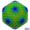
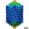
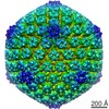
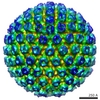
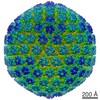
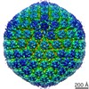
| Title | Bacillus phage PBS1 | |||||||||
 Map data Map data | Bacillus phage PBS1 | |||||||||
 Sample Sample |
| |||||||||
| Biological species |  Bacillus phage PBS1 (virus) Bacillus phage PBS1 (virus) | |||||||||
| Method |  single particle reconstruction / cryo EM / Resolution: 10.0 Å single particle reconstruction / cryo EM / Resolution: 10.0 Å | |||||||||
 Authors Authors | Hua J / Huet A / Lopez CA / Toropova K / Pope WH / Duda RL / Hendrix RW / Conway JF | |||||||||
 Citation Citation | Journal: mBio / Year: 2017 Title: Capsids and Genomes of Jumbo-Sized Bacteriophages Reveal the Evolutionary Reach of the HK97 Fold. Authors: Jianfei Hua / Alexis Huet / Carlos A Lopez / Katerina Toropova / Welkin H Pope / Robert L Duda / Roger W Hendrix / James F Conway /  Abstract: Large icosahedral viruses that infect bacteria represent an extreme of the coevolution of capsids and the genomes they accommodate. One subset of these large viruses is the jumbophages, tailed phages ...Large icosahedral viruses that infect bacteria represent an extreme of the coevolution of capsids and the genomes they accommodate. One subset of these large viruses is the jumbophages, tailed phages with double-stranded DNA genomes of at least 200,000 bp. We explored the mechanism leading to increased capsid and genome sizes by characterizing structures of several jumbophage capsids and the DNA packaged within them. Capsid structures determined for six jumbophages were consistent with the canonical phage HK97 fold, and three had capsid geometries with novel triangulation numbers (T=25, T=28, and T=52). Packaged DNA (chromosome) sizes were larger than the genome sizes, indicating that all jumbophages use a head-full DNA packaging mechanism. For two phages (PAU and G), the sizes appeared very much larger than their genome length. We used two-dimensional DNA gel electrophoresis to show that these two DNAs migrated abnormally due to base modifications and to allow us to calculate their actual chromosome sizes. Our results support a ratchet model of capsid and genome coevolution whereby mutations lead to increased capsid volume and allow the acquisition of additional genes. Once the added genes and larger capsid are established, mutations that restore the smaller size are disfavored. A large family of viruses share the same fold of the capsid protein as bacteriophage HK97, a virus that infects bacteria. Members of this family use different numbers of the capsid protein to build capsids of different sizes. Here, we examined the structures of extremely large capsids and measured their DNA content relative to the sequenced genome lengths, aiming to understand the process that increases size. We concluded that mutational changes leading to larger capsids become locked in by subsequent changes to the genome organization. | |||||||||
| History |
|
- Structure visualization
| Movie |
 Movie viewer Movie viewer |
|---|---|
| Structure viewer | EM map: SurfViewMolmilJmol/JSmol |
| Supplemental images |

- Downloads & links
-EMDB archive
| Map data | emd_8486.map.gz | 335.2 MB | EMDB map data format | |
|---|---|---|---|---|
| Header (meta data) | emd-8486-v30.xmlemd-8486.xml | 12.4 KB 12.4 KB | Display Display | EMDB header |
| FSC (resolution estimation) | emd_8486_fsc.xml | 9.4 KB | Display | FSC data file |
| Images | 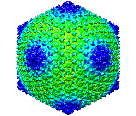 emd_8486.png emd_8486.png | 273.4 KB | ||
| Archive directory |  http://ftp.pdbj.org/pub/emdb/structures/EMD-8486ftp://ftp.pdbj.org/pub/emdb/structures/EMD-8486 http://ftp.pdbj.org/pub/emdb/structures/EMD-8486ftp://ftp.pdbj.org/pub/emdb/structures/EMD-8486 | HTTPS FTP |
-Related structure data
-Links
| EMDB pages | EMDB (EBI/PDBe) / EMDataResource |
|---|
-Map
| File | Download / File: emd_8486.map.gz / Format: CCP4 / Size: 828.1 MB / Type: IMAGE STORED AS FLOATING POINT NUMBER (4 BYTES) | ||||||||||||||||||||||||||||||||||||||||||||||||||||||||||||||||||||
|---|---|---|---|---|---|---|---|---|---|---|---|---|---|---|---|---|---|---|---|---|---|---|---|---|---|---|---|---|---|---|---|---|---|---|---|---|---|---|---|---|---|---|---|---|---|---|---|---|---|---|---|---|---|---|---|---|---|---|---|---|---|---|---|---|---|---|---|---|---|
| Annotation | Bacillus phage PBS1 | ||||||||||||||||||||||||||||||||||||||||||||||||||||||||||||||||||||
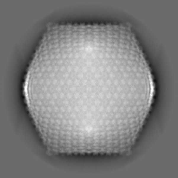
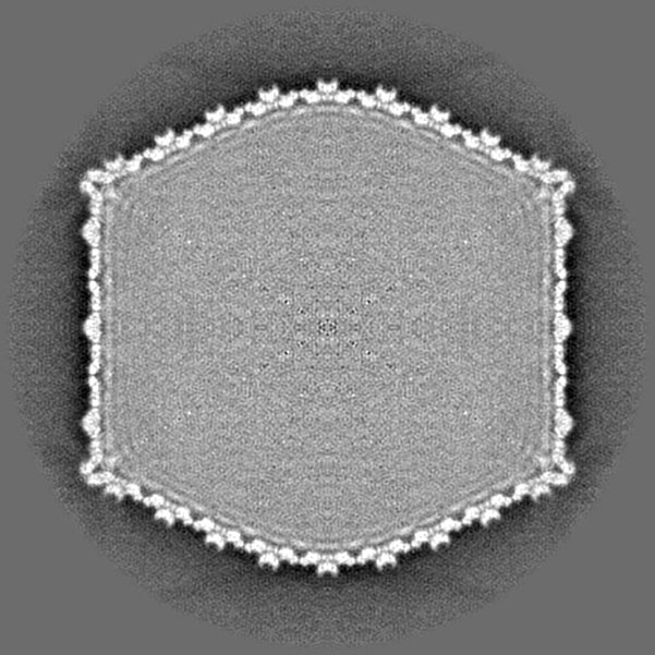
| Projections & slices | Image control
Images are generated by Spider. | ||||||||||||||||||||||||||||||||||||||||||||||||||||||||||||||||||||
| Voxel size | X=Y=Z: 2.74 Å | ||||||||||||||||||||||||||||||||||||||||||||||||||||||||||||||||||||
| Density |
| ||||||||||||||||||||||||||||||||||||||||||||||||||||||||||||||||||||
| Symmetry | Space group: 1 | ||||||||||||||||||||||||||||||||||||||||||||||||||||||||||||||||||||
| Details | EMDB XML:
CCP4 map header:
| ||||||||||||||||||||||||||||||||||||||||||||||||||||||||||||||||||||
 Z (Sec.)
 Y (Row.)
 X (Col.)


-Supplemental data
- Sample components
-Entire : Bacillus phage PBS1
| Entire | Name: Bacillus phage PBS1 (virus) |
|---|---|
| Components |
|
-Supramolecule #1: Bacillus phage PBS1
| Supramolecule | Name: Bacillus phage PBS1 / type: virus / ID: 1 / Parent: 0 / Macromolecule list: #1 / NCBI-ID: 10683 / Sci species name: Bacillus phage PBS1 / Virus type: VIRION / Virus isolate: OTHER / Virus enveloped: No / Virus empty: No |
|---|---|
| Host (natural) | Organism:  Bacillus subtilis (bacteria) Bacillus subtilis (bacteria) |
| Virus shell | Shell ID: 1 / Name: capsid / Diameter: 1300.0 Å / T number (triangulation number): 27 |
-Experimental details
-Structure determination
| Method | cryo EM |
|---|---|
 Processing Processing | single particle reconstruction |
| Aggregation state | particle |
-Sample preparation
| Concentration | 1 mg/mL | |||||||||
|---|---|---|---|---|---|---|---|---|---|---|
| Buffer | pH: 7.9 Component:
| |||||||||
| Grid | Material: COPPER / Mesh: 400 / Support film - Material: CARBON / Support film - topology: HOLEY / Pretreatment - Type: GLOW DISCHARGE | |||||||||
| Vitrification | Cryogen name: ETHANE-PROPANE / Chamber humidity: 90 % / Chamber temperature: 293 K / Instrument: FEI VITROBOT MARK II |
- Electron microscopy
| Microscope | FEI POLARA 300 |
|---|---|
| Electron beam | Acceleration voltage: 300 kV / Electron source: FIELD EMISSION GUN |
| Electron optics | Illumination mode: FLOOD BEAM / Imaging mode: BRIGHT FIELDBright-field microscopy / Cs: 2.7 mm / Nominal defocus max: 5.0 µm / Nominal defocus min: 1.0 µm / Nominal magnification: 78000 |
| Sample stage | Specimen holder model: GATAN 910 MULTI-SPECIMEN SINGLE TILT CRYO TRANSFER HOLDER Cooling holder cryogen: NITROGEN |
| Image recording | Film or detector model: FEI FALCON II (4k x 4k) / Digitization - Sampling interval: 14.0 µm / Digitization - Frames/image: 2-9 / Number real images: 3000 / Average exposure time: 2.0 sec. / Average electron dose: 30.0 e/Å2 |
| Experimental equipment |  Model: Tecnai Polara / Image courtesy: FEI Company |
-Image processing
| Particle selection | Number selected: 2495 |
|---|---|
| CTF correction | Software - Name: CTFFIND3 |
| Initial angle assignment | Type: COMMON LINE / Software - Name: Auto3DEM |
| Final angle assignment | Type: COMMON LINE / Software - Name: Auto3DEM |
| Final reconstruction | Applied symmetry - Point group: I (icosahedral) / Resolution.type: BY AUTHOR / Resolution: 10.0 Å / Resolution method: FSC 0.5 CUT-OFF / Software - Name: Auto3DEM / Number images used: 2000 |
| FSC plot (resolution estimation) | 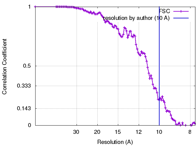 |
-Atomic model buiding 1
| Refinement | Space: REAL / Protocol: RIGID BODY FIT |
|---|
