 Movie
Movie Controller

[English] 日本語
 Yorodumi
- EMDB-7040: Tectonic conformational changes of a coronavirus spike glycoprote... -
+ Open data

- Basic information
| Entry | Database: EMDB / ID: EMD-7040 | |||||||||
|---|---|---|---|---|---|---|---|---|---|---|
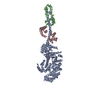
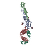
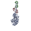
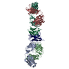
| Title | Tectonic conformational changes of a coronavirus spike glycoprotein promote membrane fusion | |||||||||
 Map data Map data | Tectonic conformational changes of a coronavirus spike glycoprotein promote membrane fusion | |||||||||
 Sample Sample |
| |||||||||
| Function / homology |  Function and homology information Function and homology informationendocytosis involved in viral entry into host cell / host cell Golgi apparatus / host cell endoplasmic reticulum-Golgi intermediate compartment membrane / receptor-mediated virion attachment to host cell / fusion of virus membrane with host plasma membrane / fusion of virus membrane with host endosome membrane /  viral envelope / host cell plasma membrane / virion membrane / membrane / identical protein binding viral envelope / host cell plasma membrane / virion membrane / membrane / identical protein bindingSimilarity search - Function | |||||||||
| Biological species |  Murine hepatitis virus / Murine coronavirus Murine hepatitis virus / Murine coronavirus | |||||||||
| Method | single particle reconstruction / cryo EM / Resolution: 4.1 Å | |||||||||
 Authors Authors | Walls AC / Tortorici MA / Snijder J / Xiong X / Bosch BJ / Rey FA / Veesler D | |||||||||
| Funding support |  United States, 1 items United States, 1 items
| |||||||||
 Citation Citation | Journal: Proc Natl Acad Sci U S A / Year: 2017 Title: Tectonic conformational changes of a coronavirus spike glycoprotein promote membrane fusion. Authors: Alexandra C Walls / M Alejandra Tortorici / Joost Snijder / Xiaoli Xiong / Berend-Jan Bosch / Felix A Rey / David Veesler /   Abstract: The tremendous pandemic potential of coronaviruses was demonstrated twice in the past few decades by two global outbreaks of deadly pneumonia. The coronavirus spike (S) glycoprotein initiates ...The tremendous pandemic potential of coronaviruses was demonstrated twice in the past few decades by two global outbreaks of deadly pneumonia. The coronavirus spike (S) glycoprotein initiates infection by promoting fusion of the viral and cellular membranes through conformational changes that remain largely uncharacterized. Here we report the cryoEM structure of a coronavirus S glycoprotein in the postfusion state, showing large-scale secondary, tertiary, and quaternary rearrangements compared with the prefusion trimer and rationalizing the free-energy landscape of this conformational machine. We also biochemically characterized the molecular events associated with refolding of the metastable prefusion S glycoprotein to the postfusion conformation using limited proteolysis, mass spectrometry, and single-particle EM. The observed similarity between postfusion coronavirus S and paramyxovirus F structures demonstrates that a conserved refolding trajectory mediates entry of these viruses and supports the evolutionary relatedness of their fusion subunits. Finally, our data provide a structural framework for understanding the mode of neutralization of antibodies targeting the fusion machinery and for engineering next-generation subunit vaccines or inhibitors against this medically important virus family. | |||||||||
| History |
|
- Structure visualization
| Movie |
Movie viewer |
|---|---|
| Structure viewer | EM map: SurfViewMolmilJmol/JSmol |
| Supplemental images |

- Downloads & links
-EMDB archive
| Map data | emd_7040.map.gz | 7.3 MB | EMDB map data format | |
|---|---|---|---|---|
| Header (meta data) | emd-7040-v30.xmlemd-7040.xml | 12.4 KB 12.4 KB | Display Display | EMDB header |
| Images | 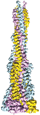 emd_7040.png emd_7040.png | 78.5 KB | ||
| Archive directory |  http://ftp.pdbj.org/pub/emdb/structures/EMD-7040ftp://ftp.pdbj.org/pub/emdb/structures/EMD-7040 http://ftp.pdbj.org/pub/emdb/structures/EMD-7040ftp://ftp.pdbj.org/pub/emdb/structures/EMD-7040 | HTTPS FTP |
-Related structure data
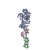
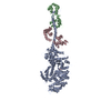
| Related structure data | 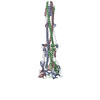 6b3oMC M: atomic model generated by this map C: citing same article ( |
|---|---|
| Similar structure data |
-Links
| EMDB pages | EMDB (EBI/PDBe) / EMDataResource |
|---|
-Map
| File | Download / File: emd_7040.map.gz / Format: CCP4 / Size: 125 MB / Type: IMAGE STORED AS FLOATING POINT NUMBER (4 BYTES) | ||||||||||||||||||||||||||||||||||||||||||||||||||||||||||||
|---|---|---|---|---|---|---|---|---|---|---|---|---|---|---|---|---|---|---|---|---|---|---|---|---|---|---|---|---|---|---|---|---|---|---|---|---|---|---|---|---|---|---|---|---|---|---|---|---|---|---|---|---|---|---|---|---|---|---|---|---|---|
| Annotation | Tectonic conformational changes of a coronavirus spike glycoprotein promote membrane fusion | ||||||||||||||||||||||||||||||||||||||||||||||||||||||||||||
| Voxel size | X=Y=Z: 1.36 Å | ||||||||||||||||||||||||||||||||||||||||||||||||||||||||||||
| Density |
| ||||||||||||||||||||||||||||||||||||||||||||||||||||||||||||
| Symmetry | Space group: 1 | ||||||||||||||||||||||||||||||||||||||||||||||||||||||||||||
| Details | EMDB XML:
CCP4 map header:
| ||||||||||||||||||||||||||||||||||||||||||||||||||||||||||||
-Supplemental data
- Sample components
-Entire : Mouse hepatitis virus spike glycoprotein (S2 subunit) in the post...
| Entire | Name: Mouse hepatitis virus spike glycoprotein (S2 subunit) in the postfusion conformation |
|---|---|
| Components |
|
-Supramolecule #1: Mouse hepatitis virus spike glycoprotein (S2 subunit) in the post...
| Supramolecule | Name: Mouse hepatitis virus spike glycoprotein (S2 subunit) in the postfusion conformation type: complex / ID: 1 / Parent: 0 / Macromolecule list: all |
|---|---|
| Source (natural) | Organism: Murine hepatitis virus |
| Recombinant expression | Organism:  Drosophila melanogaster (fruit fly) Drosophila melanogaster (fruit fly) |
| Molecular weight | Theoretical: 180 KDa |
-Macromolecule #1: Spike glycoprotein
| Macromolecule | Name: Spike glycoprotein / type: protein_or_peptide / ID: 1 / Number of copies: 3 / Enantiomer: LEVO |
|---|---|
| Source (natural) | Organism: Murine coronavirus / Strain: A59 |
| Molecular weight | Theoretical: 66.199094 KDa |
| Recombinant expression | Organism: Drosophila melanogaster (fruit fly) |
| Sequence | String: MKLCILLAVV AFVGLSLGRS LASVSTGYRL TTFEPYTPML VNDSVQSVDG LYEMQIPTNF TIGHHEEFIQ TRSPKVTIDC AAFVCGDNT ACRQQLVEYG SFCVNVNAIL NEVNNLLDNM QLQVASALMQ GVTISSRLPD GISGPIDDIN FSPLLGCIGS T CAEDGNGP ...String: MKLCILLAVV AFVGLSLGRS LASVSTGYRL TTFEPYTPML VNDSVQSVDG LYEMQIPTNF TIGHHEEFIQ TRSPKVTIDC AAFVCGDNT ACRQQLVEYG SFCVNVNAIL NEVNNLLDNM QLQVASALMQ GVTISSRLPD GISGPIDDIN FSPLLGCIGS T CAEDGNGP SAIRGRSAIE DLLFDKVKLS DVGFVEAYNN CTGGQEVRDL LCVQSFNGIK VLPPVLSESQ ISGYTTGATA AA MFPPWSA AAGVPFSLSV QYRINGLGVT MNVLSENQKM IASAFNNALG AIQDGFDATN SALGKIQSVV NANAEALNNL LNQ LSNRFG AISASLQEIL TRLEAVEAKA QIDRLINGRL TALNAYISKQ LSDSTLIKVS AAQAIEKVNE CVKSQTTRIN FCGN GNHIL SLVQNAPYGL YFIHFSYVPI SFTTANVSPG LCISGDRGLA PKAGYFVQDD GEWKFTGSSY YYPEPITDKN SVIMS SCAV NYTKAPEVFL NTSIPNPPDF KEELDKWFKN QTSIAPDLSL DFEKLNVTLL DLTYEMNRIQ DAIKKLNESY INLIKR MKQ IEDKIEEIES KQKKIENEIA RIKKIKLVPR GSLEWSHPQF EK |
-Experimental details
-Structure determination
| Method | cryo EM |
|---|---|
 Processing Processing | single particle reconstruction |
| Aggregation state | particle |
-Sample preparation
| Buffer | pH: 7.5 |
|---|---|
| Grid | Model: C-flat / Material: COPPER / Mesh: 400 / Support film - Material: CARBON / Support film - topology: HOLEY / Pretreatment - Type: GLOW DISCHARGE |
| Vitrification | Cryogen name: ETHANE |
- Electron microscopy
| Microscope | FEI TITAN KRIOS |
|---|---|
| Electron beam | Acceleration voltage: 300 kV / Electron source: FIELD EMISSION GUN |
| Electron optics | Illumination mode: FLOOD BEAM / Imaging mode: BRIGHT FIELDBright-field microscopy |
| Image recording | Film or detector model: GATAN K2 SUMMIT (4k x 4k) / Detector mode: COUNTING / Average electron dose: 60.0 e/Å2 |
| Experimental equipment |  Model: Titan Krios / Image courtesy: FEI Company |
-Image processing
| CTF correction | Software - Name: RELION (ver. 2.0.3) |
|---|---|
| Initial angle assignment | Type: PROJECTION MATCHING / Software - Name: RELION (ver. 2.0.3) |
| Final angle assignment | Type: PROJECTION MATCHING / Software - Name: RELION (ver. 2.0.3) |
| Final reconstruction | Applied symmetry - Point group: C3 (3 fold cyclic) / Resolution.type: BY AUTHOR / Resolution: 4.1 Å / Resolution method: FSC 0.143 CUT-OFF / Software - Name: RELION (ver. 2.0.3) / Number images used: 106000 |
-Atomic model buiding 1
| Refinement | Space: REAL / Protocol: AB INITIO MODEL |
|---|---|
| Output model | PDB-6b3o: |
