 Movie
Movie Controller

[English] 日本語
 Yorodumi
- PDB-6bfu: Glycan shield and fusion activation of a deltacoronavirus spike g... -
+ Open data

- Basic information
| Entry | Database: PDB / ID: 6bfu | |||||||||
|---|---|---|---|---|---|---|---|---|---|---|
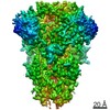
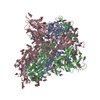
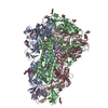
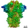
| Title | Glycan shield and fusion activation of a deltacoronavirus spike glycoprotein fine-tuned for enteric infections | |||||||||
 Components Components | Spike protein | |||||||||
 Keywords Keywords | VIRAL PROTEIN / coronavirus spike glycoprotein / porcine deltacoronavirus / viral fusion protein / glycan shield / Structural Genomics / Seattle Structural Genomics Center for Infectious Disease / SSGCID | |||||||||
| Function / homology |  Function and homology information Function and homology information: / endocytosis involved in viral entry into host cell / receptor-mediated virion attachment to host cell / membrane => GO:0016020 / fusion of virus membrane with host plasma membrane / fusion of virus membrane with host endosome membrane / viral envelope / virion membraneSimilarity search - Function | |||||||||
| Biological species |  Porcine deltacoronavirus Porcine deltacoronavirus | |||||||||
| Method | ELECTRON MICROSCOPY / single particle reconstruction / cryo EM / Resolution: 3.5 Å | |||||||||
 Authors Authors | Xiong, X. / Tortorici, M.A. / Snijder, S. / Yoshioka, C. / Walls, A.C. / Li, W. / Seattle Structural Genomics Center for Infectious Disease (SSGCID) / McGuire, A.T. / Rey, F.A. / Bosch, B.J. / Veesler, D. | |||||||||
| Funding support |  United States, 1items United States, 1items
| |||||||||
 Citation Citation | Journal: J Virol / Year: 2018 Title: Glycan Shield and Fusion Activation of a Deltacoronavirus Spike Glycoprotein Fine-Tuned for Enteric Infections. Authors: Xiaoli Xiong / M Alejandra Tortorici / Joost Snijder / Craig Yoshioka / Alexandra C Walls / Wentao Li / Andrew T McGuire / Félix A Rey / Berend-Jan Bosch / David Veesler /   Abstract: Coronaviruses recently emerged as major human pathogens causing outbreaks of severe acute respiratory syndrome and Middle East respiratory syndrome. They utilize the spike (S) glycoprotein anchored ...Coronaviruses recently emerged as major human pathogens causing outbreaks of severe acute respiratory syndrome and Middle East respiratory syndrome. They utilize the spike (S) glycoprotein anchored in the viral envelope to mediate host attachment and fusion of the viral and cellular membranes to initiate infection. The S protein is a major determinant of the zoonotic potential of coronaviruses and is also the main target of the host humoral immune response. We report here the 3.5-Å-resolution cryo-electron microscopy structure of the S glycoprotein trimer from the pathogenic porcine deltacoronavirus (PDCoV), which belongs to the recently identified genus. Structural and glycoproteomics data indicate that the glycans of PDCoV S are topologically conserved compared with the human respiratory coronavirus NL63 S, resulting in similar surface areas being shielded from neutralizing antibodies and implying that both viruses are under comparable immune pressure in their respective hosts. The structure further reveals a shortened S' activation loop, containing a reduced number of basic amino acids, which participates in rendering the spike largely protease resistant. This property distinguishes PDCoV S from recently characterized betacoronavirus S proteins and suggests that the S protein of enterotropic PDCoV has evolved to tolerate the protease-rich environment of the small intestine and to fine-tune its fusion activation to avoid premature triggering and reduction of infectivity. Coronaviruses use transmembrane S glycoprotein trimers to promote host attachment and fusion of the viral and cellular membranes. We determined a near-atomic-resolution cryo-electron microscopy structure of the S ectodomain trimer from the pathogenic PDCoV, which is responsible for diarrhea in piglets and has had devastating consequences for the swine industry worldwide. Structural and glycoproteomics data reveal that PDCoV S is decorated with 78 N-linked glycans obstructing the protein surface to limit accessibility to neutralizing antibodies in a way reminiscent of what has recently been described for a human respiratory coronavirus. PDCoV S is largely protease resistant, which distinguishes it from most other characterized coronavirus S glycoproteins and suggests that enteric coronaviruses have evolved to fine-tune fusion activation in the protease-rich environment of the small intestine of infected hosts. | |||||||||
| History |
|
- Structure visualization
| Movie |
Movie viewer |
|---|---|
| Structure viewer | Molecule: MolmilJmol/JSmol |
- Downloads & links
-Download
| PDBx/mmCIF format | 6bfu.cif.gz | 600 KB | Display | PDBx/mmCIF format |
|---|---|---|---|---|
| PDB format | pdb6bfu.ent.gz | 503.9 KB | Display | PDB format |
| PDBx/mmJSON format | 6bfu.json.gz | Tree view | PDBx/mmJSON format | |
| Others |  Other downloads Other downloads |
-Validation report
| Arichive directory | https://data.pdbj.org/pub/pdb/validation_reports/bf/6bfuftp://data.pdbj.org/pub/pdb/validation_reports/bf/6bfu | HTTPS FTP |
|---|
-Related structure data
| Related structure data | 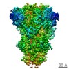 7094MC M: map data used to model this data C: citing same article ( |
|---|---|
| Similar structure data |


-Links
 PDBj
PDBj- Assembly
| Deposited unit | 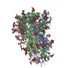
|
|---|---|
| 1 |
|
-Components
-Protein , 1 types, 3 molecules ABC
| #1: Protein | / spike glycoprotein Mass: 112514.773 Da / Num. of mol.: 3 Source method: isolated from a genetically manipulated source Source: (gene. exp.) Porcine deltacoronavirus / Production host:  Drosophila melanogaster (fruit fly) / References: UniProt: A0A140ES81, UniProt: A0A140ESF1*PLUS Drosophila melanogaster (fruit fly) / References: UniProt: A0A140ES81, UniProt: A0A140ESF1*PLUS |
|---|
-Sugars , 5 types, 63 molecules 
| #2: Polysaccharide | alpha-D-mannopyranose-(1-2)-alpha-D-mannopyranose-(1-3)-[alpha-D-mannopyranose-(1-6)]beta-D- ...alpha-D-mannopyranose-(1-2)-alpha-D-mannopyranose-(1-3)-[alpha-D-mannopyranose-(1-6)]beta-D-mannopyranose-(1-4)-2-acetamido-2-deoxy-beta-D-glucopyranose-(1-4)-2-acetamido-2-deoxy-beta-D-glucopyranose / Mass: 1072.964 Da / Num. of mol.: 9Source method: isolated from a genetically manipulated source #3: Polysaccharide | beta-D-mannopyranose-(1-4)-2-acetamido-2-deoxy-beta-D-glucopyranose-(1-4)-2-acetamido-2-deoxy-beta- ...beta-D-mannopyranose-(1-4)-2-acetamido-2-deoxy-beta-D-glucopyranose-(1-4)-2-acetamido-2-deoxy-beta-D-glucopyranose / Mass: 586.542 Da / Num. of mol.: 21Source method: isolated from a genetically manipulated source #4: Polysaccharide | 2-acetamido-2-deoxy-beta-D-glucopyranose-(1-4)-2-acetamido-2-deoxy-beta-D-glucopyranose / Mass: 424.401 Da / Num. of mol.: 6Source method: isolated from a genetically manipulated source #5: Polysaccharide | / Mass: 910.823 Da / Num. of mol.: 3Source method: isolated from a genetically manipulated source #6: Sugar | ChemComp-NAG / N-Acetylglucosamine Type: D-saccharide, beta linking / Mass: 221.208 Da / Num. of mol.: 24 Type: D-saccharide, beta linking / Mass: 221.208 Da / Num. of mol.: 24Source method: isolated from a genetically manipulated source Formula: C8H15NO6 |
|---|
-Experimental details
-Experiment
| Experiment | Method: ELECTRON MICROSCOPY |
|---|---|
| EM experiment | Aggregation state: PARTICLE / 3D reconstruction method: single particle reconstruction |
- Sample preparation
| Component | Name: Porcine deltacoronavirus spike glycoprotein / Type: COMPLEX / Entity ID: #1 / Source: RECOMBINANT |
|---|---|
| Molecular weight | Experimental value: NO |
| Source (natural) | Organism: Porcine deltacoronavirus |
| Source (recombinant) | Organism: Drosophila melanogaster (fruit fly) |
| Buffer solution | pH: 8 |
| Specimen | Conc.: 0.5 mg/ml / Embedding applied: NO / Shadowing applied: NO / Staining applied: NO / Vitrification applied: YES |
| Vitrification | Cryogen name: ETHANE |
- Electron microscopy imaging
| Experimental equipment |  Model: Titan Krios / Image courtesy: FEI Company |
|---|---|
| Microscopy | Model: FEI TITAN KRIOS |
| Electron gun | Electron source: FIELD EMISSION GUN / Accelerating voltage: 300 kV / Illumination mode: FLOOD BEAM |
| Electron lens | Mode: BRIGHT FIELDBright-field microscopy |
| Image recording | Electron dose: 40 e/Å2 / Film or detector model: GATAN K2 SUMMIT (4k x 4k) |
- Processing
| EM software |
| |||||||||
|---|---|---|---|---|---|---|---|---|---|---|
| CTF correction | Type: PHASE FLIPPING AND AMPLITUDE CORRECTION | |||||||||
| Symmetry | Point symmetry: C3 (3 fold cyclic) | |||||||||
| 3D reconstruction | Resolution: 3.5 Å / Resolution method: FSC 0.143 CUT-OFF / Num. of particles: 455710 / Symmetry type: POINT |
