 Movie
Movie Controller

[English] 日本語
 Yorodumi
- PDB-6b1t: Improved cryoEM structure of human adenovirus type 5 with atomic ... -
+ Open data

- Basic information
| Entry | Database: PDB / ID: 6b1t | ||||||||||||||||||||||||
|---|---|---|---|---|---|---|---|---|---|---|---|---|---|---|---|---|---|---|---|---|---|---|---|---|---|
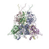
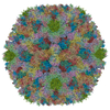
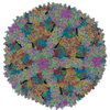
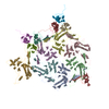
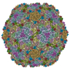
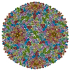
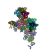
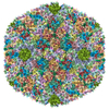
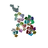
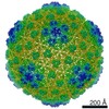
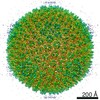
| Title | Improved cryoEM structure of human adenovirus type 5 with atomic details of minor proteins VI and VII | ||||||||||||||||||||||||
 Components Components |
| ||||||||||||||||||||||||
 Keywords Keywords |  VIRUS / human adenovirus / cement protein / dsDNA genome packaging / genome-capsid co-assembly VIRUS / human adenovirus / cement protein / dsDNA genome packaging / genome-capsid co-assembly | ||||||||||||||||||||||||
| Function / homology |  Function and homology information Function and homology informationhexon binding / protein transport along microtubule / viral capsid, decoration / T=25 icosahedral viral capsid / lysis of host organelle involved in viral entry into host cell / viral procapsid / host cell nucleolus / microtubule-dependent intracellular transport of viral material towards nucleus / viral release from host cell / viral life cycle ...hexon binding / protein transport along microtubule / viral capsid, decoration / T=25 icosahedral viral capsid / lysis of host organelle involved in viral entry into host cell / viral procapsid / host cell nucleolus / microtubule-dependent intracellular transport of viral material towards nucleus / viral release from host cell / viral life cycle / viral penetration into host nucleus / viral capsid / clathrin-dependent endocytosis of virus by host cell / host cell cytoplasm / symbiont entry into host cell / host cell nucleus / structural molecule activity / virion attachment to host cell / DNA bindingSimilarity search - Function | ||||||||||||||||||||||||
| Biological species |   Human adenovirus C serotype 5 Human adenovirus C serotype 5 | ||||||||||||||||||||||||
| Method | ELECTRON MICROSCOPY / single particle reconstruction / cryo EM / Resolution: 3.2 Å | ||||||||||||||||||||||||
 Authors Authors | Dai, X.H. / Wu, L. / Sun, R. / Zhou, Z.H. | ||||||||||||||||||||||||
| Funding support |  United States, 7items United States, 7items
| ||||||||||||||||||||||||
 Citation Citation | Journal: J Virol / Year: 2017 Title: Atomic Structures of Minor Proteins VI and VII in Human Adenovirus. Authors: Xinghong Dai / Lily Wu / Ren Sun / Z Hong Zhou / Abstract: Human adenoviruses (Ad) are double-stranded DNA (dsDNA) viruses associated with infectious diseases, but they are better known as tools for gene delivery and oncolytic anticancer therapy. Atomic ...Human adenoviruses (Ad) are double-stranded DNA (dsDNA) viruses associated with infectious diseases, but they are better known as tools for gene delivery and oncolytic anticancer therapy. Atomic structures of Ad provide the basis for the development of antivirals and for engineering efforts toward more effective applications. Since 2010, atomic models of human Ad5 have been derived independently from photographic film cryo-electron microscopy (cryo-EM) and X-ray crystallography studies, but discrepancies exist concerning the assignment of cement proteins IIIa, VIII, and IX. To clarify these discrepancies, we employed the technology of direct electron counting to obtain a cryo-EM structure of human Ad5 at 3.2-Å resolution. Our improved structure unambiguously confirms our previous cryo-EM models of proteins IIIa, VIII, and IX and explains the likely cause of conflict in the crystallography models. The improved structure also allows the identification of three new components in the cavity of hexon-the cleaved N terminus of precursor protein VI (pVIn), the cleaved N terminus of precursor protein VII (pVIIn2), and mature protein VI. The binding of pVIIn2-and, by extension, that of genome-condensing pVII-to hexons is consistent with the previously proposed dsDNA genome-capsid coassembly for adenoviruses, which resembles that of single-stranded RNA (ssRNA) viruses but differs from the well-established mechanism of pumping dsDNA into a preformed protein capsid exemplified by tailed bacteriophages and herpesviruses. Adenovirus is a double-edged sword to humans: it is a widespread pathogen but can be used as a bioengineering tool for anticancer and gene therapies. The atomic structure of the virus provides the basis for antiviral and application developments, but conflicting atomic models for the important cement proteins IIIa, VIII, and IX from conventional/film cryo-EM and X-ray crystallography studies have caused confusion. Using cutting-edge cryo-EM technology with electron counting, we improved the structure of human adenovirus type 5 and confirmed our previous models of cement proteins IIIa, VIII, and IX, thus clarifying the inconsistent structures. The improved structure also reveals atomic details of membrane-lytic protein VI and genome-condensing protein VII and supports the previously proposed genome-capsid coassembly mechanism for adenoviruses. | ||||||||||||||||||||||||
| History |
|
- Structure visualization
| Movie |
Movie viewer |
|---|---|
| Structure viewer | Molecule: MolmilJmol/JSmol |
- Downloads & links
-Download
| PDBx/mmCIF format | 6b1t.cif.gz | 2.1 MB | Display | PDBx/mmCIF format |
|---|---|---|---|---|
| PDB format | pdb6b1t.ent.gz | Display | PDB format | |
| PDBx/mmJSON format | 6b1t.json.gz | Tree view | PDBx/mmJSON format | |
| Others |  Other downloads Other downloads |
-Validation report
| Arichive directory | https://data.pdbj.org/pub/pdb/validation_reports/b1/6b1tftp://data.pdbj.org/pub/pdb/validation_reports/b1/6b1t | HTTPS FTP |
|---|
-Related structure data
| Related structure data | 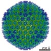 7034MC M: map data used to model this data C: citing same article ( |
|---|---|
| Similar structure data |
-Links
 PDBj
PDBj


- Assembly
| Deposited unit | 
|
|---|---|
| 1 | x 60
|
| 2 |
|
| 3 | x 5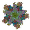
|
| 4 | x 6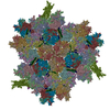
|
| 5 | 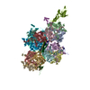
|
| Symmetry | Point symmetry: (Schoenflies symbol: I (icosahedral)) |
-Components
-Protein , 4 types, 18 molecules ABCDEFGHIJKLMQRSTX
| #1: Protein | / CP-H / Protein II Mass: 108107.617 Da / Num. of mol.: 12 / Source method: isolated from a natural source / Source: (natural) Human adenovirus C serotype 5 / References: UniProt: P04133#2: Protein | | / CP-P / Penton base protein / Protein IIIMass: 63356.602 Da / Num. of mol.: 1 / Source method: isolated from a natural source / Source: (natural) Human adenovirus C serotype 5 / References: UniProt: P12538#5: Protein | Mass: 14468.134 Da / Num. of mol.: 4 / Source method: isolated from a natural source / Source: (natural) Human adenovirus C serotype 5 / References: UniProt: P03281#8: Protein | | Mass: 23460.559 Da / Num. of mol.: 1 / Source method: isolated from a natural source / Source: (natural) Human adenovirus C serotype 5 / References: UniProt: P24937 |
|---|
-Pre-hexon-linking protein ... , 2 types, 3 molecules NOP
| #3: Protein | Mass: 65322.805 Da / Num. of mol.: 1 / Source method: isolated from a natural source / Source: (natural) Human adenovirus C serotype 5 / References: UniProt: P12537 |
|---|---|
| #4: Protein | Mass: 24710.590 Da / Num. of mol.: 2 / Source method: isolated from a natural source / Source: (natural) Human adenovirus C serotype 5 / References: UniProt: P24936 |
-Protein/peptide , 2 types, 4 molecules UVYW
| #6: Protein/peptide | Mass: 3584.953 Da / Num. of mol.: 3 / Source method: isolated from a natural source / Source: (natural) Human adenovirus C serotype 5 / References: UniProt: P24937#7: Protein/peptide | | Mass: 1198.438 Da / Num. of mol.: 1 / Source method: isolated from a natural source / Source: (natural) Human adenovirus C serotype 5 / References: UniProt: P68951 |
|---|
-Experimental details
-Experiment
| Experiment | Method: ELECTRON MICROSCOPY |
|---|---|
| EM experiment | Aggregation state: PARTICLE / 3D reconstruction method: single particle reconstruction |
- Sample preparation
| Component | Name: Human adenovirus 5Adenoviridae / Type: VIRUS / Details: Cultured in HEK293T cells and purified from media / Entity ID: all / Source: NATURAL | ||||||||||||||||||||
|---|---|---|---|---|---|---|---|---|---|---|---|---|---|---|---|---|---|---|---|---|---|
| Molecular weight | Value: 150 MDa / Experimental value: NO | ||||||||||||||||||||
| Source (natural) | Organism: Human adenovirus C serotype 5 | ||||||||||||||||||||
| Details of virus | Empty: NO / Enveloped: NO / Isolate: STRAIN / Type: VIRION | ||||||||||||||||||||
| Natural host | Organism: Homo sapiens | ||||||||||||||||||||
| Virus shell | Name: capsid / Diameter: 900 nm / Triangulation number (T number): 25 | ||||||||||||||||||||
| Buffer solution | pH: 7.4 | ||||||||||||||||||||
| Buffer component |
| ||||||||||||||||||||
| Specimen | Embedding applied: NO / Shadowing applied: NO / Staining applied: NO / Vitrification applied: YES / Details: Purified virion | ||||||||||||||||||||
| Specimen support | Grid material: COPPER / Grid mesh size: 200 divisions/in. / Grid type: Quantifoil R2/1 | ||||||||||||||||||||
| Vitrification | Instrument: FEI VITROBOT MARK II / Cryogen name: ETHANE / Humidity: 100 % / Chamber temperature: 293 K |
- Electron microscopy imaging
| Experimental equipment |  Model: Titan Krios / Image courtesy: FEI Company |
|---|---|
| Microscopy | Model: FEI TITAN KRIOS |
| Electron gun | Electron source: FIELD EMISSION GUN / Accelerating voltage: 300 kV / Illumination mode: FLOOD BEAM |
| Electron lens | Mode: BRIGHT FIELDBright-field microscopy / Nominal magnification: 81000 X / Nominal defocus max: 2000 nm / Nominal defocus min: 2000 nm / Cs: 2.7 mm / C2 aperture diameter: 70 µm / Alignment procedure: BASIC |
| Specimen holder | Cryogen: NITROGEN / Specimen holder model: FEI TITAN KRIOS AUTOGRID HOLDER / Temperature (min): 80 K |
| Image recording | Average exposure time: 9 sec. / Electron dose: 25 e/Å2 / Detector mode: SUPER-RESOLUTION / Film or detector model: GATAN K2 SUMMIT (4k x 4k) / Num. of grids imaged: 1 / Num. of real images: 5608 |
| EM imaging optics | Energyfilter name: Gatan Image Filter / Energyfilter upper: 20 eV / Energyfilter lower: 0 eV |
| Image scans | Sampling size: 2.5 µm / Width: 7676 / Height: 7420 / Movie frames/image: 32 / Used frames/image: 2-32 |
- Processing
| Software | Name: PHENIX / Version: dev_2875: / Classification: refinement | ||||||||||||||||||||||||
|---|---|---|---|---|---|---|---|---|---|---|---|---|---|---|---|---|---|---|---|---|---|---|---|---|---|
| EM software |
| ||||||||||||||||||||||||
| CTF correction | Type: PHASE FLIPPING AND AMPLITUDE CORRECTION | ||||||||||||||||||||||||
| Particle selection | Num. of particles selected: 96375 | ||||||||||||||||||||||||
| Symmetry | Point symmetry: C1 (asymmetric) | ||||||||||||||||||||||||
| 3D reconstruction | Resolution: 3.2 Å / Resolution method: FSC 0.143 CUT-OFF / Num. of particles: 53000 / Algorithm: FOURIER SPACE / Num. of class averages: 1 / Symmetry type: POINT | ||||||||||||||||||||||||
| Atomic model building | Protocol: RIGID BODY FIT / Space: REAL / Target criteria: Correlation coefficient | ||||||||||||||||||||||||
| Refine LS restraints |
|
