+Open data
-Basic information
Entry | Database: EMDB / ID: EMD-6685 | |||||||||
---|---|---|---|---|---|---|---|---|---|---|
Title | Structure of Japanese encephalitis virus | |||||||||
Map data | Cryo-EM map for the JEV particle | |||||||||
Sample |
| |||||||||
Function / homology | Function and homology information symbiont-mediated suppression of host JAK-STAT cascade via inhibition of host TYK2 activity / flavivirin / symbiont-mediated suppression of host JAK-STAT cascade via inhibition of STAT2 activity / symbiont-mediated suppression of host JAK-STAT cascade via inhibition of STAT1 activity / viral capsid / nucleoside-triphosphate phosphatase / double-stranded RNA binding / mRNA (guanine-N7)-methyltransferase / methyltransferase cap1 / clathrin-dependent endocytosis of virus by host cell ...symbiont-mediated suppression of host JAK-STAT cascade via inhibition of host TYK2 activity / flavivirin / symbiont-mediated suppression of host JAK-STAT cascade via inhibition of STAT2 activity / symbiont-mediated suppression of host JAK-STAT cascade via inhibition of STAT1 activity / viral capsid / nucleoside-triphosphate phosphatase / double-stranded RNA binding / mRNA (guanine-N7)-methyltransferase / methyltransferase cap1 / clathrin-dependent endocytosis of virus by host cell / mRNA (nucleoside-2'-O-)-methyltransferase activity / mRNA 5'-cap (guanine-N7-)-methyltransferase activity / host cell surface / RNA helicase activity / membrane => GO:0016020 / host cell endoplasmic reticulum membrane / host cell perinuclear region of cytoplasm / protein dimerization activity / RNA helicase / induction by virus of host autophagy / RNA-directed RNA polymerase / viral RNA genome replication / RNA-dependent RNA polymerase activity / serine-type endopeptidase activity / fusion of virus membrane with host endosome membrane / viral envelope / symbiont-mediated suppression of host type I interferon-mediated signaling pathway / host cell nucleus / structural molecule activity / virion attachment to host cell / virion membrane / ATP hydrolysis activity / proteolysis / extracellular region / ATP binding / membrane / metal ion binding Similarity search - Function | |||||||||
Biological species | Japanese encephalitis virus | |||||||||
Method | single particle reconstruction / cryo EM / Resolution: 4.3 Å | |||||||||
Authors | Wang X / Zhu L / Li S / Yuan S / Qin C / Fry EE / Stuart ID / Rao Z | |||||||||
Funding support | China, 1 items
| |||||||||
Citation | 
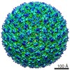
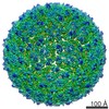
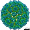
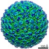
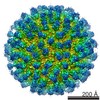
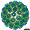
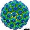
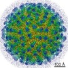
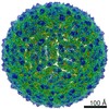
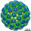
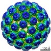
Journal: Nat Commun / Year: 2017 Title: Near-atomic structure of Japanese encephalitis virus reveals critical determinants of virulence and stability. Authors: Xiangxi Wang / Shi-Hua Li / Ling Zhu / Qing-Gong Nian / Shuai Yuan / Qiang Gao / Zhongyu Hu / Qing Ye / Xiao-Feng Li / Dong-Yang Xie / Neil Shaw / Junzhi Wang / Thomas S Walter / Juha T ...Authors: Xiangxi Wang / Shi-Hua Li / Ling Zhu / Qing-Gong Nian / Shuai Yuan / Qiang Gao / Zhongyu Hu / Qing Ye / Xiao-Feng Li / Dong-Yang Xie / Neil Shaw / Junzhi Wang / Thomas S Walter / Juha T Huiskonen / Elizabeth E Fry / Cheng-Feng Qin / David I Stuart / Zihe Rao / Abstract: Although several different flaviviruses may cause encephalitis, Japanese encephalitis virus is the most significant, being responsible for thousands of deaths each year in Asia. The structural and ...Although several different flaviviruses may cause encephalitis, Japanese encephalitis virus is the most significant, being responsible for thousands of deaths each year in Asia. The structural and molecular basis of this encephalitis is not fully understood. Here, we report the cryo-electron microscopy structure of mature Japanese encephalitis virus at near-atomic resolution, which reveals an unusual "hole" on the surface, surrounded by five encephalitic-specific motifs implicated in receptor binding. Glu138 of E, which is highly conserved in encephalitic flaviviruses, maps onto one of these motifs and is essential for binding to neuroblastoma cells, with the E138K mutation abrogating the neurovirulence and neuroinvasiveness of Japanese encephalitis virus in mice. We also identify structural elements modulating viral stability, notably Gln264 of E, which, when replaced by His264 strengthens a hydrogen-bonding network, leading to a more stable virus. These studies unveil determinants of neurovirulence and stability in Japanese encephalitis virus, opening up new avenues for therapeutic interventions against neurotropic flaviviruses.Japanese encephalitis virus (JEV) is a Flavivirus responsible for thousands of deaths every year for which there are no specific anti-virals. Here, Wang et al. report the cryo-EM structure of mature JEV at near-atomic resolution and identify structural elements that modulate stability and virulence. | |||||||||
History |
|
-Structure visualization
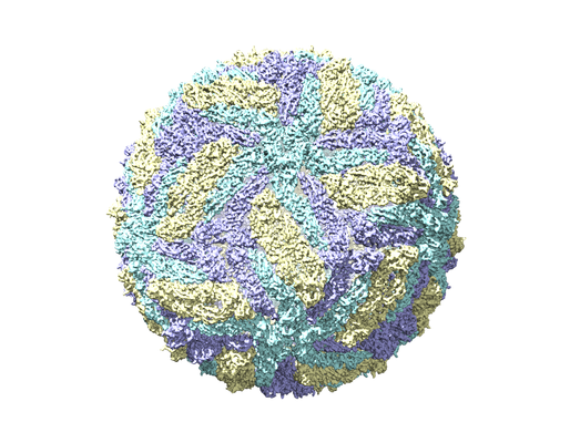
Movie |
Movie viewer |
---|---|
Structure viewer | EM map: SurfViewMolmilJmol/JSmol |
Supplemental images |
-Downloads & links
-EMDB archive
Map data | emd_6685.map.gz | 757.9 MB | EMDB map data format | |
---|---|---|---|---|
Header (meta data) | emd-6685-v30.xml emd-6685.xml | 16.6 KB 16.6 KB | Display Display | EMDB header |
Images | emd_6685.png | 217.6 KB | ||
Archive directory | http://ftp.pdbj.org/pub/emdb/structures/EMD-6685 ftp://ftp.pdbj.org/pub/emdb/structures/EMD-6685 | HTTPS FTP |
-Related structure data
Related structure data | 5wsnMC M: atomic model generated by this map C: citing same article (ref.) |
---|---|
Similar structure data |
-Links
EMDB pages | EMDB (EBI/PDBe) / EMDataResource |
---|---|
Related items in Molecule of the Month |
-Map
File | Download / File: emd_6685.map.gz / Format: CCP4 / Size: 824 MB / Type: IMAGE STORED AS FLOATING POINT NUMBER (4 BYTES) | ||||||||||||||||||||||||||||||||||||||||||||||||||||||||||||
---|---|---|---|---|---|---|---|---|---|---|---|---|---|---|---|---|---|---|---|---|---|---|---|---|---|---|---|---|---|---|---|---|---|---|---|---|---|---|---|---|---|---|---|---|---|---|---|---|---|---|---|---|---|---|---|---|---|---|---|---|---|
Annotation | Cryo-EM map for the JEV particle | ||||||||||||||||||||||||||||||||||||||||||||||||||||||||||||
Voxel size | X=Y=Z: 1.35 Å | ||||||||||||||||||||||||||||||||||||||||||||||||||||||||||||
Density |
| ||||||||||||||||||||||||||||||||||||||||||||||||||||||||||||
Symmetry | Space group: 1 | ||||||||||||||||||||||||||||||||||||||||||||||||||||||||||||
Details | EMDB XML:
CCP4 map header:
|
-Supplemental data
-Sample components
-Entire : Japanese encephalitis virus
Entire | Name: Japanese encephalitis virus |
---|---|
Components |
|
-Supramolecule #1: Japanese encephalitis virus
Supramolecule | Name: Japanese encephalitis virus / type: virus / ID: 1 / Parent: 0 / Macromolecule list: all / NCBI-ID: 11072 / Sci species name: Japanese encephalitis virus / Sci species strain: P3 / Virus type: VIRION / Virus isolate: STRAIN / Virus enveloped: Yes / Virus empty: No |
---|---|
Host (natural) | Organism: Homo sapiens (human) |
Host system | Organism: Chlorocebus aethiops (grivet) / Recombinant cell: vero / Recombinant plasmid: no plasmids |
Molecular weight | Theoretical: 11.8 MDa |
Virus shell | Shell ID: 1 / Name: Envelope protein / Diameter: 500.0 Å / T number (triangulation number): 3 |
-Macromolecule #1: E protein
Macromolecule | Name: E protein / type: protein_or_peptide / ID: 1 / Number of copies: 3 / Enantiomer: LEVO |
---|---|
Source (natural) | Organism: Japanese encephalitis virus / Strain: P3 / Organ: Homo sapiens |
Molecular weight | Theoretical: 53.508684 KDa |
Recombinant expression | Organism: Chlorocebus aethiops (grivet) |
Sequence | String: FNCLGMGNRD FIEGASGATW VDLVLEGDSC LTIMANDKPT LDVRMINIEA SQLAEVRSYC YHASVTDIST VARCPMTGEA HNEKRADSS YVCKQGFTDR GWGNGCGLFG KGSIDTCAKF SCTSKAIGRT IQPENIKYEV GIFVHGTTTS ENHGNYSAQV G ASQAAKFT ...String: FNCLGMGNRD FIEGASGATW VDLVLEGDSC LTIMANDKPT LDVRMINIEA SQLAEVRSYC YHASVTDIST VARCPMTGEA HNEKRADSS YVCKQGFTDR GWGNGCGLFG KGSIDTCAKF SCTSKAIGRT IQPENIKYEV GIFVHGTTTS ENHGNYSAQV G ASQAAKFT VTPNAPSITL KLGDYGEVTL DCEPRSGLNT EAFYVMTVGS KSFLVHREWF HDLALPWTPP SSTAWRNREL LM EFEEAHA TKQSVVALGS QEGGLHQALA GAIVVEYSSS VKLTSGHLKC RLKMDKLALK GTTYGMCTGK FSFAKNPADT GHG TVVIEL SYSGSDGPCK IPIVSVASLN DMTPVGRLVT VNPFVATSSA NSKVLVEMEP PFGDSYIVVG RGDKQINHHW HKAG STLGK AFLTTLKGAQ RLAALGDTAW DFGSIGGVFN SIGKAVHQVF GGAFRTLFGG MSWITQGLMG ALLLWMGVNA RDRSI ALAF LATGGVLLFL ATNVHA |
-Macromolecule #2: M protein
Macromolecule | Name: M protein / type: protein_or_peptide / ID: 2 / Number of copies: 3 / Enantiomer: LEVO |
---|---|
Source (natural) | Organism: Japanese encephalitis virus / Strain: P3 / Organ: Homo sapiens |
Molecular weight | Theoretical: 8.250488 KDa |
Recombinant expression | Organism: Chlorocebus aethiops (grivet) |
Sequence | String: SVSVQTHGES SLVNKTETWL DSTKATRYLM KTENWIIRNP GYAFLAAVLG WMLGSNNGQR VVFTILLLLV APAY |
-Experimental details
-Structure determination
Method | cryo EM |
---|---|
Processing | single particle reconstruction |
Aggregation state | particle |
-Sample preparation
Concentration | 2.5 mg/mL |
---|---|
Buffer | pH: 7.4 / Details: PBS buffer |
Grid | Model: C-flat / Material: COPPER / Mesh: 400 / Pretreatment - Type: GLOW DISCHARGE |
Vitrification | Cryogen name: ETHANE / Chamber humidity: 95 % / Chamber temperature: 298 K / Instrument: FEI VITROBOT MARK III / Details: blot for 3 seconds before plunging. |
-Electron microscopy
Microscope | FEI POLARA 300 |
---|---|
Electron beam | Acceleration voltage: 300 kV / Electron source: FIELD EMISSION GUN |
Electron optics | C2 aperture diameter: 100.0 µm / Calibrated defocus max: 3.0 µm / Calibrated defocus min: 1.2 µm / Illumination mode: FLOOD BEAM / Imaging mode: BRIGHT FIELDBright-field microscopy / Cs: 2.0 mm / Nominal defocus max: 3.0 µm / Nominal defocus min: 1.2 µm |
Sample stage | Specimen holder model: OTHER |
Image recording | Film or detector model: GATAN K2 SUMMIT (4k x 4k) / Detector mode: COUNTING / Digitization - Dimensions - Width: 3710 pixel / Digitization - Dimensions - Height: 3710 pixel / Digitization - Frames/image: 2-18 / Number grids imaged: 5 / Number real images: 2500 / Average exposure time: 1.5 sec. / Average electron dose: 1.2 e/Å2 |
Experimental equipment | Model: Tecnai Polara / Image courtesy: FEI Company |
-Image processing
Particle selection | Number selected: 22000 |
---|---|
CTF correction | Software - Name: Gctf (ver. 5.0) |
Startup model | Type of model: EMDB MAP EMDB ID: |
Initial angle assignment | Type: PROJECTION MATCHING / Software - Name: RELION |
Final 3D classification | Number classes: 150 / Avg.num./class: 120 / Software - Name: RELION (ver. 1.3) |
Final angle assignment | Type: RANDOM ASSIGNMENT / Software - Name: RELION (ver. 1.3) |
Final reconstruction | Number classes used: 120 / Applied symmetry - Point group: I (icosahedral) / Resolution.type: BY AUTHOR / Resolution: 4.3 Å / Resolution method: FSC 0.143 CUT-OFF / Software - Name: RELION (ver. 1.3) / Number images used: 15260 |