 Movie
Movie Controller


+ Open data

- Basic information
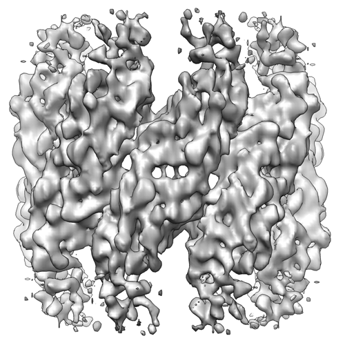
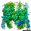
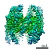
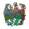
| Entry | Database: EMDB / ID: EMD-5258 | |||||||||
|---|---|---|---|---|---|---|---|---|---|---|
| Title | Lidless D386A Mm-cpn in the pre-hydrolysis ATP-bound state | |||||||||
 Map data Map data | This is the density map of lidless D386A Mm-cpn in the pre-hydrolysis ATP-bound state. | |||||||||
 Sample Sample |
| |||||||||
 Keywords Keywords | Mm-cpn /  Chaperonin / ATP-bound Chaperonin / ATP-bound | |||||||||
| Function / homology |  Function and homology information Function and homology informationATP-dependent protein folding chaperone / unfolded protein binding / ATP hydrolysis activity / ATP binding / identical protein bindingSimilarity search - Function | |||||||||
| Biological species |  Methanococcus maripaludis (archaea) Methanococcus maripaludis (archaea) | |||||||||
| Method | single particle reconstruction / cryo EM / Resolution: 8.0 Å | |||||||||
 Authors Authors | Zhang J / Ma B / DiMaio F / Douglas NR / Joachimiak L / Baker D / Frydman J / Levitt M / Chiu W | |||||||||
 Citation Citation | Journal: Structure / Year: 2011 Title: Cryo-EM structure of a group II chaperonin in the prehydrolysis ATP-bound state leading to lid closure. Authors: Junjie Zhang / Boxue Ma / Frank DiMaio / Nicholai R Douglas / Lukasz A Joachimiak / David Baker / Judith Frydman / Michael Levitt / Wah Chiu /  Abstract: Chaperonins are large ATP-driven molecular machines that mediate cellular protein folding. Group II chaperonins use their "built-in lid" to close their central folding chamber. Here we report the ...Chaperonins are large ATP-driven molecular machines that mediate cellular protein folding. Group II chaperonins use their "built-in lid" to close their central folding chamber. Here we report the structure of an archaeal group II chaperonin in its prehydrolysis ATP-bound state at subnanometer resolution using single particle cryo-electron microscopy (cryo-EM). Structural comparison of Mm-cpn in ATP-free, ATP-bound, and ATP-hydrolysis states reveals that ATP binding alone causes the chaperonin to close slightly with a ∼45° counterclockwise rotation of the apical domain. The subsequent ATP hydrolysis drives each subunit to rock toward the folding chamber and to close the lid completely. These motions are attributable to the local interactions of specific active site residues with the nucleotide, the tight couplings between the apical and intermediate domains within the subunit, and the aligned interactions between two subunits across the rings. This mechanism of structural changes in response to ATP is entirely different from those found in group I chaperonins. | |||||||||
| History |
|
- Structure visualization
| Movie |
Movie viewer |
|---|---|
| Structure viewer | EM map: SurfViewMolmilJmol/JSmol |
| Supplemental images |
- Downloads & links
-EMDB archive
| Map data | emd_5258.map.gz | 3.2 MB | EMDB map data format | |
|---|---|---|---|---|
| Header (meta data) | emd-5258-v30.xmlemd-5258.xml | 9.5 KB 9.5 KB | Display Display | EMDB header |
| Images |  emd_5258_1.png emd_5258_1.png | 219.5 KB | ||
| Archive directory |  http://ftp.pdbj.org/pub/emdb/structures/EMD-5258ftp://ftp.pdbj.org/pub/emdb/structures/EMD-5258 http://ftp.pdbj.org/pub/emdb/structures/EMD-5258ftp://ftp.pdbj.org/pub/emdb/structures/EMD-5258 | HTTPS FTP |
-Related structure data
| Related structure data |  3j02MC  3j03C M: atomic model generated by this map C: citing same article ( |
|---|---|
| Similar structure data |
-Links
| EMDB pages | EMDB (EBI/PDBe) / EMDataResource |
|---|---|
| Related items in Molecule of the Month |

-Map
| File | Download / File: emd_5258.map.gz / Format: CCP4 / Size: 51.5 MB / Type: IMAGE STORED AS FLOATING POINT NUMBER (4 BYTES) | ||||||||||||||||||||||||||||||||||||||||||||||||||||||||||||||||||||
|---|---|---|---|---|---|---|---|---|---|---|---|---|---|---|---|---|---|---|---|---|---|---|---|---|---|---|---|---|---|---|---|---|---|---|---|---|---|---|---|---|---|---|---|---|---|---|---|---|---|---|---|---|---|---|---|---|---|---|---|---|---|---|---|---|---|---|---|---|---|
| Annotation | This is the density map of lidless D386A Mm-cpn in the pre-hydrolysis ATP-bound state. | ||||||||||||||||||||||||||||||||||||||||||||||||||||||||||||||||||||
| Voxel size | X=Y=Z: 1.42 Å | ||||||||||||||||||||||||||||||||||||||||||||||||||||||||||||||||||||
| Density |
| ||||||||||||||||||||||||||||||||||||||||||||||||||||||||||||||||||||
| Symmetry | Space group: 1 | ||||||||||||||||||||||||||||||||||||||||||||||||||||||||||||||||||||
| Details | EMDB XML:
CCP4 map header:
| ||||||||||||||||||||||||||||||||||||||||||||||||||||||||||||||||||||
-Supplemental data
- Sample components
-Entire : Lidless D386A Mm-cpn variant
| Entire | Name: Lidless D386A Mm-cpn variant |
|---|---|
| Components |
|
-Supramolecule #1000: Lidless D386A Mm-cpn variant
| Supramolecule | Name: Lidless D386A Mm-cpn variant / type: sample / ID: 1000 / Number unique components: 1 |
|---|---|
| Molecular weight | Experimental: 1 MDa / Theoretical: 1 MDa |
-Macromolecule #1: Methanococcus maripaludis chaperonin
| Macromolecule | Name: Methanococcus maripaludis chaperonin / type: protein_or_peptide / ID: 1 / Name.synonym: Mm-cpn / Details: incubated with 1mM ATP / Number of copies: 1 / Oligomeric state: 16-mer / Recombinant expression: Yes |
|---|---|
| Source (natural) | Organism: Methanococcus maripaludis (archaea) |
| Molecular weight | Experimental: 1 MDa / Theoretical: 1 MDa |
| Recombinant expression | Organism:  Escherichia coli (E. coli) Escherichia coli (E. coli) |
-Experimental details
-Structure determination
| Method | cryo EM |
|---|---|
 Processing Processing | single particle reconstruction |
| Aggregation state | particle |
-Sample preparation
| Vitrification | Cryogen name: ETHANE / Chamber humidity: 100 % / Instrument: OTHER / Details: Vitrification instrument: vitrobot |
|---|
- Electron microscopy
| Microscope | JEOL 2200FS |
|---|---|
| Electron beam | Acceleration voltage: 200 kV / Electron source: FIELD EMISSION GUN |
| Electron optics | Calibrated magnification: 112000 / Illumination mode: FLOOD BEAM / Imaging mode: BRIGHT FIELDBright-field microscopy / Nominal defocus max: 2.5 µm / Nominal defocus min: 1.0 µm / Nominal magnification: 80000 |
| Specialist optics | Energy filter - Name: in-column omega energy filter / Energy filter - Lower energy threshold: 0.0 eV / Energy filter - Upper energy threshold: 10.0 eV |
| Sample stage | Specimen holder: Gatan side entry / Specimen holder model: GATAN LIQUID NITROGEN |
| Date | Sep 8, 2009 |
| Image recording | Category: CCD / Film or detector model: GENERIC GATAN (4k x 4k) / Number real images: 64 / Average electron dose: 20 e/Å2 |
-Image processing
| CTF correction | Details: Each CCD image |
|---|---|
| Final reconstruction | Algorithm: OTHER / Resolution.type: BY AUTHOR / Resolution: 8.0 Å / Resolution method: FSC 0.5 CUT-OFF / Software - Name: EMAN / Number images used: 12761 |
