 Movie
Movie Controller


+ Open data

- Basic information
| Entry | Database: EMDB / ID: EMD-3875 | ||||||||||||
|---|---|---|---|---|---|---|---|---|---|---|---|---|---|
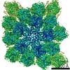
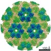
| Title | The structure of Marburg virus nucleocapsid from virions | ||||||||||||
 Map data Map data | Subtomogram averaging of Marburg virus nucleocapsid within intact virions | ||||||||||||
 Sample Sample |
| ||||||||||||
| Biological species |   Marburg virus - Musoke, Kenya, 1980 / Marburg virus - Angola / Lake Victoria marburgvirus - Popp Marburg virus - Musoke, Kenya, 1980 / Marburg virus - Angola / Lake Victoria marburgvirus - Popp | ||||||||||||
| Method | subtomogram averaging / cryo EM / Resolution: 9.1 Å | ||||||||||||
 Authors Authors | Wan W / Kolesnikova L / Clarke M / Koehler A / Noda T / Becker S / Briggs JAG | ||||||||||||
| Funding support |  Germany, 3 items Germany, 3 items
| ||||||||||||
 Citation Citation | Journal: Nature / Year: 2017 Title: Structure and assembly of the Ebola virus nucleocapsid. Authors: William Wan / Larissa Kolesnikova / Mairi Clarke / Alexander Koehler / Takeshi Noda / Stephan Becker / John A G Briggs /   Abstract: Ebola and Marburg viruses are filoviruses: filamentous, enveloped viruses that cause haemorrhagic fever. Filoviruses are within the order Mononegavirales, which also includes rabies virus, measles ...Ebola and Marburg viruses are filoviruses: filamentous, enveloped viruses that cause haemorrhagic fever. Filoviruses are within the order Mononegavirales, which also includes rabies virus, measles virus, and respiratory syncytial virus. Mononegaviruses have non-segmented, single-stranded negative-sense RNA genomes that are encapsidated by nucleoprotein and other viral proteins to form a helical nucleocapsid. The nucleocapsid acts as a scaffold for virus assembly and as a template for genome transcription and replication. Insights into nucleoprotein-nucleoprotein interactions have been derived from structural studies of oligomerized, RNA-encapsidating nucleoprotein, and cryo-electron microscopy of nucleocapsid or nucleocapsid-like structures. There have been no high-resolution reconstructions of complete mononegavirus nucleocapsids. Here we apply cryo-electron tomography and subtomogram averaging to determine the structure of Ebola virus nucleocapsid within intact viruses and recombinant nucleocapsid-like assemblies. These structures reveal the identity and arrangement of the nucleocapsid components, and suggest that the formation of an extended α-helix from the disordered carboxy-terminal region of nucleoprotein-core links nucleoprotein oligomerization, nucleocapsid condensation, RNA encapsidation, and accessory protein recruitment. | ||||||||||||
| History |
|
- Structure visualization
| Movie |
 Movie viewer Movie viewer |
|---|---|
| Structure viewer | EM map: SurfViewMolmilJmol/JSmol |
| Supplemental images |

- Downloads & links
-EMDB archive
| Map data | emd_3875.map.gz | 24.2 MB | EMDB map data format | |
|---|---|---|---|---|
| Header (meta data) | emd-3875-v30.xmlemd-3875.xml | 17.4 KB 17.4 KB | Display Display | EMDB header |
| Images | 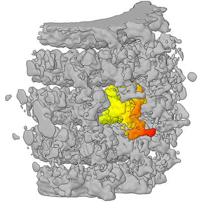 emd_3875.png emd_3875.png | 169.4 KB | ||
| Archive directory |  http://ftp.pdbj.org/pub/emdb/structures/EMD-3875ftp://ftp.pdbj.org/pub/emdb/structures/EMD-3875 http://ftp.pdbj.org/pub/emdb/structures/EMD-3875ftp://ftp.pdbj.org/pub/emdb/structures/EMD-3875 | HTTPS FTP |
-Related structure data
| Related structure data |  3869C  3870C  3871C 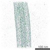 3872C 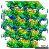 3873C 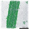 3874C 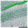 3876C 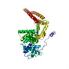 6ehlC 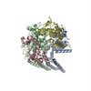 6ehmC C: citing same article ( |
|---|---|
| Similar structure data |


-Links
| EMDB pages | EMDB (EBI/PDBe) / EMDataResource |
|---|
-Map
| File | Download / File: emd_3875.map.gz / Format: CCP4 / Size: 27 MB / Type: IMAGE STORED AS FLOATING POINT NUMBER (4 BYTES) | ||||||||||||||||||||||||||||||||||||||||||||||||||||||||||||||||||||
|---|---|---|---|---|---|---|---|---|---|---|---|---|---|---|---|---|---|---|---|---|---|---|---|---|---|---|---|---|---|---|---|---|---|---|---|---|---|---|---|---|---|---|---|---|---|---|---|---|---|---|---|---|---|---|---|---|---|---|---|---|---|---|---|---|---|---|---|---|---|
| Annotation | Subtomogram averaging of Marburg virus nucleocapsid within intact virions | ||||||||||||||||||||||||||||||||||||||||||||||||||||||||||||||||||||
| Voxel size | X=Y=Z: 1.78 Å | ||||||||||||||||||||||||||||||||||||||||||||||||||||||||||||||||||||
| Density |
| ||||||||||||||||||||||||||||||||||||||||||||||||||||||||||||||||||||
| Symmetry | Space group: 1 | ||||||||||||||||||||||||||||||||||||||||||||||||||||||||||||||||||||
| Details | EMDB XML:
CCP4 map header:
| ||||||||||||||||||||||||||||||||||||||||||||||||||||||||||||||||||||
-Supplemental data
- Sample components
-Entire : Lake Victoria marburgvirus - Popp
| Entire | Name: Lake Victoria marburgvirus - Popp |
|---|---|
| Components |
|
-Supramolecule #1: Lake Victoria marburgvirus - Popp
| Supramolecule | Name: Lake Victoria marburgvirus - Popp / type: virus / ID: 1 / Parent: 0 / Macromolecule list: all Details: Virus was isolated from infected Huh-7 cells. Purified viruses were fixed with paraformaldehyde. NCBI-ID: 33728 / Sci species name: Lake Victoria marburgvirus - Popp / Virus type: VIRION / Virus isolate: STRAIN / Virus enveloped: Yes / Virus empty: No |
|---|---|
| Host system | Organism:  Homo sapiens (human) / Recombinant cell: HEK 293T Homo sapiens (human) / Recombinant cell: HEK 293T |
| Virus shell | Shell ID: 1 / Name: Nucleocapsid / Diameter: 310.0 Å |
-Macromolecule #1: Marburg nucleoprotein
| Macromolecule | Name: Marburg nucleoprotein / type: protein_or_peptide / ID: 1 / Enantiomer: LEVO |
|---|---|
| Source (natural) | Organism: Marburg virus - Musoke, Kenya, 1980 |
| Sequence | String: MDLHSLLELG TKPTAPHVRN KKVILFDTNH QVSICNQIID AINSGIDLGD LLEGGLLTLC VEHYYNSDK DKFNTSPVAK YLRDAGYEFD VIKNADATRF LDVSPNEPHY SPLILALKTL E STESQRGR IGLFLSFCSL FLPKLVVGDR ASIEKALRQV TVHQEQGIVT ...String: MDLHSLLELG TKPTAPHVRN KKVILFDTNH QVSICNQIID AINSGIDLGD LLEGGLLTLC VEHYYNSDK DKFNTSPVAK YLRDAGYEFD VIKNADATRF LDVSPNEPHY SPLILALKTL E STESQRGR IGLFLSFCSL FLPKLVVGDR ASIEKALRQV TVHQEQGIVT YPNHWLTTGH MK VIFGILR SSFILKFVLI HQGVNLVTGH DAYDSIISNS VGQTRFSGLL IVKTVLEFIL QKT DSGVTL HPLVRTSKVK NEVASFKQAL SNLARHGEYA PFARVLNLSG INNLEHGLYP QLSA IALGV ATAHGSTLAG VNVGEQYQQL REAAHDAEVK LQRRHEHQEI QAIAEDDEER KILEQ FHLQ KTEITHSQTL AVLSQKREKL ARLAAEIENN IVEDQGFKQS QNRVSQSFLN DPTPVE VTV QARPMNRPTA LPPPVDDKIE HESTEDSSSS SSFVDLNDPF ALLNEDEDTL DDSVMIP GT TSREFQGIPE PPRQSQDLNN SQGKQEDEST NRIKKQFLRY QELPPVQEDD ESEYTTDS Q ESIDQPGSDN EQGVDLPPPP LYAQEKRQDP IQHPAANPQD PFGSIGDVNG DILEPIRSP SSPSAPQEDT RMREAYELSP DFTNDEDNQQ NWPQRVVTKK GRTFLYPNDL LQTNPPESLI TALVEEYQN PVSAKELQAD WPDMSFDERR HVAMNL |
-Macromolecule #2: Marburg VP24
| Macromolecule | Name: Marburg VP24 / type: protein_or_peptide / ID: 2 / Enantiomer: LEVO |
|---|---|
| Source (natural) | Organism: Marburg virus - Musoke, Kenya, 1980 |
| Sequence | String: MAELSTRYNL PANVTENSIN LDLNSTARWI KEPSVGGWTV KWGNFVFHIP NTGMTLLHHL KSNFVVPEW QQTRNLFSHL FKNPKSTIIE PFLALRILLG VALKDQELQQ SLIPGFRSIV H MLSEWLLL EVTSAIHISP NLLGIYLTSD MFKILMAGVK NFFNKMFTLH ...String: MAELSTRYNL PANVTENSIN LDLNSTARWI KEPSVGGWTV KWGNFVFHIP NTGMTLLHHL KSNFVVPEW QQTRNLFSHL FKNPKSTIIE PFLALRILLG VALKDQELQQ SLIPGFRSIV H MLSEWLLL EVTSAIHISP NLLGIYLTSD MFKILMAGVK NFFNKMFTLH VVNDHGKPSS IE IKLTGQQ IIITRVNMGF LVEVRRIDIE PCCGETVLSE SVVFGLVAEA VLREHSQMEK GQP LNLTQY MNSKIAI |
-Macromolecule #3: Marburg VP35
| Macromolecule | Name: Marburg VP35 / type: protein_or_peptide / ID: 3 / Enantiomer: LEVO |
|---|---|
| Source (natural) | Organism: Marburg virus - Angola |
| Sequence | String: MWDSSYMQQV SEGLMTGKVP IDQVFGANPL EKLYKRRKPK GTVGLQCSPC LMSKATSTDD IIWDQLIVK RTLADLLIPI NRQISDIQST LSEVTTRVHE IERQLHEITP VLKMGRTLEA I SKGMSEML AKYDHLVIST GRTTAPAAAF DAYLNEHGVP PPQPAIFKDL ...String: MWDSSYMQQV SEGLMTGKVP IDQVFGANPL EKLYKRRKPK GTVGLQCSPC LMSKATSTDD IIWDQLIVK RTLADLLIPI NRQISDIQST LSEVTTRVHE IERQLHEITP VLKMGRTLEA I SKGMSEML AKYDHLVIST GRTTAPAAAF DAYLNEHGVP PPQPAIFKDL GVAQQACSKG TM VKNATTD AADKMSKVLE LSEETFSKPN LSAKDLALLL FTHLPGNNTP FHILAQVLSK IAY KSGKSG AFLDAFHQIL SEGENAQAAL TRLSRTFDAF LGVVPPVIRV KNFQTVPRPS QKSL RAVPP NPTIDKGWVC VYSSEQGETR ALKI |
-Experimental details
-Structure determination
| Method | cryo EM |
|---|---|
 Processing Processing | subtomogram averaging |
| Aggregation state | helical array |
-Sample preparation
| Buffer | pH: 7.4 Details: Virus was purified into Dulbecco's modified Eagle's medium (DMEM) with 4% paraformaldehyde |
|---|---|
| Grid | Model: C-flat 2/1 3C / Material: COPPER / Mesh: 300 / Support film - Material: CARBON / Support film - topology: HOLEY / Support film - Film thickness: 20.0 nm / Pretreatment - Type: GLOW DISCHARGE / Pretreatment - Atmosphere: AIR / Pretreatment - Pressure: 0.039 kPa |
| Vitrification | Cryogen name: ETHANE / Chamber humidity: 95 % / Instrument: FEI VITROBOT MARK II |
- Electron microscopy
| Microscope | FEI TITAN KRIOS |
|---|---|
| Electron beam | Acceleration voltage: 300 kV / Electron source: FIELD EMISSION GUN |
| Electron optics | C2 aperture diameter: 50.0 µm / Illumination mode: FLOOD BEAM / Imaging mode: BRIGHT FIELDBright-field microscopy / Cs: 2.7 mm / Nominal defocus max: 4.5 µm / Nominal defocus min: 2.0 µm / Nominal magnification: 81000 |
| Specialist optics | Energy filter - Name: GIF Quantum LS / Energy filter - Lower energy threshold: -10 eV / Energy filter - Upper energy threshold: 10 eV |
| Sample stage | Specimen holder model: FEI TITAN KRIOS AUTOGRID HOLDER / Cooling holder cryogen: NITROGEN |
| Image recording | Film or detector model: GATAN K2 QUANTUM (4k x 4k) / Detector mode: SUPER-RESOLUTION / Digitization - Dimensions - Width: 3708 pixel / Digitization - Dimensions - Height: 3708 pixel / Digitization - Frames/image: 1-5 / Average exposure time: 1.0 sec. / Average electron dose: 2.9 e/Å2 |
| Experimental equipment |  Model: Titan Krios / Image courtesy: FEI Company |
-Image processing
| Extraction | Number tomograms: 73 / Number images used: 65800 / Reference model: None Software:
Details: Points along the helical axis were manually placed to define a spline. A cylindrical grid as defined at a given radius from the spline; grid spacing was chosen to provide ~4x oversampling. | ||||||
|---|---|---|---|---|---|---|---|
| CTF correction | Software:
Details: CTF amplitude correction was performed during the wedge-weighted subtomogram averaging step. | ||||||
| Final angle assignment | Type: OTHER / Software - Name: TOM Details: Iterative angular search was performed via maximization of a modified constrained cross-correlation function. | ||||||
| Final reconstruction | Number classes used: 1 / Applied symmetry - Point group: C1 (asymmetric) / Algorithm: BACK PROJECTION / Resolution.type: BY AUTHOR / Resolution: 9.1 Å / Resolution method: FSC 0.143 CUT-OFF / Software - Name: TOM Details: Local resolution was estimated using moving window FSC calculations. Resolution varies from 9.1 to 13.8 Angstroms. Number subtomograms used: 1 | ||||||
| Details | Frames were aligned using K2Align software, based off the MotionCorr algorithm. Tomograms were reconstructed with IMOD, using stripwise CTF-correction and weighted back projection. Subtomogram averaging was performed using scripts derived from TOM, AV3, and DYNAMO. |
