 Movie
Movie Controller

[English] 日本語
 Yorodumi
- EMDB-3822: Structure of the full-length African cichlid nackednavirus icosah... -
+ Open data

- Basic information
| Entry | Database: EMDB / ID: EMD-3822 | |||||||||
|---|---|---|---|---|---|---|---|---|---|---|
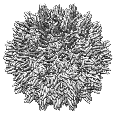
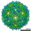
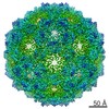
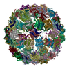
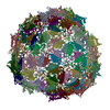
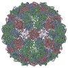
| Title | Structure of the full-length African cichlid nackednavirus icosahedral capsid | |||||||||
 Map data Map data | Structure of the full-length African cichlid nackednavirus capsid. | |||||||||
 Sample Sample |
| |||||||||
| Biological species |  Retro-transcribing viruses Retro-transcribing viruses | |||||||||
| Method |  single particle reconstruction / cryo EM / Resolution: 8.0 Å single particle reconstruction / cryo EM / Resolution: 8.0 Å | |||||||||
 Authors Authors | Mattei S / Briggs JAG / Seitz S | |||||||||
 Citation Citation | Journal: Cell Host Microbe / Year: 2017 Title: Deciphering the Origin and Evolution of Hepatitis B Viruses by Means of a Family of Non-enveloped Fish Viruses. Authors: Chris Lauber / Stefan Seitz / Simone Mattei / Alexander Suh / Jürgen Beck / Jennifer Herstein / Jacob Börold / Walter Salzburger / Lars Kaderali / John A G Briggs / Ralf Bartenschlager /     Abstract: Hepatitis B viruses (HBVs), which are enveloped viruses with reverse-transcribed DNA genomes, constitute the family Hepadnaviridae. An outstanding feature of HBVs is their streamlined genome ...Hepatitis B viruses (HBVs), which are enveloped viruses with reverse-transcribed DNA genomes, constitute the family Hepadnaviridae. An outstanding feature of HBVs is their streamlined genome organization with extensive gene overlap. Remarkably, the ∼1,100 bp open reading frame (ORF) encoding the envelope proteins is fully nested within the ORF of the viral replicase P. Here, we report the discovery of a diversified family of fish viruses, designated nackednaviruses, which lack the envelope protein gene, but otherwise exhibit key characteristics of HBVs including genome replication via protein-primed reverse-transcription and utilization of structurally related capsids. Phylogenetic reconstruction indicates that these two virus families separated more than 400 million years ago before the rise of tetrapods. We show that HBVs are of ancient origin, descending from non-enveloped progenitors in fishes. Their envelope protein gene emerged de novo, leading to a major transition in viral lifestyle, followed by co-evolution with their hosts over geologic eras. | |||||||||
| History |
|
- Structure visualization
| Movie |
 Movie viewer Movie viewer |
|---|---|
| Structure viewer | EM map: SurfViewMolmilJmol/JSmol |
| Supplemental images |
- Downloads & links
-EMDB archive
| Map data | emd_3822.map.gz | 471.6 MB | EMDB map data format | |
|---|---|---|---|---|
| Header (meta data) | emd-3822-v30.xmlemd-3822.xml | 12.3 KB 12.3 KB | Display Display | EMDB header |
| Images |  emd_3822.png emd_3822.png | 106.6 KB | ||
| Archive directory |  http://ftp.pdbj.org/pub/emdb/structures/EMD-3822ftp://ftp.pdbj.org/pub/emdb/structures/EMD-3822 http://ftp.pdbj.org/pub/emdb/structures/EMD-3822ftp://ftp.pdbj.org/pub/emdb/structures/EMD-3822 | HTTPS FTP |
-Related structure data


-Links
| EMDB pages | EMDB (EBI/PDBe) / EMDataResource |
|---|
-Map
| File | Download / File: emd_3822.map.gz / Format: CCP4 / Size: 512 MB / Type: IMAGE STORED AS FLOATING POINT NUMBER (4 BYTES) | ||||||||||||||||||||||||||||||||||||||||||||||||||||||||||||
|---|---|---|---|---|---|---|---|---|---|---|---|---|---|---|---|---|---|---|---|---|---|---|---|---|---|---|---|---|---|---|---|---|---|---|---|---|---|---|---|---|---|---|---|---|---|---|---|---|---|---|---|---|---|---|---|---|---|---|---|---|---|
| Annotation | Structure of the full-length African cichlid nackednavirus capsid. | ||||||||||||||||||||||||||||||||||||||||||||||||||||||||||||
| Voxel size | X=Y=Z: 1.08 Å | ||||||||||||||||||||||||||||||||||||||||||||||||||||||||||||
| Density |
| ||||||||||||||||||||||||||||||||||||||||||||||||||||||||||||
| Symmetry | Space group: 1 | ||||||||||||||||||||||||||||||||||||||||||||||||||||||||||||
| Details | EMDB XML:
CCP4 map header:
| ||||||||||||||||||||||||||||||||||||||||||||||||||||||||||||
-Supplemental data
- Sample components
-Entire : Retro-transcribing viruses
| Entire | Name: Retro-transcribing viruses |
|---|---|
| Components |
|
-Supramolecule #1: Retro-transcribing viruses
| Supramolecule | Name: Retro-transcribing viruses / type: virus / ID: 1 / Parent: 0 / Details: Full-length capsid (aa1-174) / NCBI-ID: 35268 / Sci species name: Retro-transcribing viruses / Virus type: VIRION / Virus isolate: STRAIN / Virus enveloped: No / Virus empty: Yes |
|---|---|
| Host (natural) | Organism:  Ophthalmotilapia ventralis (fish) Ophthalmotilapia ventralis (fish) |
| Host system | Organism:  Escherichia coli (E. coli) Escherichia coli (E. coli) |
| Virus shell | Shell ID: 1 / Name: capsid / Diameter: 320.0 Å / T number (triangulation number): 3 |
-Experimental details
-Structure determination
| Method | cryo EM |
|---|---|
 Processing Processing | single particle reconstruction |
| Aggregation state | particle |
-Sample preparation
| Buffer | pH: 7.2 |
|---|---|
| Grid | Model: Protochips C-Flat / Material: COPPER / Mesh: 200 / Pretreatment - Type: GLOW DISCHARGE / Pretreatment - Atmosphere: AIR / Details: 20 mA |
| Vitrification | Cryogen name: ETHANE / Chamber humidity: 100 % / Chamber temperature: 288 K / Instrument: FEI VITROBOT MARK II |
- Electron microscopy
| Microscope | FEI TITAN KRIOS |
|---|---|
| Electron beam | Acceleration voltage: 300 kV / Electron source: FIELD EMISSION GUN |
| Electron optics | Illumination mode: FLOOD BEAM / Imaging mode: BRIGHT FIELDBright-field microscopy / Cs: 2.7 mm / Nominal defocus max: 4.0 µm / Nominal defocus min: 1.0 µm |
| Sample stage | Specimen holder model: FEI TITAN KRIOS AUTOGRID HOLDER / Cooling holder cryogen: NITROGEN |
| Image recording | Film or detector model: FEI FALCON II (4k x 4k) / Detector mode: INTEGRATING / Digitization - Dimensions - Width: 4096 pixel / Digitization - Dimensions - Height: 4096 pixel / Digitization - Sampling interval: 14.0 µm / Digitization - Frames/image: 1-7 / Number grids imaged: 1 / Number real images: 1548 / Average electron dose: 25.0 e/Å2 |
| Experimental equipment |  Model: Titan Krios / Image courtesy: FEI Company |
-Image processing
| Particle selection | Number selected: 53431 Details: manual picking of a few hundred for template production, then automated |
|---|---|
| CTF correction | Software - Name: CTFFIND (ver. 3) |
| Startup model | Type of model: NONE Details: A spherical density with diameter of 40 nm was provided as an initial model |
| Initial angle assignment | Type: PROJECTION MATCHING / Software - Name: RELION (ver. 1.2) |
| Final 3D classification | Number classes: 20 / Software - Name: RELION (ver. 1.2) Details: 2D classification was performed using first IMAGIC and then RELION 1.2 to select regular, intact particles and remove disrupted ones. |
| Final angle assignment | Type: PROJECTION MATCHING / Software - Name: RELION (ver. 1.2) |
| Final reconstruction | Applied symmetry - Point group: I (icosahedral) / Algorithm: BACK PROJECTION / Resolution.type: BY AUTHOR / Resolution: 8.0 Å / Resolution method: FSC 0.143 CUT-OFF / Software - Name: RELION (ver. 1.2) / Number images used: 2547 |
