 Movie
Movie Controller


+ Open data

- Basic information
| Entry | Database: EMDB / ID: EMD-3604 | |||||||||
|---|---|---|---|---|---|---|---|---|---|---|
| Title | Structure of the C. crescentus S-layer | |||||||||
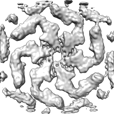
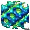
 Map data Map data | Sub-tomogram averaging of the Caulobacter crescentus S-layer | |||||||||
 Sample Sample |
| |||||||||
| Function / homology | RsaA N-terminal domain / RTX calcium-binding nonapeptide repeat / RTX calcium-binding nonapeptide repeat (4 copies) / Serralysin-like metalloprotease, C-terminal /  calcium ion binding / S-layer protein rsaA calcium ion binding / S-layer protein rsaA Function and homology information Function and homology information | |||||||||
| Biological species |  Caulobacter crescentus NA1000 (bacteria) Caulobacter crescentus NA1000 (bacteria) | |||||||||
| Method | subtomogram averaging / cryo EM / Resolution: 7.4 Å | |||||||||
 Authors Authors | Bharat TA / Hagen WJ / Briggs JA / Lowe J | |||||||||
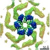
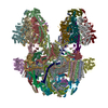
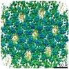
 Citation Citation | Journal: Nat Microbiol / Year: 2017 Title: Structure of the hexagonal surface layer on Caulobacter crescentus cells. Authors: Tanmay A M Bharat / Danguole Kureisaite-Ciziene / Gail G Hardy / Ellen W Yu / Jessica M Devant / Wim J H Hagen / Yves V Brun / John A G Briggs / Jan Löwe /    Abstract: Many prokaryotic cells are encapsulated by a surface layer (S-layer) consisting of repeating units of S-layer proteins. S-layer proteins are a diverse class of molecules found in Gram-positive and ...Many prokaryotic cells are encapsulated by a surface layer (S-layer) consisting of repeating units of S-layer proteins. S-layer proteins are a diverse class of molecules found in Gram-positive and Gram-negative bacteria and most archaea. S-layers protect cells from the outside, provide mechanical stability and also play roles in pathogenicity. In situ structural information about this highly abundant class of proteins is scarce, so atomic details of how S-layers are arranged on the surface of cells have remained elusive. Here, using purified Caulobacter crescentus' sole S-layer protein RsaA, we obtained a 2.7 Å X-ray structure that shows the hexameric S-layer lattice. We also solved a 7.4 Å structure of the S-layer through electron cryotomography and sub-tomogram averaging of cell stalks. The X-ray structure was docked unambiguously into the electron cryotomography map, resulting in a pseudo-atomic-level description of the in vivo S-layer, which agrees completely with the atomic X-ray lattice model. The cellular S-layer atomic structure shows that the S-layer is porous, with a largest gap dimension of 27 Å, and is stabilized by multiple Ca ions bound near the interfaces. This study spans different spatial scales from atoms to cells by combining X-ray crystallography with electron cryotomography and sub-nanometre-resolution sub-tomogram averaging. | |||||||||
| History |
|
- Structure visualization
| Movie |
Movie viewer |
|---|---|
| Structure viewer | EM map: SurfViewMolmilJmol/JSmol |
| Supplemental images |
- Downloads & links
-EMDB archive
| Map data | emd_3604.map.gz | 27.8 MB | EMDB map data format | |
|---|---|---|---|---|
| Header (meta data) | emd-3604-v30.xmlemd-3604.xml | 14.3 KB 14.3 KB | Display Display | EMDB header |
| Images |  emd_3604.png emd_3604.png | 97.3 KB | ||
| Archive directory |  http://ftp.pdbj.org/pub/emdb/structures/EMD-3604ftp://ftp.pdbj.org/pub/emdb/structures/EMD-3604 http://ftp.pdbj.org/pub/emdb/structures/EMD-3604ftp://ftp.pdbj.org/pub/emdb/structures/EMD-3604 | HTTPS FTP |
-Related structure data
| Related structure data | 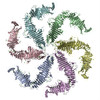 5n97MC 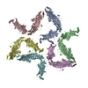 5n8pC M: atomic model generated by this map C: citing same article ( |
|---|---|
| Similar structure data |


-Links
| EMDB pages | EMDB (EBI/PDBe) / EMDataResource |
|---|
-Map
| File | Download / File: emd_3604.map.gz / Format: CCP4 / Size: 30.5 MB / Type: IMAGE STORED AS FLOATING POINT NUMBER (4 BYTES) | ||||||||||||||||||||||||||||||||||||||||||||||||||||||||||||
|---|---|---|---|---|---|---|---|---|---|---|---|---|---|---|---|---|---|---|---|---|---|---|---|---|---|---|---|---|---|---|---|---|---|---|---|---|---|---|---|---|---|---|---|---|---|---|---|---|---|---|---|---|---|---|---|---|---|---|---|---|---|
| Annotation | Sub-tomogram averaging of the Caulobacter crescentus S-layer | ||||||||||||||||||||||||||||||||||||||||||||||||||||||||||||
| Voxel size | X=Y=Z: 1.35 Å | ||||||||||||||||||||||||||||||||||||||||||||||||||||||||||||
| Density |
| ||||||||||||||||||||||||||||||||||||||||||||||||||||||||||||
| Symmetry | Space group: 1 | ||||||||||||||||||||||||||||||||||||||||||||||||||||||||||||
| Details | EMDB XML:
CCP4 map header:
| ||||||||||||||||||||||||||||||||||||||||||||||||||||||||||||
-Supplemental data
- Sample components
-Entire : Caulobacter crescentus S-layer
| Entire | Name: Caulobacter crescentus S-layer |
|---|---|
| Components |
|
-Supramolecule #1: Caulobacter crescentus S-layer
| Supramolecule | Name: Caulobacter crescentus S-layer / type: organelle_or_cellular_component / ID: 1 / Parent: 0 / Macromolecule list: #1 / Details: Caulobacter crescentus S-layer |
|---|---|
| Source (natural) | Organism: Caulobacter crescentus NA1000 (bacteria) / Strain: YB2811 / Location in cell: extra-cellular |
-Macromolecule #1: S-layer protein rsaA
| Macromolecule | Name: S-layer protein rsaA / type: protein_or_peptide / ID: 1 / Number of copies: 6 / Enantiomer: LEVO |
|---|---|
| Source (natural) | Organism: Caulobacter crescentus NA1000 (bacteria) |
| Molecular weight | Theoretical: 73.159305 KDa |
| Sequence | String: GSTLSLTTGT DTLTGTANND TFVAGEVAGA ATLTVGDTLS GGAGTDVLNW VQAAAVTALP TGVTISGIET MNVTSGAAIT LNTSSGVTG LTALNTNTSG AAQTVTAGAG QNLTATTAAQ AANNVAVDGG ANVTVASTGV TSGTTTVGAN SAASGTVSVS V ANSSTTTT ...String: GSTLSLTTGT DTLTGTANND TFVAGEVAGA ATLTVGDTLS GGAGTDVLNW VQAAAVTALP TGVTISGIET MNVTSGAAIT LNTSSGVTG LTALNTNTSG AAQTVTAGAG QNLTATTAAQ AANNVAVDGG ANVTVASTGV TSGTTTVGAN SAASGTVSVS V ANSSTTTT GAIAVTGGTA VTVAQTAGNA VNTTLTQADV TVTGNSSTTA VTVTQTAAAT AGATVAGRVN GAVTITDSAA AS ATTAGKI ATVTLGSFGA ATIDSSALTT VNLSGTGTSL GIGRGALTAT PTANTLTLNV NGLTTTGAIT DSEAAADDGF TTI NIAGST ASSTIASLVA ADATTLNISG DARVTITSHT AAALTGITVT NSVGATLGAE LATGLVFTGG AGADSILLGA TTKA IVMGA GDDTVTVSSA TLGAGGSVNG GDGTDVLVAN VNGSSFSADP AFGGFETLRV AGAAAQGSHN ANGFTALQLG ATAGA TTFT NVAVNVGLTV LAAPTGTTTV TLANATGTSD VFNLTLSSSA ALAAGTVALA GVETVNIAAT DTNTTAHVDT LTLQAT SAK SIVVTGNAGL NLTNTGNTAV TSFDASAVTG TGSAVTFVSA NTTVGEVVTI RGGAGADSLT GSATANDTII GGAGADT LV YTGGTDTFTG GTGADIFDIN AIGTSTAFVT ITDAAVGDKL DLVGISTNGA IADGAFGAAV TLGAAATLAQ YLDAAAAG D GSGTSVAKWF QFGGDTYVVV DSSAGATFVS GADAVIKLTG LVTLTTSAFA TEVLTLA |
-Macromolecule #2: CALCIUM ION
| Macromolecule | Name: CALCIUM ION / type: ligand / ID: 2 / Number of copies: 114 / Formula: CA |
|---|---|
| Molecular weight | Theoretical: 40.078 Da |
-Experimental details
-Structure determination
| Method | cryo EM |
|---|---|
 Processing Processing | subtomogram averaging |
| Aggregation state | particle |
-Sample preparation
| Concentration | 1 mg/mL |
|---|---|
| Buffer | pH: 7 / Details: PYE medium |
| Grid | Model: Quantifoil R2/2 / Material: COPPER/RHODIUM / Mesh: 200 / Support film - Material: CARBON / Support film - topology: HOLEY ARRAY / Pretreatment - Type: GLOW DISCHARGE / Pretreatment - Atmosphere: AIR |
| Vitrification | Cryogen name: ETHANE / Chamber humidity: 100 % / Chamber temperature: 283.15 K / Instrument: FEI VITROBOT MARK IV / Details: 1.5 s blot. |
| Details | Caulobacter crescentus stalk |
- Electron microscopy
| Microscope | FEI TITAN KRIOS |
|---|---|
| Electron beam | Acceleration voltage: 300 kV / Electron source: FIELD EMISSION GUN |
| Electron optics | C2 aperture diameter: 70.0 µm / Calibrated defocus max: 5.0 µm / Calibrated defocus min: 2.0 µm / Calibrated magnification: 105000 / Illumination mode: FLOOD BEAM / Imaging mode: BRIGHT FIELDBright-field microscopy / Cs: 2.7 mm / Nominal defocus max: 5.0 µm / Nominal defocus min: 2.0 µm / Nominal magnification: 105000 |
| Specialist optics | Energy filter - Name: GIF Quantum / Energy filter - Lower energy threshold: 0 eV / Energy filter - Upper energy threshold: 20 eV |
| Sample stage | Specimen holder model: FEI TITAN KRIOS AUTOGRID HOLDER / Cooling holder cryogen: NITROGEN |
| Temperature | Min: 70.0 K / Max: 70.0 K |
| Image recording | Film or detector model: GATAN K2 SUMMIT (4k x 4k) / Detector mode: SUPER-RESOLUTION / Number grids imaged: 1 / Average exposure time: 1.0 sec. / Average electron dose: 3.4 e/Å2 / Details: Dose symmetric tilt scheme (Hagen et al) |
| Experimental equipment |  Model: Titan Krios / Image courtesy: FEI Company |
-Image processing
| Crystal parameters | Unit cell - A: 220 Å / Unit cell - B: 220 Å / Unit cell - C: 220 Å / Unit cell - C sampling length: 10 Å / Unit cell - γ: 1 ° / Plane group: P 1 21 |
|---|---|
| Extraction | Number tomograms: 110 / Number images used: 51866 / Reference model: Ab initio / Method: RELION / Software - Name: RELION (ver. 1.4) / Details: RELION subtomogram averaging |
| CTF correction | Details: Following Schur et al, 2016 |
| Final angle assignment | Type: OTHER / Details: Following Schur et al, 2016 |
| Final reconstruction | Number classes used: 1 / Algorithm: FOURIER SPACE / Resolution.type: BY AUTHOR / Resolution: 7.4 Å / Resolution method: FSC 0.143 CUT-OFF / Number subtomograms used: 51866 |
-Atomic model buiding 1
| Refinement | Space: REAL / Protocol: RIGID BODY FIT / Target criteria: Cross-correlation coefficient |
|---|---|
| Output model | PDB-5n97: |
