 Movie
Movie Controller

[English] 日本語
 Yorodumi
- EMDB-3583: Cryo-EM structure of a Separase-Securin complex from Caenorhabdit... -
+ Open data

- Basic information
| Entry | Database: EMDB / ID: EMD-3583 | |||||||||
|---|---|---|---|---|---|---|---|---|---|---|
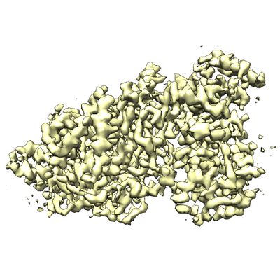
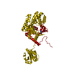
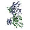
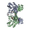
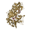
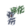
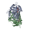
| Title | Cryo-EM structure of a Separase-Securin complex from Caenorhabditis elegans at 3.8 A resolution | |||||||||
 Map data Map data | Map of the C. elegans Separase-Securin complex. Manual B-factor sharpening of 140 applied. | |||||||||
 Sample Sample |
| |||||||||
| Function / homology |  Function and homology information Function and homology informationSeparation of Sister Chromatids / separase-securin complex / eggshell formation /  metaphase plate / regulation of nematode larval development / separase / cortical granule exocytosis / maintenance of meiotic sister chromatid cohesion / meiotic chromosome separation / maintenance of mitotic sister chromatid cohesion ...Separation of Sister Chromatids / separase-securin complex / eggshell formation / metaphase plate / regulation of nematode larval development / separase / cortical granule exocytosis / maintenance of meiotic sister chromatid cohesion / meiotic chromosome separation / maintenance of mitotic sister chromatid cohesion / polarity specification of anterior/posterior axis / polar body extrusion after meiotic divisions / cortical granule / regulation of centriole-centriole cohesion / mitotic sister chromatid separation / regulation of exocytosis / multicellular organismal reproductive process / meiotic spindle / regulation of locomotion / centrosome localization / cleavage furrow / centrosome duplication / mitotic cytokinesis / condensed chromosome / condensed nuclear chromosome / spindle microtubule / protein localization / mitotic spindle / spindle / protein processing / nuclear envelope / chromosome / cell cortex / midbody / protease binding / protein stabilization / cysteine-type endopeptidase activity / centrosome / ubiquitin protein ligase binding / nucleus / cytoplasm metaphase plate / regulation of nematode larval development / separase / cortical granule exocytosis / maintenance of meiotic sister chromatid cohesion / meiotic chromosome separation / maintenance of mitotic sister chromatid cohesion ...Separation of Sister Chromatids / separase-securin complex / eggshell formation / metaphase plate / regulation of nematode larval development / separase / cortical granule exocytosis / maintenance of meiotic sister chromatid cohesion / meiotic chromosome separation / maintenance of mitotic sister chromatid cohesion / polarity specification of anterior/posterior axis / polar body extrusion after meiotic divisions / cortical granule / regulation of centriole-centriole cohesion / mitotic sister chromatid separation / regulation of exocytosis / multicellular organismal reproductive process / meiotic spindle / regulation of locomotion / centrosome localization / cleavage furrow / centrosome duplication / mitotic cytokinesis / condensed chromosome / condensed nuclear chromosome / spindle microtubule / protein localization / mitotic spindle / spindle / protein processing / nuclear envelope / chromosome / cell cortex / midbody / protease binding / protein stabilization / cysteine-type endopeptidase activity / centrosome / ubiquitin protein ligase binding / nucleus / cytoplasmSimilarity search - Function | |||||||||
| Biological species |  Caenorhabditis elegans (invertebrata) Caenorhabditis elegans (invertebrata) | |||||||||
| Method | single particle reconstruction / cryo EM / Resolution: 3.8 Å | |||||||||
 Authors Authors | Boland A / Martin TG / Zhang Z / Yang J / Bai XC / Chang L / Scheres SHW / Barford D | |||||||||
 Citation Citation | Journal: Nat Struct Mol Biol / Year: 2017 Title: Cryo-EM structure of a metazoan separase-securin complex at near-atomic resolution. Authors: Andreas Boland / Thomas G Martin / Ziguo Zhang / Jing Yang / Xiao-Chen Bai / Leifu Chang / Sjors H W Scheres / David Barford /  Abstract: Separase is a caspase-family protease that initiates chromatid segregation by cleaving the kleisin subunits (Scc1 and Rec8) of cohesin, and regulates centrosome duplication and mitotic spindle ...Separase is a caspase-family protease that initiates chromatid segregation by cleaving the kleisin subunits (Scc1 and Rec8) of cohesin, and regulates centrosome duplication and mitotic spindle function through cleavage of kendrin and Slk19. To understand the mechanisms of securin regulation of separase, we used single-particle cryo-electron microscopy (cryo-EM) to determine a near-atomic-resolution structure of the Caenorhabditis elegans separase-securin complex. Separase adopts a triangular-shaped bilobal architecture comprising an N-terminal tetratricopeptide repeat (TPR)-like α-solenoid domain docked onto the conserved C-terminal protease domain. Securin engages separase in an extended antiparallel conformation, interacting with both lobes. It inhibits separase by interacting with the catalytic site through a pseudosubstrate mechanism, thus revealing that in the inhibited separase-securin complex, the catalytic site adopts a conformation compatible with substrate binding. Securin is protected from cleavage because an aliphatic side chain at the P1 position represses protease activity by disrupting the organization of catalytic site residues. | |||||||||
| History |
|
- Structure visualization
| Movie |
Movie viewer |
|---|---|
| Structure viewer | EM map: SurfViewMolmilJmol/JSmol |
| Supplemental images |
- Downloads & links
-EMDB archive
| Map data | emd_3583.map.gz | 1.6 MB | EMDB map data format | |
|---|---|---|---|---|
| Header (meta data) | emd-3583-v30.xmlemd-3583.xml | 15.8 KB 15.8 KB | Display Display | EMDB header |
| Images |  emd_3583.png emd_3583.png | 153 KB | ||
| Archive directory |  http://ftp.pdbj.org/pub/emdb/structures/EMD-3583ftp://ftp.pdbj.org/pub/emdb/structures/EMD-3583 http://ftp.pdbj.org/pub/emdb/structures/EMD-3583ftp://ftp.pdbj.org/pub/emdb/structures/EMD-3583 | HTTPS FTP |
-Related structure data
| Related structure data |  5mz6MC  3584C M: atomic model generated by this map C: citing same article ( |
|---|---|
| Similar structure data |

-Links
| EMDB pages | EMDB (EBI/PDBe) / EMDataResource |
|---|
-Map
| File | Download / File: emd_3583.map.gz / Format: CCP4 / Size: 22.2 MB / Type: IMAGE STORED AS FLOATING POINT NUMBER (4 BYTES) | ||||||||||||||||||||||||||||||||||||||||||||||||||||||||||||
|---|---|---|---|---|---|---|---|---|---|---|---|---|---|---|---|---|---|---|---|---|---|---|---|---|---|---|---|---|---|---|---|---|---|---|---|---|---|---|---|---|---|---|---|---|---|---|---|---|---|---|---|---|---|---|---|---|---|---|---|---|---|
| Annotation | Map of the C. elegans Separase-Securin complex. Manual B-factor sharpening of 140 applied. | ||||||||||||||||||||||||||||||||||||||||||||||||||||||||||||
| Voxel size | X=Y=Z: 1.43 Å | ||||||||||||||||||||||||||||||||||||||||||||||||||||||||||||
| Density |
| ||||||||||||||||||||||||||||||||||||||||||||||||||||||||||||
| Symmetry | Space group: 1 | ||||||||||||||||||||||||||||||||||||||||||||||||||||||||||||
| Details | EMDB XML:
CCP4 map header:
| ||||||||||||||||||||||||||||||||||||||||||||||||||||||||||||
-Supplemental data
- Sample components
-Entire : Caenorhabditis elegans Separase-Securin complex
| Entire | Name: Caenorhabditis elegans Separase-Securin complex |
|---|---|
| Components |
|
-Supramolecule #1: Caenorhabditis elegans Separase-Securin complex
| Supramolecule | Name: Caenorhabditis elegans Separase-Securin complex / type: complex / ID: 1 / Parent: 0 / Macromolecule list: all Details: C. elegans Separase-Securin complex at 3.8 Angstrom resolution refined with Relion. |
|---|---|
| Source (natural) | Organism: Caenorhabditis elegans (invertebrata) |
| Recombinant expression | Organism:  Trichoplusia ni (cabbage looper) Trichoplusia ni (cabbage looper) |
| Molecular weight | Theoretical: 170 KDa |
-Macromolecule #1: SEParase
| Macromolecule | Name: SEParase / type: protein_or_peptide / ID: 1 / Number of copies: 1 / Enantiomer: LEVO |
|---|---|
| Source (natural) | Organism: Caenorhabditis elegans (invertebrata) |
| Molecular weight | Theoretical: 144.315984 KDa |
| Recombinant expression | Organism: Trichoplusia ni (cabbage looper) |
| Sequence | String: MKITNKSVDK QHIEKLDELR KNVSCTVIGF AEQTAELQQE ISELFIAEFG VNGPIDMNSL SKLARITSYY ASSEYFQGLA KYQRTACKM FITWQTLRKE AMECRSKDRE IFASIPAKLC FFYFYNGELC RAVVCLLDYI DLSDDTLAKE AALRWLMFLG E TELIEKKL ...String: MKITNKSVDK QHIEKLDELR KNVSCTVIGF AEQTAELQQE ISELFIAEFG VNGPIDMNSL SKLARITSYY ASSEYFQGLA KYQRTACKM FITWQTLRKE AMECRSKDRE IFASIPAKLC FFYFYNGELC RAVVCLLDYI DLSDDTLAKE AALRWLMFLG E TELIEKKL KTWKMDKSSK DMFSATEFAM NYLKKSEYRV EMLEKLMKLR DKVKSDPTRS FSRYELASYV SWLCSTLSNV PV GSALREC EFPDRVSHIQ EAALKSDSLV RNRIPGLASS QFDNSVNASI WPFLDGHQED SNYYVHIGST IAWHFEMRRE CAL VNVTTA QTRDSMSAMI LNLRVALKSA SFFRVLQTTN TLAYYSSIIE EAGSEKNAKL MRVSCVNLLS SNPIIVRCST PKET GATSR AHTPMAGSSV SEKQNTMRPD LADLLGDLEL LDEQSFHPIT RSCVCNVCTI YPLHSSFAAE YMMSYAIHSD FSQLS IKHF NDEFARIRER GMSSQVLMHR DSSVRPRPNI IQNEIFGMCV IRWLTKKLDS KESADEDTME IFNNALKIVR YLQQRT TDM ILAVTQLGRQ LEFPMECNYS WMRPTIRKPR VKATIDCAVD ILRAVSPFGR RPKVEKLEKN LQPFDKERFE KVRLAMR NE MNHYGHILYR EWRCRLFAYV GRTSRDPWEA AYAWAESTQI GARNAVQSRL EKCKRGLVTM SGHDRFKTCV QSMPDEMT L VQIAMADDKT IYLVKLHADR DPIIMPLAHY SQAVELMDKF TFLLDEDEMI AKYPGDITPE EFWKRRKIVD GRMMTFVDE VQKHFLGVAA SLLMPSGQLG PKAAELAIKI HKLSKGGLLL GEAKEMVYQS KLMDAKSWEA LILRFCEMRT TDEKFKSFLP LMHRNSVEV MNQDDSIVTE KKYTYLVICP HLSQFCWERL PIFDEYPYVG RQVSIHSTFS QLEAMKSQEK QIPLQIDVQN A YYILDPDN NLGETQKRMV EYINKFNWEG TVGSAPKSNE ISAALSQRDA FFFIGHGSGS SVMPRSVLKQ STCNAISLLM GC GSVRTIP QALGFDGKTA ILDYAMAKCP LIVGCLWTVT DGEIDRFLIR MIDDCFEDSK SLTGIDKLRQ LSEAMHEARS KAR LKYLTG AAVVMYGLPV VAKQTTPFVE KDQRNLPQTP KTSARTSMRM ETVPKTPKQE FVTSKSVPMT PIFSNNENKS PSRA RMPSR VLKTPRQVKT FQEEDDEAPK RSTTRQLKPL VAPPIPATPT TRTTRSSART PSRSRNL |
-Macromolecule #2: Interactor of FizzY protein
| Macromolecule | Name: Interactor of FizzY protein / type: protein_or_peptide / ID: 2 / Number of copies: 1 / Enantiomer: LEVO |
|---|---|
| Source (natural) | Organism: Caenorhabditis elegans (invertebrata) |
| Molecular weight | Theoretical: 27.027646 KDa |
| Recombinant expression | Organism: Trichoplusia ni (cabbage looper) |
| Sequence | String: MEDLNFEERG STQIPASLQQ HFSAKLGRQN ELEKTPSRGG LGLVVNSSKT PGGKSLQSLA SACKVPPSTK KNTIPIAFEC YEDETDDQI ADVATIKKTE KHPCSPIDTA NRCETFDSLA ADIEDDMLNL EDQDVVLSED RPYGDVIDPA ESEAEALAEL G VEEWDSYP ...String: MEDLNFEERG STQIPASLQQ HFSAKLGRQN ELEKTPSRGG LGLVVNSSKT PGGKSLQSLA SACKVPPSTK KNTIPIAFEC YEDETDDQI ADVATIKKTE KHPCSPIDTA NRCETFDSLA ADIEDDMLNL EDQDVVLSED RPYGDVIDPA ESEAEALAEL G VEEWDSYP PIDPASRIGD DFNYVLRTED FAEEGDVKLE ETRHRTVIAD IDEVKMSKAE RNELFSMLAD DLDSYDLLAE EA NLPL |
-Experimental details
-Structure determination
| Method | cryo EM |
|---|---|
 Processing Processing | single particle reconstruction |
| Aggregation state | particle |
-Sample preparation
| Concentration | 0.02 mg/mL |
|---|---|
| Buffer | pH: 7.8 |
| Grid | Model: Quantifoil R1.2/1.3 / Material: GOLD / Mesh: 300 / Support film - Material: GRAPHENE OXIDE / Support film - topology: HOLEY ARRAY / Pretreatment - Type: GLOW DISCHARGE / Pretreatment - Atmosphere: AIR Details: Glow discharging was performed before applying graphene oxide solution onto the grid. Grid was washed three times, dried and sample was applied. For detailed information see: https: ...Details: Glow discharging was performed before applying graphene oxide solution onto the grid. Grid was washed three times, dried and sample was applied. For detailed information see: https://figshare.com/articles/Graphene_Oxide_Grid_Preparation/3178669 |
| Vitrification | Cryogen name: ETHANE / Chamber humidity: 100 % / Chamber temperature: 277 K / Details: Custom Manual Plunger. |
| Details | Monodisperse sample |
- Electron microscopy
| Microscope | FEI TITAN KRIOS |
|---|---|
| Electron beam | Acceleration voltage: 300 kV / Electron source: FIELD EMISSION GUN |
| Electron optics | C2 aperture diameter: 70.0 µm / Illumination mode: FLOOD BEAM / Imaging mode: BRIGHT FIELDBright-field microscopy / Cs: 2.7 mm |
| Specialist optics | Energy filter - Name: GIF / Energy filter - Lower energy threshold: -20 eV / Energy filter - Upper energy threshold: 20 eV |
| Sample stage | Specimen holder model: FEI TITAN KRIOS AUTOGRID HOLDER / Cooling holder cryogen: NITROGEN |
| Image recording | Film or detector model: GATAN K2 SUMMIT (4k x 4k) / Detector mode: SUPER-RESOLUTION / Digitization - Dimensions - Width: 3710 pixel / Digitization - Dimensions - Height: 3710 pixel / Average exposure time: 0.8 sec. / Average electron dose: 1.25 e/Å2 |
| Experimental equipment |  Model: Titan Krios / Image courtesy: FEI Company |
-Image processing
| CTF correction | Software - Name: Gctf |
|---|---|
| Startup model | Type of model: NONE / Details: Initial model was created using SIMPLE PRIME |
| Initial angle assignment | Type: ANGULAR RECONSTITUTION / Software - Name: RELION (ver. 2.0) |
| Final angle assignment | Type: ANGULAR RECONSTITUTION / Software - Name: RELION (ver. 2.0) |
| Final reconstruction | Applied symmetry - Point group: C1 (asymmetric) / Resolution.type: BY AUTHOR / Resolution: 3.8 Å / Resolution method: FSC 0.143 CUT-OFF / Software - Name: RELION (ver. 2.0) / Number images used: 103696 |
-Atomic model buiding 1
| Refinement | Space: REAL / Protocol: AB INITIO MODEL |
|---|---|
| Output model | PDB-5mz6: |
