 Movie
Movie Controller

[English] 日本語
 Yorodumi
- EMDB-1680: Macromolecular crystal data phased by negative staining electron ... -
+ Open data

- Basic information
| Entry | Database: EMDB / ID: EMD-1680 | |||||||||
|---|---|---|---|---|---|---|---|---|---|---|
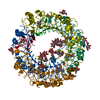
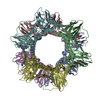
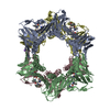
| Title | Macromolecular crystal data phased by negative staining electron microscopy reconstructions | |||||||||
 Map data Map data | Macromolecular crystal data phased by negative staining electron microscopy reconstructions as a proof of principle | |||||||||
 Sample Sample |
| |||||||||
 Keywords Keywords |  Negative staining / electron microscopy / reconstructions / phasing Negative staining / electron microscopy / reconstructions / phasing | |||||||||
| Biological species |  Candida albicans (yeast) Candida albicans (yeast) | |||||||||
| Method | single particle reconstruction / negative staining / Resolution: 15.0 Å | |||||||||
 Authors Authors | Trapani S / Schoehn G / Navaza J / Abergel C | |||||||||
 Citation Citation | Journal: Acta Crystallogr D Biol Crystallogr / Year: 2010 Title: Macromolecular crystal data phased by negative-stained electron-microscopy reconstructions. Authors: Stefano Trapani / Guy Schoehn / Jorge Navaza / Chantal Abergel /  Abstract: The combination of transmission electron microscopy with X-ray diffraction data is usually limited to relatively large particles. Here, the approach is continued one step further by utilizing ...The combination of transmission electron microscopy with X-ray diffraction data is usually limited to relatively large particles. Here, the approach is continued one step further by utilizing negative staining, a technique that is of wider applicability than cryo-electron microscopy, to produce models of medium-size proteins suitable for molecular replacement. The technique was used to solve the crystal structure of the dodecameric type II dehydroquinase enzyme from Candida albicans (approximately 190 kDa) and that of the orthologous Streptomyces coelicolor protein. | |||||||||
| History |
|
- Structure visualization
| Movie |
 Movie viewer Movie viewer |
|---|---|
| Structure viewer | EM map: SurfViewMolmilJmol/JSmol |
| Supplemental images |

- Downloads & links
-EMDB archive
| Map data | emd_1680.map.gz | 2 MB | EMDB map data format | |
|---|---|---|---|---|
| Header (meta data) | emd-1680-v30.xmlemd-1680.xml | 8.9 KB 8.9 KB | Display Display | EMDB header |
| Images | 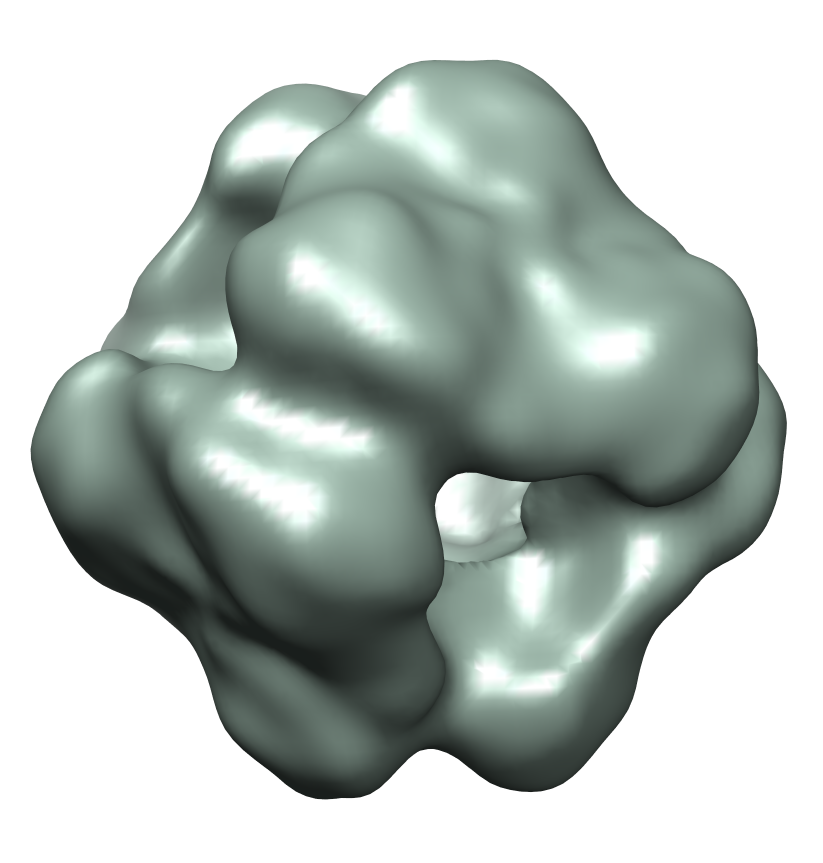 emd-1680.png emd-1680.png | 278.6 KB | ||
| Archive directory |  http://ftp.pdbj.org/pub/emdb/structures/EMD-1680ftp://ftp.pdbj.org/pub/emdb/structures/EMD-1680 http://ftp.pdbj.org/pub/emdb/structures/EMD-1680ftp://ftp.pdbj.org/pub/emdb/structures/EMD-1680 | HTTPS FTP |
-Related structure data

-Links
| EMDB pages | EMDB (EBI/PDBe) / EMDataResource |
|---|
-Map
| File | Download / File: emd_1680.map.gz / Format: CCP4 / Size: 2.5 MB / Type: IMAGE STORED AS FLOATING POINT NUMBER (4 BYTES) | ||||||||||||||||||||||||||||||||||||||||||||||||||||||||||||||||||||
|---|---|---|---|---|---|---|---|---|---|---|---|---|---|---|---|---|---|---|---|---|---|---|---|---|---|---|---|---|---|---|---|---|---|---|---|---|---|---|---|---|---|---|---|---|---|---|---|---|---|---|---|---|---|---|---|---|---|---|---|---|---|---|---|---|---|---|---|---|---|
| Annotation | Macromolecular crystal data phased by negative staining electron microscopy reconstructions as a proof of principle | ||||||||||||||||||||||||||||||||||||||||||||||||||||||||||||||||||||
| Voxel size | X=Y=Z: 1.4 Å | ||||||||||||||||||||||||||||||||||||||||||||||||||||||||||||||||||||
| Density |
| ||||||||||||||||||||||||||||||||||||||||||||||||||||||||||||||||||||
| Symmetry | Space group: 1 | ||||||||||||||||||||||||||||||||||||||||||||||||||||||||||||||||||||
| Details | EMDB XML:
CCP4 map header:
| ||||||||||||||||||||||||||||||||||||||||||||||||||||||||||||||||||||
-Supplemental data
- Sample components
-Entire : Type-II dehydroquinase (DHQ) from Candida albicans (CaDHQ)
| Entire | Name: Type-II dehydroquinase (DHQ) from Candida albicans (CaDHQ) |
|---|---|
| Components |
|
-Supramolecule #1000: Type-II dehydroquinase (DHQ) from Candida albicans (CaDHQ)
| Supramolecule | Name: Type-II dehydroquinase (DHQ) from Candida albicans (CaDHQ) type: sample / ID: 1000 / Oligomeric state: Dodecamer / Number unique components: 1 |
|---|---|
| Molecular weight | Experimental: 200 KDa |
-Macromolecule #1: CaDHQ
| Macromolecule | Name: CaDHQ / type: protein_or_peptide / ID: 1 / Name.synonym: CaDHQ / Number of copies: 12 / Oligomeric state: Dodecamer / Recombinant expression: Yes |
|---|---|
| Source (natural) | Organism: Candida albicans (yeast) |
| Molecular weight | Theoretical: 17 KDa |
| Recombinant expression | Organism:  Escherichia coli (E. coli) Escherichia coli (E. coli) |
-Experimental details
-Structure determination
| Method | negative staining |
|---|---|
 Processing Processing | single particle reconstruction |
| Aggregation state | particle |
-Sample preparation
| Concentration | 0.05 mg/mL |
|---|---|
| Buffer | pH: 7.4 / Details: 20mM NaCl, 10mM Tris-HCL |
| Staining | Type: NEGATIVE Details: Negative staining using 1% methylamine vanadate, CH3NH2VO3 |
| Grid | Details: 400 mesk copper grid |
| Vitrification | Cryogen name: NONE / Instrument: OTHER / Details: Negative staining at room temperature |
- Electron microscopy
| Microscope | JEOL 1200EXII |
|---|---|
| Electron beam | Acceleration voltage: 100 kV / Electron source: TUNGSTEN HAIRPIN |
| Electron optics | Illumination mode: OTHER / Imaging mode: BRIGHT FIELDBright-field microscopy / Nominal defocus max: 2.5 µm / Nominal defocus min: 1.0 µm / Nominal magnification: 50000 |
| Sample stage | Specimen holder: Eucentric / Specimen holder model: JEOL |
| Alignment procedure | Legacy - Astigmatism: Objective lens astigmatism was corrected at 100,000 times magnification |
| Details | Low dose negative staining |
| Date | Dec 12, 2006 |
| Image recording | Category: FILM / Film or detector model: KODAK SO-163 FILM / Digitization - Scanner: ZEISS SCAI / Digitization - Sampling interval: 7 µm / Number real images: 8 / Bits/pixel: 8 |
-Image processing
| CTF correction | Details: Each negative |
|---|---|
| Final two d classification | Number classes: 114 |
| Final reconstruction | Applied symmetry - Point group: T (tetrahedral) / Algorithm: OTHER / Resolution.type: BY AUTHOR / Resolution: 15.0 Å / Resolution method: FSC 0.5 CUT-OFF / Software - Name: Spider Details: 7200 particles included in the reconstruction (out of 12000). 432 symmetry imposed Number images used: 7200 |
