 Movie
Movie Controller

[English] 日本語
 Yorodumi
- EMDB-1391: Structured mRNAs regulate translation initiation by binding to th... -
+ Open data

- Basic information
| Entry | Database: EMDB / ID: EMD-1391 | |||||||||
|---|---|---|---|---|---|---|---|---|---|---|
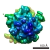
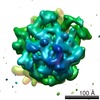
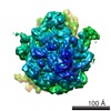
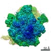
| Title | Structured mRNAs regulate translation initiation by binding to the platform of the ribosome. | |||||||||
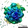
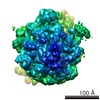
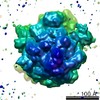
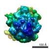
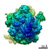
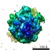
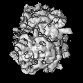
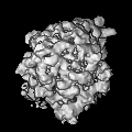
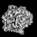
 Map data Map data | Cryo-EM map of the E.coli 70S pre-initiation complex elucidating the entrapment mechanism by the auto-regulatory S15-rpsOmRNA. | |||||||||
 Sample Sample |
| |||||||||
| Function / homology |  Function and homology information Function and homology informationmRNA base-pairing translational repressor activity / small ribosomal subunit rRNA binding /  ribosomal small subunit assembly / cytosolic small ribosomal subunit / cytoplasmic translation / rRNA binding / structural constituent of ribosome / translation / cytosol / cytoplasm ribosomal small subunit assembly / cytosolic small ribosomal subunit / cytoplasmic translation / rRNA binding / structural constituent of ribosome / translation / cytosol / cytoplasmSimilarity search - Function | |||||||||
| Biological species |  Escherichia coli (E. coli) Escherichia coli (E. coli) | |||||||||
| Method | single particle reconstruction / cryo EM / negative staining / Resolution: 10.0 Å | |||||||||
 Authors Authors | Marzi S | |||||||||
 Citation Citation | Journal: Cell / Year: 2007 Title: Structured mRNAs regulate translation initiation by binding to the platform of the ribosome. Authors: Stefano Marzi / Alexander G Myasnikov / Alexander Serganov / Chantal Ehresmann / Pascale Romby / Marat Yusupov / Bruno P Klaholz /  Abstract: Gene expression can be regulated at the level of initiation of protein biosynthesis via structural elements present at the 5' untranslated region of mRNAs. These folded mRNA segments may bind to the ...Gene expression can be regulated at the level of initiation of protein biosynthesis via structural elements present at the 5' untranslated region of mRNAs. These folded mRNA segments may bind to the ribosome, thus blocking translation until the mRNA unfolds. Here, we report a series of cryo-electron microscopy snapshots of ribosomal complexes directly visualizing either the mRNA structure blocked by repressor protein S15 or the unfolded, active mRNA. In the stalled state, the folded mRNA prevents the start codon from reaching the peptidyl-tRNA (P) site inside the ribosome. Upon repressor release, the mRNA unfolds and moves into the mRNA channel allowing translation initiation. A comparative structure and sequence analysis suggests the existence of a universal stand-by site on the ribosome (the 30S platform) dedicated for binding regulatory 5' mRNA elements. Different types of mRNA structures may be accommodated during translation preinitiation and regulate gene expression by transiently stalling the ribosome. | |||||||||
| History |
|
- Structure visualization
| Movie |
Movie viewer |
|---|---|
| Structure viewer | EM map: SurfViewMolmilJmol/JSmol |
| Supplemental images |

- Downloads & links
-EMDB archive
| Map data | emd_1391.map.gz | 1.8 MB | EMDB map data format | |
|---|---|---|---|---|
| Header (meta data) | emd-1391-v30.xmlemd-1391.xml | 11.7 KB 11.7 KB | Display Display | EMDB header |
| Images |  1391.gif 1391.gif | 29 KB | ||
| Archive directory |  http://ftp.pdbj.org/pub/emdb/structures/EMD-1391ftp://ftp.pdbj.org/pub/emdb/structures/EMD-1391 http://ftp.pdbj.org/pub/emdb/structures/EMD-1391ftp://ftp.pdbj.org/pub/emdb/structures/EMD-1391 | HTTPS FTP |
-Related structure data
| Related structure data |  2vazMC M: atomic model generated by this map C: citing same article ( |
|---|---|
| Similar structure data |
-Links
| EMDB pages | EMDB (EBI/PDBe) / EMDataResource |
|---|---|
| Related items in Molecule of the Month |


-Map
| File | Download / File: emd_1391.map.gz / Format: CCP4 / Size: 6.4 MB / Type: IMAGE STORED AS FLOATING POINT NUMBER (4 BYTES) | ||||||||||||||||||||||||||||||||||||||||||||||||||||||||||||||||||||
|---|---|---|---|---|---|---|---|---|---|---|---|---|---|---|---|---|---|---|---|---|---|---|---|---|---|---|---|---|---|---|---|---|---|---|---|---|---|---|---|---|---|---|---|---|---|---|---|---|---|---|---|---|---|---|---|---|---|---|---|---|---|---|---|---|---|---|---|---|---|
| Annotation | Cryo-EM map of the E.coli 70S pre-initiation complex elucidating the entrapment mechanism by the auto-regulatory S15-rpsOmRNA. | ||||||||||||||||||||||||||||||||||||||||||||||||||||||||||||||||||||
| Projections & slices | Image control
Images are generated by Spider. | ||||||||||||||||||||||||||||||||||||||||||||||||||||||||||||||||||||
| Voxel size |
| ||||||||||||||||||||||||||||||||||||||||||||||||||||||||||||||||||||
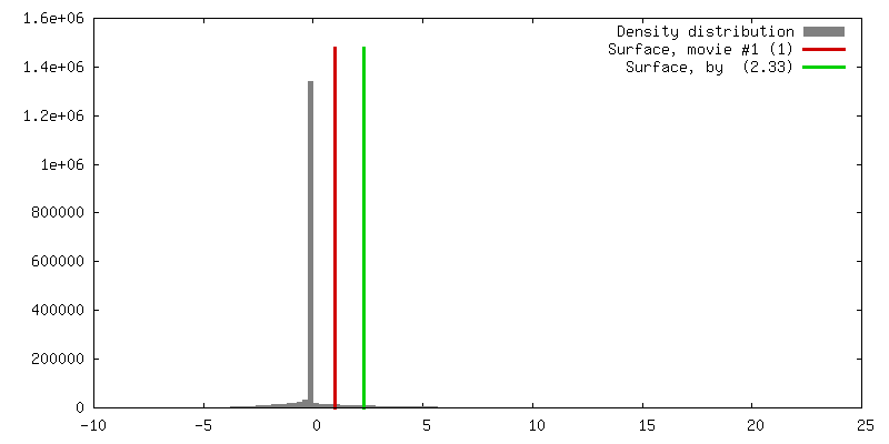
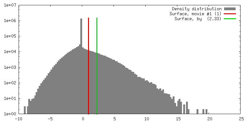
| Density |
| ||||||||||||||||||||||||||||||||||||||||||||||||||||||||||||||||||||
| Symmetry | Space group: 1 | ||||||||||||||||||||||||||||||||||||||||||||||||||||||||||||||||||||
| Details | EMDB XML:
CCP4 map header:
| ||||||||||||||||||||||||||||||||||||||||||||||||||||||||||||||||||||
 Z (Sec.)
 Y (Row.)
 X (Col.)


-Supplemental data
- Sample components
-Entire : 70S pre-initiation complex entrapped by S15-rpsOmRNA
| Entire | Name: 70S pre-initiation complex entrapped by S15-rpsOmRNA |
|---|---|
| Components |
|
-Supramolecule #1000: 70S pre-initiation complex entrapped by S15-rpsOmRNA
| Supramolecule | Name: 70S pre-initiation complex entrapped by S15-rpsOmRNA / type: sample / ID: 1000 Details: 70S E. coli ribosome complexed with S15-rpsOmRNA,stalling Translation Initiation at the pre-initiation step. The structure reveals the molecular details of the Entrapment mechanism. Oligomeric state: monomer / Number unique components: 3 |
|---|---|
| Molecular weight | Theoretical: 2.5 MDa |
-Supramolecule #1: 70S
| Supramolecule | Name: 70S / type: complex / ID: 1 / Name.synonym: 70S / Details: E. coli 70S ribosome / Recombinant expression: No / Ribosome-details: ribosome-prokaryote: ALL |
|---|---|
| Source (natural) | Organism: Escherichia coli (E. coli) |
-Macromolecule #1: rpsO messanger RNA
| Macromolecule | Name: rpsO messanger RNA / type: rna / ID: 1 / Name.synonym: S15mRNA / Classification: OTHER / Structure: SINGLE STRANDED / Synthetic?: Yes |
|---|---|
| Source (natural) | Organism: Escherichia coli (E. coli) |
| Sequence | String: GGGCGAAUUC GAGCUCGGUA CCCAACGUCG CGUAAAUUGU UUAACACUUU GCGUAACGUA CACUGGGAUC GCUGAAUUAG AGAUCGGCGU CCUUUCAUUC UAUAUACUAA GGAGGUUAAA AUGUCUCUAA GUACUGAAGC AACAGCUAAA AUCGUUUCUG AGUUUGGUCG ACCUGCAGGC AUGCA |
-Macromolecule #2: Ribosomal protein S15
| Macromolecule | Name: Ribosomal protein S15 / type: protein_or_peptide / ID: 2 / Name.synonym: S15 / Details: rpsO gene / Oligomeric state: monomeric / Recombinant expression: Yes |
|---|---|
| Source (natural) | Organism: Escherichia coli (E. coli) |
| Recombinant expression | Organism: Escherichia coli BL21(DE3) (bacteria) / Recombinant plasmid: pET11c |
| Sequence | GO: translation / InterPro: Ribosomal protein S15, bacterial-type |
-Experimental details
-Structure determination
| Method | negative staining, cryo EM |
|---|---|
 Processing Processing | single particle reconstruction |
| Aggregation state | particle |
-Sample preparation
| Concentration | 0.5 mg/mL |
|---|---|
| Buffer | pH: 7.5 Details: 20 mM Tris-HCl pH 7.5, 60 mM KCl, 1mM DTT, 7.5 mM MgCl2 |
| Staining | Type: NEGATIVE / Details: no staining, cryo-EM with holey carbon grids |
| Grid | Details: 300 mesh Cu/Rh |
| Vitrification | Cryogen name: ETHANE / Instrument: HOMEMADE PLUNGER / Details: Vitrification instrument: home-made cryo-plunger / Method: Blot for 2 seconds before plunging |
- Electron microscopy
| Microscope | FEI TECNAI F20 |
|---|---|
| Electron beam | Acceleration voltage: 200 kV / Electron source: FIELD EMISSION GUN |
| Electron optics | Calibrated magnification: 51484 / Illumination mode: FLOOD BEAM / Imaging mode: BRIGHT FIELDBright-field microscopy / Cs: 2 mm / Nominal defocus max: 3.5 µm / Nominal defocus min: 0.8 µm / Nominal magnification: 50000 |
| Sample stage | Specimen holder: Side entry liquid nitrogen-cooled cryo specimen holder Specimen holder model: GATAN LIQUID NITROGEN |
| Temperature | Average: 77 K |
| Alignment procedure | Legacy - Astigmatism: lens astigmatism was corrected at 50,000 times magnification |
| Date | Jan 5, 2005 |
| Image recording | Category: FILM / Film or detector model: KODAK SO-163 FILM / Digitization - Scanner: PRIMESCAN / Digitization - Sampling interval: 5 µm / Number real images: 67 / Average electron dose: 20 e/Å2 / Od range: 1.8 / Bits/pixel: 16 |
| Experimental equipment |  Model: Tecnai F20 / Image courtesy: FEI Company |
-Image processing
| CTF correction | Details: individual particles |
|---|---|
| Final angle assignment | Details: beta gamma |
| Final reconstruction | Applied symmetry - Point group: C1 (asymmetric) / Algorithm: OTHER / Resolution.type: BY AUTHOR / Resolution: 10.0 Å / Resolution method: FSC 0.143 CUT-OFF / Software - Name: IMAGIC-5 / Details: exact filtered back-projection / Number images used: 31415 |
-Atomic model buiding 1
| Initial model | (PDB ID:  2aw7 2awb |
|---|---|
| Software | Name: O |
| Details | Protocol: manual. The domains were separately fitted by manual docking using program O |
| Refinement | Protocol: RIGID BODY FIT |
| Output model | PDB-2vaz: |
