Movie
Movie Controller

[English] 日本語
 Yorodumi
- PDB-6nxe: Cryo-EM Reconstruction of Protease-Activateable Adeno-Associated ... -
+ Open data

- Basic information
| Entry | Database: PDB / ID: 6nxe | ||||||
|---|---|---|---|---|---|---|---|
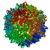
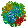
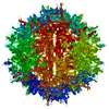
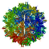
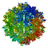
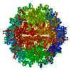
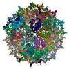
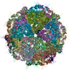
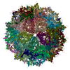
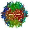
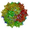
| Title | Cryo-EM Reconstruction of Protease-Activateable Adeno-Associated Virus 9 (AAV9-L001) | ||||||
 Components Components | Capsid protein VP1 | ||||||
 Keywords Keywords | VIRUS / AAV / activatable / gene therapy / provector | ||||||
| Function / homology | Phospholipase A2-like domain / Phospholipase A2-like domain / Parvovirus coat protein VP2 / Parvovirus coat protein VP1/VP2 / Parvovirus coat protein VP2 / Capsid/spike protein, ssDNA virus / T=1 icosahedral viral capsid / structural molecule activity / Capsid protein VP1 Function and homology information Function and homology information | ||||||
| Biological species |   Adeno-associated virus Adeno-associated virus | ||||||
| Method | ELECTRON MICROSCOPY / single particle reconstruction / cryo EM / Resolution: 3.16 Å | ||||||
 Authors Authors | Bennett, A.B. / Agbandje-Mckenna, M. | ||||||
| Funding support |  United States, 1items United States, 1items
| ||||||
 Citation Citation | Journal: Mol Ther / Year: 2019 Title: Protease-Activatable Adeno-Associated Virus Vector for Gene Delivery to Damaged Heart Tissue. Authors: Caitlin M Guenther / Mitchell J Brun / Antonette D Bennett / Michelle L Ho / Weitong Chen / Banghe Zhu / Michael Lam / Momona Yamagami / Sunkuk Kwon / Nilakshee Bhattacharya / Duncan Sousa / ...Authors: Caitlin M Guenther / Mitchell J Brun / Antonette D Bennett / Michelle L Ho / Weitong Chen / Banghe Zhu / Michael Lam / Momona Yamagami / Sunkuk Kwon / Nilakshee Bhattacharya / Duncan Sousa / Annicka C Evans / Julie Voss / Eva M Sevick-Muraca / Mavis Agbandje-McKenna / Junghae Suh / Abstract: Adeno-associated virus (AAV) has emerged as a promising gene delivery vector because of its non-pathogenicity, simple structure and genome, and low immunogenicity compared to other viruses. However, ...Adeno-associated virus (AAV) has emerged as a promising gene delivery vector because of its non-pathogenicity, simple structure and genome, and low immunogenicity compared to other viruses. However, its adoption as a safe and effective delivery vector for certain diseases relies on altering its tropism to deliver transgenes to desired cell populations. To this end, we have developed a protease-activatable AAV vector, named provector, that responds to elevated extracellular protease activity commonly found in diseased tissue microenvironments. The AAV9-based provector is initially inactive, but then it can be switched on by matrix metalloproteinases (MMP)-2 and -9. Cryo-electron microscopy and image reconstruction reveal that the provector capsid is structurally similar to that of AAV9, with a flexible peptide insertion at the top of the 3-fold protrusions. In an in vivo model of myocardial infarction (MI), the provector is able to deliver transgenes site specifically to high-MMP-activity regions of the damaged heart, with concomitant decreased delivery to many off-target organs, including the liver. The AAV provector may be useful in the future for enhanced delivery of transgenes to sites of cardiac damage. | ||||||
| History |
|
- Structure visualization
| Movie |
Movie viewer |
|---|---|
| Structure viewer | Molecule: MolmilJmol/JSmol |
- Downloads & links
-Download
| PDBx/mmCIF format | 6nxe.cif.gz | 5.2 MB | Display | PDBx/mmCIF format |
|---|---|---|---|---|
| PDB format | pdb6nxe.ent.gz | Display | PDB format | |
| PDBx/mmJSON format | 6nxe.json.gz | Tree view | PDBx/mmJSON format | |
| Others |  Other downloads Other downloads |
-Validation report
| Arichive directory | https://data.pdbj.org/pub/pdb/validation_reports/nx/6nxeftp://data.pdbj.org/pub/pdb/validation_reports/nx/6nxe | HTTPS FTP |
|---|
-Related structure data
| Related structure data | 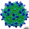 0535MC M: map data used to model this data C: citing same article ( |
|---|---|
| Similar structure data |

-Links
 PDBj
PDBj


- Assembly
| Deposited unit | 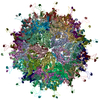
|
|---|---|
| 1 |
|
-Components
| #1: Protein | Mass: 60939.168 Da / Num. of mol.: 60 / Fragment: UNP residues 219-736 Source method: isolated from a genetically manipulated source Source: (gene. exp.) Adeno-associated virus / Gene: cap / Variant: Serotype9-L001 / Cell line (production host): HEK293 / Production host:  Homo sapiens (human) / References: UniProt: Q6JC22 Homo sapiens (human) / References: UniProt: Q6JC22 |
|---|
-Experimental details
-Experiment
| Experiment | Method: ELECTRON MICROSCOPY |
|---|---|
| EM experiment | Aggregation state: PARTICLE / 3D reconstruction method: single particle reconstruction |
- Sample preparation
| Component | Name: Adeno-associated virus / Type: VIRUS / Entity ID: all / Source: RECOMBINANT |
|---|---|
| Source (natural) | Organism: Adeno-associated virus / Strain: 9-L001 |
| Source (recombinant) | Organism: Homo sapiens (human) |
| Details of virus | Empty: NO / Enveloped: NO / Isolate: SEROTYPE / Type: VIRION |
| Virus shell | Diameter: 260 nm / Triangulation number (T number): 1 |
| Buffer solution | pH: 7.4 |
| Buffer component | Name: TD Buffer / Formula: PBS-MK |
| Specimen | Conc.: 0.5 mg/ml / Embedding applied: NO / Shadowing applied: NO / Staining applied: NO / Vitrification applied: YES / Details: The sample was monodisperse. |
| Specimen support | Details: unspecified |
| Vitrification | Cryogen name: ETHANE |
- Electron microscopy imaging
| Experimental equipment |  Model: Titan Krios / Image courtesy: FEI Company |
|---|---|
| Microscopy | Model: FEI TITAN KRIOS |
| Electron gun | Electron source: FIELD EMISSION GUN / Accelerating voltage: 300 kV / Illumination mode: FLOOD BEAM |
| Electron lens | Mode: BRIGHT FIELDBright-field microscopy |
| Image recording | Electron dose: 20 e/Å2 / Film or detector model: DIRECT ELECTRON DE-20 (5k x 3k) |
- Processing
| EM software |
| ||||||||||||
|---|---|---|---|---|---|---|---|---|---|---|---|---|---|
| CTF correction | Type: PHASE FLIPPING AND AMPLITUDE CORRECTION | ||||||||||||
| 3D reconstruction | Resolution: 3.16 Å / Resolution method: FSC 0.143 CUT-OFF / Num. of particles: 114044 / Symmetry type: POINT | ||||||||||||
| Atomic model building | Space: REAL | ||||||||||||
| Atomic model building | PDB-ID: 3UX1 Pdb chain-ID: A / Accession code: 3UX1 / Source name: PDB / Type: experimental model |