 Movie
Movie Controller

[English] 日本語
 Yorodumi
- PDB-5a9k: Structural basis for DNA strand separation by a hexameric replica... -
+ Open data

- Basic information
| Entry | Database: PDB / ID: 5a9k | ||||||
|---|---|---|---|---|---|---|---|
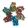
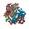
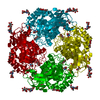
| Title | Structural basis for DNA strand separation by a hexameric replicative helicase | ||||||
 Components Components | REPLICATION PROTEIN E1 DNA replication DNA replication | ||||||
 Keywords Keywords | HYDROLASE / DNA REPLICATION FORK | ||||||
| Function / homology |  Function and homology informationDNA helicase activity / DNA helicase / DNA replication / host cell nucleus / ATP hydrolysis activity / DNA binding / ATP binding Function and homology informationDNA helicase activity / DNA helicase / DNA replication / host cell nucleus / ATP hydrolysis activity / DNA binding / ATP bindingSimilarity search - Function | ||||||
| Biological species |  BOVINE PAPILLOMAVIRUS BOVINE PAPILLOMAVIRUS | ||||||
| Method | ELECTRON MICROSCOPY / single particle reconstruction / negative staining / Resolution: 19 Å | ||||||
 Authors Authors | Chaban, Y. / Stead, J.A. / Ryzhenkova, K. / Whelan, F. / Lamber, K. / Antson, F. / Sanders, C.M. / Orlova, E.V. | ||||||
 Citation Citation | Journal: Nucleic Acids Res / Year: 2015 Title: Structural basis for DNA strand separation by a hexameric replicative helicase. Authors: Yuriy Chaban / Jonathan A Stead / Ksenia Ryzhenkova / Fiona Whelan / Ekaterina P Lamber / Alfred Antson / Cyril M Sanders / Elena V Orlova /  Abstract: Hexameric helicases are processive DNA unwinding machines but how they engage with a replication fork during unwinding is unknown. Using electron microscopy and single particle analysis we determined ...Hexameric helicases are processive DNA unwinding machines but how they engage with a replication fork during unwinding is unknown. Using electron microscopy and single particle analysis we determined structures of the intact hexameric helicase E1 from papillomavirus and two complexes of E1 bound to a DNA replication fork end-labelled with protein tags. By labelling a DNA replication fork with streptavidin (dsDNA end) and Fab (5' ssDNA) we located the positions of these labels on the helicase surface, showing that at least 10 bp of dsDNA enter the E1 helicase via a side tunnel. In the currently accepted 'steric exclusion' model for dsDNA unwinding, the active 3' ssDNA strand is pulled through a central tunnel of the helicase motor domain as the dsDNA strands are wedged apart outside the protein assembly. Our structural observations together with nuclease footprinting assays indicate otherwise: strand separation is taking place inside E1 in a chamber above the helicase domain and the 5' passive ssDNA strands exits the assembly through a separate tunnel opposite to the dsDNA entry point. Our data therefore suggest an alternative to the current general model for DNA unwinding by hexameric helicases. | ||||||
| History |
|
- Structure visualization
| Movie |
Movie viewer |
|---|---|
| Structure viewer | Molecule: MolmilJmol/JSmol |
- Downloads & links
-Download
| PDBx/mmCIF format | 5a9k.cif.gz | 317.1 KB | Display | PDBx/mmCIF format |
|---|---|---|---|---|
| PDB format | pdb5a9k.ent.gz | 265.4 KB | Display | PDB format |
| PDBx/mmJSON format | 5a9k.json.gz | Tree view | PDBx/mmJSON format | |
| Others |  Other downloads Other downloads |
-Validation report
| Arichive directory | https://data.pdbj.org/pub/pdb/validation_reports/a9/5a9kftp://data.pdbj.org/pub/pdb/validation_reports/a9/5a9k | HTTPS FTP |
|---|
-Related structure data
| Related structure data |  3087MC 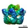 3088C 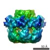 3089C 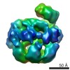 3090C  3091C  5a9l 5a9m 5a9n 5a9o M: map data used to model this data C: citing same article ( |
|---|---|
| Similar structure data |


-Links
 PDBj
PDBj

- Assembly
| Deposited unit | 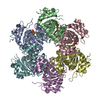
|
|---|---|
| 1 |
|
-Components
| #1: Protein | DNA replication / 3.6.4.12 / ATP-DEPENDENT HELICASE E1 Mass: 34645.016 Da / Num. of mol.: 6 / Fragment: HELICASE DOMAIN, RESIDUES 301-605 Source method: isolated from a genetically manipulated source Source: (gene. exp.) BOVINE PAPILLOMAVIRUS / Organ: NUCLEUSCell nucleus / Plasmid: PET11C / Production host:  ESCHERICHIA COLI (E. coli) / Strain (production host): BL21(DE3) / References: UniProt: P03116, DNA helicase ESCHERICHIA COLI (E. coli) / Strain (production host): BL21(DE3) / References: UniProt: P03116, DNA helicase#2: Chemical | ChemComp-PO4 / Phosphate  Mass: 94.971 Da / Num. of mol.: 9 / Source method: obtained synthetically / Formula: PO4 Mass: 94.971 Da / Num. of mol.: 9 / Source method: obtained synthetically / Formula: PO4#3: Chemical |   Mass: 24.305 Da / Num. of mol.: 3 / Source method: obtained synthetically / Formula: Mg Mass: 24.305 Da / Num. of mol.: 3 / Source method: obtained synthetically / Formula: Mg#4: Water | ChemComp-HOH / | Water Mass: 18.015 Da / Num. of mol.: 18 / Source method: isolated from a natural source / Formula: H2O Mass: 18.015 Da / Num. of mol.: 18 / Source method: isolated from a natural source / Formula: H2O |
|---|
-Experimental details
-Experiment
| Experiment | Method: ELECTRON MICROSCOPY |
|---|---|
| EM experiment | Aggregation state: PARTICLE / 3D reconstruction method: single particle reconstruction |
- Sample preparation
| Component | Name: HELICASE DOMAIN OF THE FULL-LENGTH E1 HELICASE FROM BOVINE PAPILLOMAVIRUS Type: VIRUS |
|---|---|
| Buffer solution | Name: 10 MM TRIS-CL PH 8.0, 225 MM NACL, 2 MM DTT, 0.1 MM PMSF, 0.1 MM EDTA pH: 8 Details: 10 MM TRIS-CL PH 8.0, 225 MM NACL, 2 MM DTT, 0.1 MM PMSF, 0.1 MM EDTA |
| Specimen | Conc.: 3 mg/ml / Embedding applied: NO / Shadowing applied: NO / Staining applied: YES / Vitrification applied: NO |
| EM staining | Type: NEGATIVE / Material: uranyl acetate |
| Specimen support | Details: CARBON |
- Electron microscopy imaging
| Experimental equipment |  Model: Tecnai F20 / Image courtesy: FEI Company |
|---|---|
| Microscopy | Model: FEI TECNAI F20 / Date: Feb 18, 2010 |
| Electron gun | Electron source: FIELD EMISSION GUN / Accelerating voltage: 200 kV / Illumination mode: FLOOD BEAM |
| Electron lens | Mode: BRIGHT FIELDBright-field microscopy / Nominal magnification: 62000 X / Calibrated magnification: 67000 X / Nominal defocus max: 1500 nm / Nominal defocus min: 500 nm / Cs: 2.1 mm |
| Specimen holder | Temperature: 293 K |
| Image recording | Electron dose: 20 e/Å2 / Film or detector model: GATAN ULTRASCAN 4000 (4k x 4k) |
| Image scans | Num. digital images: 30 |
| Radiation wavelength | Relative weight: 1 |
- Processing
| EM software |
| ||||||||||||||||||||
|---|---|---|---|---|---|---|---|---|---|---|---|---|---|---|---|---|---|---|---|---|---|
| CTF correction | Details: FRAMES | ||||||||||||||||||||
| Symmetry | Point symmetry: C6 (6 fold cyclic) | ||||||||||||||||||||
| 3D reconstruction | Method: FILTERED BACK PROJECTION / Resolution: 19 Å / Num. of particles: 8265 / Actual pixel size: 1.6 Å Magnification calibration: CROSS- -CORRELATION DENSITIES WITHIN SPHERICAL SHELL Details: SUBMISSION BASED ON EXPERIMENTAL DATA FROM EMDB EMD-3087. (DEPOSITION ID: 13536). Symmetry type: POINT | ||||||||||||||||||||
| Atomic model building | Protocol: OTHER / Space: REAL / Target criteria: Cross-correlation coefficient Details: METHOD--LOCAL CORRELATION REFINEMENT PROTOCOL--X-RAY | ||||||||||||||||||||
| Atomic model building | PDB-ID: 2V9P | ||||||||||||||||||||
| Refinement | Highest resolution: 19 Å | ||||||||||||||||||||
| Refinement step | Cycle: LAST / Highest resolution: 19 Å
|

