 Movie
Movie Controller

[English] 日本語
 Yorodumi
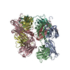
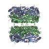
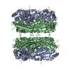
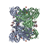
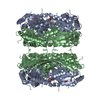
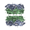
- PDB-4d1k: Cryo-electron microscopy of tubular arrays of HIV-1 Gag resolves ... -
+ Open data

- Basic information
| Entry | Database: PDB / ID: 4d1k | ||||||
|---|---|---|---|---|---|---|---|
| Title | Cryo-electron microscopy of tubular arrays of HIV-1 Gag resolves structures essential for immature virus assembly. | ||||||
 Components Components | GAG PROTEIN HIV-1 protease HIV-1 protease | ||||||
 Keywords Keywords | VIRAL PROTEIN / CAPSID / SP1 / HELICAL RECONSTRUCTION | ||||||
| Function / homology | gag protein p24 N-terminal domain / viral process / Retroviral nucleocapsid Gag protein p24, C-terminal domain / Gag protein p24 C-terminal domain / viral capsid / Retrovirus capsid, C-terminal / Retrovirus capsid, N-terminal / Gag protein Function and homology information Function and homology information | ||||||
| Biological species |   HUMAN IMMUNODEFICIENCY VIRUS 1 HUMAN IMMUNODEFICIENCY VIRUS 1 | ||||||
| Method | ELECTRON MICROSCOPY / helical reconstruction / cryo EM / Resolution: 9.4 Å | ||||||
 Authors Authors | Bharat, T.A.M. / Castillo-Menendez, L.R. / Hagen, W.J.H. / Lux, V. / Igonet, S. / Schorb, M. / Schur, F.K.M. / Krauesslich, H.G. / Briggs, J.A.G. | ||||||
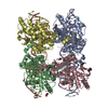
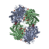
 Citation Citation | Journal: Proc Natl Acad Sci U S A / Year: 2014 Title: Cryo-electron microscopy of tubular arrays of HIV-1 Gag resolves structures essential for immature virus assembly. Authors: Tanmay A M Bharat / Luis R Castillo Menendez / Wim J H Hagen / Vanda Lux / Sebastien Igonet / Martin Schorb / Florian K M Schur / Hans-Georg Kräusslich / John A G Briggs /    Abstract: The assembly of HIV-1 is mediated by oligomerization of the major structural polyprotein, Gag, into a hexameric protein lattice at the plasma membrane of the infected cell. This leads to budding and ...The assembly of HIV-1 is mediated by oligomerization of the major structural polyprotein, Gag, into a hexameric protein lattice at the plasma membrane of the infected cell. This leads to budding and release of progeny immature virus particles. Subsequent proteolytic cleavage of Gag triggers rearrangement of the particles to form mature infectious virions. Obtaining a structural model of the assembled lattice of Gag within immature virus particles is necessary to understand the interactions that mediate assembly of HIV-1 particles in the infected cell, and to describe the substrate that is subsequently cleaved by the viral protease. An 8-Å resolution structure of an immature virus-like tubular array assembled from a Gag-derived protein of the related retrovirus Mason-Pfizer monkey virus (M-PMV) has previously been reported, and a model for the arrangement of the HIV-1 capsid (CA) domains has been generated based on homology to this structure. Here we have assembled tubular arrays of a HIV-1 Gag-derived protein with an immature-like arrangement of the C-terminal CA domains and have solved their structure by using hybrid cryo-EM and tomography analysis. The structure reveals the arrangement of the C-terminal domain of CA within an immature-like HIV-1 Gag lattice, and provides, to our knowledge, the first high-resolution view of the region immediately downstream of CA, which is essential for assembly, and is significantly different from the respective region in M-PMV. Our results reveal a hollow column of density for this region in HIV-1 that is compatible with the presence of a six-helix bundle at this position. | ||||||
| History |
|
- Structure visualization
| Movie |
Movie viewer |
|---|---|
| Structure viewer | Molecule: MolmilJmol/JSmol |
- Downloads & links
-Download
| PDBx/mmCIF format | 4d1k.cif.gz | 144.9 KB | Display | PDBx/mmCIF format |
|---|---|---|---|---|
| PDB format | pdb4d1k.ent.gz | 79.2 KB | Display | PDB format |
| PDBx/mmJSON format | 4d1k.json.gz | Tree view | PDBx/mmJSON format | |
| Others |  Other downloads Other downloads |
-Validation report
| Arichive directory | https://data.pdbj.org/pub/pdb/validation_reports/d1/4d1kftp://data.pdbj.org/pub/pdb/validation_reports/d1/4d1k | HTTPS FTP |
|---|
-Related structure data
| Related structure data |  2638MC 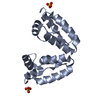 4cocC 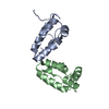 4copC C: citing same article ( M: map data used to model this data |
|---|---|
| Similar structure data |
-Links
 PDBj
PDBj
- Assembly
| Deposited unit | 
|
|---|---|
| 1 |
|
-Components
| #1: Protein | HIV-1 protease Mass: 24421.988 Da / Num. of mol.: 6 / Fragment: CAPSID, RESIDUES 1-219 / Mutation: YES Source method: isolated from a genetically manipulated source Details: 10NM PROTEIN-A CONJUGATED GOLD PARTICLES WERE ADDED TO THE SAMPLE. PROTEIN CONSTRUCT CONSISTS OF CA TO NC DOMAINS WITH MUTATION IN POSITION Y169. Source: (gene. exp.) HUMAN IMMUNODEFICIENCY VIRUS 1 / Production host:  ESCHERICHIA COLI (E. coli) / Strain (production host): BL21(DE3) / References: UniProt: Q5D0H3 ESCHERICHIA COLI (E. coli) / Strain (production host): BL21(DE3) / References: UniProt: Q5D0H3Sequence details | THE PROTEIN DEPOSITED IS A MUTANT AT POSITION Y169. | |
|---|
-Experimental details
-Experiment
| Experiment | Method: ELECTRON MICROSCOPY |
|---|---|
| EM experiment | Aggregation state: HELICAL ARRAY / 3D reconstruction method: helical reconstruction |
- Sample preparation
| Component | Name: HIV-1 CANC Y169 MUTANT PROTEIN ASSEMBLED WITH 73MER DNA INTO TUBULAR PARTICLES Type: COMPLEX |
|---|---|
| Buffer solution | Name: 30 MM MES, 1 MM EDTA, 1 MM DTT, 0.5 M NACL / pH: 6 / Details: 30 MM MES, 1 MM EDTA, 1 MM DTT, 0.5 M NACL |
| Specimen | Conc.: 2 mg/ml / Embedding applied: NO / Shadowing applied: NO / Staining applied: NO / Vitrification applied: YES |
| Specimen support | Details: HOLEY CARBON |
| Vitrification | Instrument: HOMEMADE PLUNGER / Cryogen name: ETHANE Details: VITRIFICATION 1 -- CRYOGEN- ETHANE, HUMIDITY- 100, INSTRUMENT- HOMEMADE PLUNGER, |
- Electron microscopy imaging
| Experimental equipment |  Model: Titan Krios / Image courtesy: FEI Company |
|---|---|
| Microscopy | Model: FEI TITAN KRIOS / Date: Dec 25, 2012 |
| Electron gun | Electron source: FIELD EMISSION GUN / Accelerating voltage: 300 kV / Illumination mode: FLOOD BEAM |
| Electron lens | Mode: BRIGHT FIELDBright-field microscopy / Nominal defocus max: 4500 nm / Nominal defocus min: 1000 nm / Cs: 2.7 mm |
| Image recording | Electron dose: 23 e/Å2 / Film or detector model: KODAK SO-163 FILM |
| Image scans | Num. digital images: 82 |
| Radiation wavelength | Relative weight: 1 |
- Processing
| EM software |
| |||||||||||||||
|---|---|---|---|---|---|---|---|---|---|---|---|---|---|---|---|---|
| 3D reconstruction | Resolution: 9.4 Å / Nominal pixel size: 1.5 Å / Actual pixel size: 1.53 Å Details: ATOMIC MODELS WERE FITTED USING MOLECULAR MOLECULAR DYNAMICS FLEXIBLE FITTING (MDFF). THE CA-NTD ATOMIC MODEL PDB ID 2JPR AND THE CA-CTD ATOMIC MODEL PDB ID 4COP WERE USED AS STARTING MODELS. ...Details: ATOMIC MODELS WERE FITTED USING MOLECULAR MOLECULAR DYNAMICS FLEXIBLE FITTING (MDFF). THE CA-NTD ATOMIC MODEL PDB ID 2JPR AND THE CA-CTD ATOMIC MODEL PDB ID 4COP WERE USED AS STARTING MODELS. SUBMISSION BASED ON EXPERIMENTAL DATA FROM EMDB EMD-2638. (DEPOSITION ID: 12470). Symmetry type: HELICAL | |||||||||||||||
| Atomic model building | Protocol: FLEXIBLE FIT / Space: REAL / Details: METHOD--FLEXIBLE REFINEMENT PROTOCOL--X-RAY | |||||||||||||||
| Atomic model building | PDB-ID: 4COP | |||||||||||||||
| Refinement | Highest resolution: 9.4 Å | |||||||||||||||
| Refinement step | Cycle: LAST / Highest resolution: 9.4 Å
|
