Movie
Movie Controller


+ Open data

- Basic information
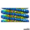
| Entry | Database: PDB / ID: 3j4f | ||||||
|---|---|---|---|---|---|---|---|
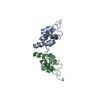
| Title | Structure of HIV-1 capsid protein by cryo-EM | ||||||
 Components Components | capsid protein Capsid Capsid | ||||||
 Keywords Keywords | VIRAL PROTEIN / HIV-1 capsid / core / all-atom model / MDFF / tubular assembly / hexamer | ||||||
| Function / homology |  Function and homology information Function and homology informationviral budding via host ESCRT complex / viral nucleocapsid / host cell cytoplasm / structural molecule activity / virion membrane / RNA binding / zinc ion binding / identical protein binding / cytoplasmSimilarity search - Function | ||||||
| Biological species |   Human immunodeficiency virus 1 Human immunodeficiency virus 1 | ||||||
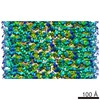
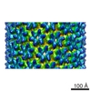
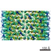
| Method | ELECTRON MICROSCOPY / helical reconstruction / cryo EM / Resolution: 8.6 Å | ||||||
 Authors Authors | Zhao, G. / Perilla, J.R. / Meng, X. / Schulten, K. / Zhang, P. | ||||||
 Citation Citation | Journal: Nature / Year: 2013 Title: Mature HIV-1 capsid structure by cryo-electron microscopy and all-atom molecular dynamics. Authors: Gongpu Zhao / Juan R Perilla / Ernest L Yufenyuy / Xin Meng / Bo Chen / Jiying Ning / Jinwoo Ahn / Angela M Gronenborn / Klaus Schulten / Christopher Aiken / Peijun Zhang /  Abstract: Retroviral capsid proteins are conserved structurally but assemble into different morphologies. The mature human immunodeficiency virus-1 (HIV-1) capsid is best described by a 'fullerene cone' model, ...Retroviral capsid proteins are conserved structurally but assemble into different morphologies. The mature human immunodeficiency virus-1 (HIV-1) capsid is best described by a 'fullerene cone' model, in which hexamers of the capsid protein are linked to form a hexagonal surface lattice that is closed by incorporating 12 capsid-protein pentamers. HIV-1 capsid protein contains an amino-terminal domain (NTD) comprising seven α-helices and a β-hairpin, a carboxy-terminal domain (CTD) comprising four α-helices, and a flexible linker with a 310-helix connecting the two structural domains. Structures of the capsid-protein assembly units have been determined by X-ray crystallography; however, structural information regarding the assembled capsid and the contacts between the assembly units is incomplete. Here we report the cryo-electron microscopy structure of a tubular HIV-1 capsid-protein assembly at 8 Å resolution and the three-dimensional structure of a native HIV-1 core by cryo-electron tomography. The structure of the tubular assembly shows, at the three-fold interface, a three-helix bundle with critical hydrophobic interactions. Mutagenesis studies confirm that hydrophobic residues in the centre of the three-helix bundle are crucial for capsid assembly and stability, and for viral infectivity. The cryo-electron-microscopy structures enable modelling by large-scale molecular dynamics simulation, resulting in all-atom models for the hexamer-of-hexamer and pentamer-of-hexamer elements as well as for the entire capsid. Incorporation of pentamers results in closer trimer contacts and induces acute surface curvature. The complete atomic HIV-1 capsid model provides a platform for further studies of capsid function and for targeted pharmacological intervention. | ||||||
| History |
|
- Structure visualization
| Movie |
Movie viewer |
|---|---|
| Structure viewer | Molecule: MolmilJmol/JSmol |
- Downloads & links
-Download
| PDBx/mmCIF format | 3j4f.cif.gz | 230.3 KB | Display | PDBx/mmCIF format |
|---|---|---|---|---|
| PDB format | pdb3j4f.ent.gz | 191.6 KB | Display | PDB format |
| PDBx/mmJSON format | 3j4f.json.gz | Tree view | PDBx/mmJSON format | |
| Others |  Other downloads Other downloads |
-Validation report
| Arichive directory | https://data.pdbj.org/pub/pdb/validation_reports/j4/3j4fftp://data.pdbj.org/pub/pdb/validation_reports/j4/3j4f | HTTPS FTP |
|---|
-Related structure data
| Related structure data | 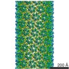 5582MC 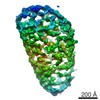 5639C  3j34C  3j3qC  3j3yC M: map data used to model this data C: citing same article ( |
|---|---|
| Similar structure data |

-Links
 PDBj
PDBj

- Assembly
| Deposited unit | 
|
|---|---|
| 1 | x 35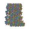
|
| 2 |
|
| 3 | 
|
| Symmetry | Helical symmetry: (Circular symmetry: 1 / Dyad axis: no / N subunits divisor: 1 / Num. of operations: 35 / Rise per n subunits: 7.247 Å / Rotation per n subunits: -31.13 °) |
-Components
| #1: Protein | Capsid Mass: 25702.490 Da / Num. of mol.: 6 / Fragment: UNP residues 133-363 / Mutation: A92E Source method: isolated from a genetically manipulated source Source: (gene. exp.) Human immunodeficiency virus 1 / Gene: gag / Plasmid: pET21 / Production host:  Escherichia coli (E. coli) / Strain (production host): Rosetta 2 (DE3) / References: UniProt: Q79791 Escherichia coli (E. coli) / Strain (production host): Rosetta 2 (DE3) / References: UniProt: Q79791 |
|---|
-Experimental details
-Experiment
| Experiment | Method: ELECTRON MICROSCOPY |
|---|---|
| EM experiment | Aggregation state: HELICAL ARRAY / 3D reconstruction method: helical reconstruction |
- Sample preparation
| Component | Name: HIV-1 capsid protein / Type: COMPLEX / Details: hexamer / Synonym: HIV CA |
|---|---|
| Molecular weight | Value: 0.025 MDa / Experimental value: NO |
| Buffer solution | Name: 1 M NaCl, 50 mM Tris-HCl / pH: 8 / Details: 1 M NaCl, 50 mM Tris-HCl |
| Specimen | Conc.: 2 mg/ml / Embedding applied: NO / Shadowing applied: NO / Staining applied: NO / Vitrification applied: YES |
| Specimen support | Details: 200 mesh quantifoil R2/1 copper grid |
| Vitrification | Instrument: HOMEMADE PLUNGER / Cryogen name: ETHANE / Temp: 95 K / Humidity: 80 % Details: With 2.5 uL sample on carbon side, add 3 uL dilution buffer (100 mM NaCl, 50 mM Tris, pH 8.0) to back side. Blot 3-5 seconds from back side and plunge into liquid ethane with a homemade plunger. Method: With 2.5 uL sample on carbon side, add 3 uL dilution buffer (100 mM NaCl, 50 mM Tris, pH 8.0) to back side. Blot 3-5 seconds from back side. |
- Electron microscopy imaging
| Experimental equipment |  Model: Tecnai Polara / Image courtesy: FEI Company |
|---|---|
| Microscopy | Model: FEI POLARA 300 / Date: Dec 11, 2010 |
| Electron gun | Electron source: FIELD EMISSION GUN / Accelerating voltage: 200 kV / Illumination mode: FLOOD BEAM |
| Electron lens | Mode: BRIGHT FIELDBright-field microscopy / Nominal magnification: 59000 X / Calibrated magnification: 58257 X / Nominal defocus max: 3500 nm / Nominal defocus min: 1000 nm / Cs: 2 mm / Camera length: 0 mm |
| Specimen holder | Specimen holder model: OTHER / Specimen holder type: Polara cartridge / Temperature: 82 K / Temperature (max): 85 K / Temperature (min): 80 K / Tilt angle max: 0 ° / Tilt angle min: 0 ° |
| Image recording | Electron dose: 15 e/Å2 / Film or detector model: KODAK SO-163 FILM |
| Image scans | Num. digital images: 27 |
| Radiation | Protocol: SINGLE WAVELENGTH / Monochromatic (M) / Laue (L): M / Scattering type: x-ray |
| Radiation wavelength | Relative weight: 1 |
- Processing
| EM software |
| ||||||||||||
|---|---|---|---|---|---|---|---|---|---|---|---|---|---|
| CTF correction | Details: each filament | ||||||||||||
| Helical symmerty | Angular rotation/subunit: 31.13 ° / Axial rise/subunit: 7.247 Å / Axial symmetry: C1 | ||||||||||||
| 3D reconstruction | Method: real space helical reconstruction / Resolution: 8.6 Å / Resolution method: FSC 0.5 CUT-OFF / Num. of particles: 3210 / Actual pixel size: 1.09 Å Details: (Helical Details: The segments were aligned and reconstructed using Frealign. Twofold symmetry was imposed using IHRSR++.) Symmetry type: HELICAL | ||||||||||||
| Atomic model building |
| ||||||||||||
| Atomic model building |
| ||||||||||||
| Refinement step | Cycle: LAST
|