 Movie
Movie Controller


+ Open data

- Basic information
| Entry | Database: PDB / ID: 1nn8 | ||||||
|---|---|---|---|---|---|---|---|
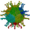
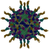
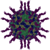
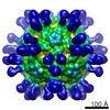
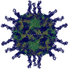
| Title | CryoEM structure of poliovirus receptor bound to poliovirus | ||||||
 Components Components |
| ||||||
 Keywords Keywords | Virus/Receptor / icosahedral virus /  picornavirus / Virus-Receptor COMPLEX picornavirus / Virus-Receptor COMPLEX | ||||||
| Function / homology |  Function and homology information Function and homology informationsusceptibility to T cell mediated cytotoxicity / susceptibility to natural killer cell mediated cytotoxicity / Nectin/Necl trans heterodimerization / positive regulation of natural killer cell mediated cytotoxicity directed against tumor cell target / symbiont-mediated suppression of host translation initiation / heterophilic cell-cell adhesion via plasma membrane cell adhesion molecules / positive regulation of natural killer cell mediated cytotoxicity / homophilic cell adhesion via plasma membrane adhesion molecules / symbiont-mediated suppression of host cytoplasmic pattern recognition receptor signaling pathway via inhibition of RIG-I activity / symbiont-mediated suppression of host cytoplasmic pattern recognition receptor signaling pathway via inhibition of MDA-5 activity ...susceptibility to T cell mediated cytotoxicity / susceptibility to natural killer cell mediated cytotoxicity / Nectin/Necl trans heterodimerization / positive regulation of natural killer cell mediated cytotoxicity directed against tumor cell target / symbiont-mediated suppression of host translation initiation / heterophilic cell-cell adhesion via plasma membrane cell adhesion molecules / positive regulation of natural killer cell mediated cytotoxicity / homophilic cell adhesion via plasma membrane adhesion molecules / symbiont-mediated suppression of host cytoplasmic pattern recognition receptor signaling pathway via inhibition of RIG-I activity / symbiont-mediated suppression of host cytoplasmic pattern recognition receptor signaling pathway via inhibition of MDA-5 activity / symbiont-mediated suppression of host cytoplasmic pattern recognition receptor signaling pathway via inhibition of MAVS activity / picornain 2A / cell adhesion molecule binding / symbiont-mediated suppression of host mRNA export from nucleus / ribonucleoside triphosphate phosphatase activity / symbiont genome entry into host cell via pore formation in plasma membrane / picornain 3C / T=pseudo3 icosahedral viral capsid / host cell cytoplasmic vesicle membrane / adherens junction / endocytosis involved in viral entry into host cell / : / Immunoregulatory interactions between a Lymphoid and a non-Lymphoid cell / nucleoside-triphosphate phosphatase / virus receptor activity / signaling receptor activity / protein complex oligomerization / monoatomic ion channel activity / RNA helicase activity / induction by virus of host autophagy / RNA-directed RNA polymerase / symbiont-mediated suppression of host gene expression / viral RNA genome replication / cysteine-type endopeptidase activity / RNA-dependent RNA polymerase activity / focal adhesion / DNA-templated transcription / host cell nucleus / structural molecule activity / virion attachment to host cell / cell surface / proteolysis / extracellular space / RNA binding / ATP binding / membrane / metal ion binding / plasma membrane / cytoplasmSimilarity search - Function | ||||||
| Biological species |  Homo sapiens (human) Homo sapiens (human)  Human poliovirus 1 Mahoney Human poliovirus 1 Mahoney | ||||||
| Method | ELECTRON MICROSCOPY / single particle reconstruction / cryo EM / Resolution: 15 Å | ||||||
 Authors Authors | He, Y. / Mueller, S. / Chipman, P.R. / Bator, C.M. / Peng, X. / Bowman, V.D. / Mukhopadhyay, S. / Wimmer, E. / Kuhn, R.J. / Rossmann, M.G. | ||||||
 Citation Citation | Journal: J Virol / Year: 2003 Title: Complexes of poliovirus serotypes with their common cellular receptor, CD155. Authors: Yongning He / Steffen Mueller / Paul R Chipman / Carol M Bator / Xiaozhong Peng / Valorie D Bowman / Suchetana Mukhopadhyay / Eckard Wimmer / Richard J Kuhn / Michael G Rossmann /  Abstract: Structures of all three poliovirus (PV) serotypes (PV1, PV2, and PV3) complexed with their cellular receptor, PV receptor (PVR or CD155), were determined by cryoelectron microscopy. Both glycosylated ...Structures of all three poliovirus (PV) serotypes (PV1, PV2, and PV3) complexed with their cellular receptor, PV receptor (PVR or CD155), were determined by cryoelectron microscopy. Both glycosylated and fully deglycosylated CD155 exhibited similar binding sites and orientations in the viral canyon for all three PV serotypes, showing that all three serotypes use a common mechanism for cell entry. Difference maps between the glycosylated and deglycosylated CD155 complexes determined the sites of the carbohydrate moieties that, in turn, helped to verify the position of the receptor relative to the viral surface. The proximity of the CD155 carbohydrate site at Asn105 to the viral surface in the receptor-virus complex suggests that it might interfere with receptor docking, an observation consistent with the properties of mutant CD155. The footprints of CD155 on PV surfaces indicate that the south rim of the canyon dominates the virus-receptor interactions and may correspond to the initial CD155 binding state of the receptor-mediated viral uncoating. In contrast, the interaction of CD155 with the north rim of the canyon, especially the region immediately outside the viral hydrophobic pocket that normally binds a cellular "pocket factor," may be critical for the release of the pocket factor, decreasing the virus stability and hence initiating uncoating. The large area of the CD155 footprint on the PV surface, in comparison with other picornavirus-receptor interactions, could be a potential limitation on the viability of PV escape mutants from antibody neutralization. Many of these are likely to have lost their ability to bind CD155, resulting in there being only three PV serotypes. | ||||||
| History |
| ||||||
| Remark 999 | Authors submitted coordinates for alpha carbons only. | ||||||
| Remark 99 | CHAINS R, S, and T REPRESENT THE DOCKING POSITIONS OF CD155 FITTED INTO PV1, PV2 and PV3 EM MAPS, ...CHAINS R, S, and T REPRESENT THE DOCKING POSITIONS OF CD155 FITTED INTO PV1, PV2 and PV3 EM MAPS, RESPECTIVELY. |
- Structure visualization
| Movie |
Movie viewer |
|---|---|
| Structure viewer | Molecule: MolmilJmol/JSmol |
- Downloads & links
-Download
| PDBx/mmCIF format | 1nn8.cif.gz | 72.1 KB | Display | PDBx/mmCIF format |
|---|---|---|---|---|
| PDB format | pdb1nn8.ent.gz | 40.1 KB | Display | PDB format |
| PDBx/mmJSON format | 1nn8.json.gz | Tree view | PDBx/mmJSON format | |
| Others |  Other downloads Other downloads |
-Validation report
| Arichive directory | https://data.pdbj.org/pub/pdb/validation_reports/nn/1nn8ftp://data.pdbj.org/pub/pdb/validation_reports/nn/1nn8 | HTTPS FTP |
|---|
-Related structure data
| Related structure data | |
|---|---|
| Similar structure data |
-Links
 PDBj
PDBj


- Assembly
| Deposited unit | 
|
|---|---|
| 1 | x 60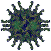
|
| 2 |
|
| 3 | x 5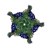
|
| 4 | x 6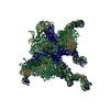
|
| 5 | 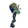
|
| Symmetry | Point symmetry: (Hermann–Mauguin notation: 532 / Schoenflies symbol: I (icosahedral)) |
-Components
-Coat protein ... , 4 types, 4 molecules 1234
| #2: Protein | Mass: 33334.301 Da / Num. of mol.: 1 Source method: isolated from a genetically manipulated source Source: (gene. exp.) Human poliovirus 1 Mahoney / Genus: Enterovirus / Species: Poliovirus / Cellular location (production host): Hela cells / Production host: Homo sapiens (human) / References: UniProt: P03300 |
|---|---|
| #3: Protein | Mass: 30075.783 Da / Num. of mol.: 1 Source method: isolated from a genetically manipulated source Source: (gene. exp.) Human poliovirus 1 Mahoney / Genus: Enterovirus / Species: Poliovirus / Cellular location (production host): Hela cells / Production host: Homo sapiens (human) / References: UniProt: P03300 |
| #4: Protein | Mass: 26235.115 Da / Num. of mol.: 1 Source method: isolated from a genetically manipulated source Source: (gene. exp.) Human poliovirus 1 Mahoney / Genus: Enterovirus / Species: Poliovirus / Cellular location (production host): Hela cells / Production host: Homo sapiens (human) / References: UniProt: P03300 |
| #5: Protein | Mass: 7393.050 Da / Num. of mol.: 1 Source method: isolated from a genetically manipulated source Source: (gene. exp.) Human poliovirus 1 Mahoney / Genus: Enterovirus / Species: Poliovirus / Cellular location (production host): Hela cells / Production host: Homo sapiens (human) / References: UniProt: P03300 |
-Protein / Non-polymers , 2 types, 4 molecules RST
| #1: Protein | CD155 / CD155 antigen / Coordinate model: Cα atoms only Mass: 32928.160 Da / Num. of mol.: 3 Source method: isolated from a genetically manipulated source Source: (gene. exp.) Homo sapiens (human) / Cellular location (production host): 293 cells / Production host: Homo sapiens (human) / References: UniProt: P15151#6: Chemical | ChemComp-MYR / | Myristic acid Mass: 228.371 Da / Num. of mol.: 1 / Source method: obtained synthetically / Formula: C14H28O2 Mass: 228.371 Da / Num. of mol.: 1 / Source method: obtained synthetically / Formula: C14H28O2 |
|---|
-Experimental details
-Experiment
| Experiment | Method: ELECTRON MICROSCOPY |
|---|---|
| EM experiment | Aggregation state: PARTICLE / 3D reconstruction method: single particle reconstruction |
- Sample preparation
| Component | Name: poliovirus receptor bound to poliovirus / Type: VIRUS |
|---|---|
| Specimen | Embedding applied: NO / Shadowing applied: NO / Staining applied: NO / Vitrification applied: YES |
| Crystal grow | *PLUS Method: electron microscopy / Details: electron microscopy |
- Electron microscopy imaging
| Microscopy | Model: FEI/PHILIPS CM300FEG/T |
|---|---|
| Electron gun | Electron source: FIELD EMISSION GUN / Accelerating voltage: 300 kV / Illumination mode: FLOOD BEAM |
| Electron lens | Mode: BRIGHT FIELDBright-field microscopy / Nominal defocus max: 3700 nm / Nominal defocus min: 1400 nm |
- Processing
| CTF correction | Details: CTF is corrected for each image | ||||||||||||
|---|---|---|---|---|---|---|---|---|---|---|---|---|---|
| Symmetry | Point symmetry: I (icosahedral) | ||||||||||||
| 3D reconstruction | Method: polar fourier transform (PFT) / Resolution: 15 Å / Num. of particles: 2022 / Nominal pixel size: 3.11 Å / Actual pixel size: 2.91 Å / Magnification calibration: 45000 Details: 4799 particles are collected, defocus range: 1.4um-3.7um Symmetry type: POINT | ||||||||||||
| Refinement step | Cycle: LAST
|
