Movie
Movie Controller

[English] 日本語
 Yorodumi
- EMDB-8582: Structure of the HIV-1 Capsid Protein and spacer peptide 1 by Cryo-EM -
+ Open data

- Basic information
| Entry | Database: EMDB / ID: EMD-8582 | |||||||||
|---|---|---|---|---|---|---|---|---|---|---|
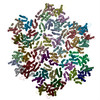
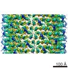
| Title | Structure of the HIV-1 Capsid Protein and spacer peptide 1 by Cryo-EM | |||||||||
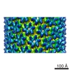
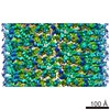
 Map data Map data | CryoEM structure of HIV-1 capsid protein and spacer peptide-1 (CA-SP1) | |||||||||
 Sample Sample |
| |||||||||
| Function / homology |  Function and homology information Function and homology informationviral process / viral nucleocapsid / host cell cytoplasm / structural molecule activity / virion membrane /  RNA binding / zinc ion binding / cytoplasm RNA binding / zinc ion binding / cytoplasmSimilarity search - Function | |||||||||
| Biological species |   Human immunodeficiency virus 1 Human immunodeficiency virus 1 | |||||||||
| Method | helical reconstruction / cryo EM / Resolution: 9.0 Å | |||||||||
 Authors Authors | Zhang P / Randall S | |||||||||
 Citation Citation | Journal: Nat Commun / Year: 2017 Title: Quenching protein dynamics interferes with HIV capsid maturation. Authors: Mingzhang Wang / Caitlin M Quinn / Juan R Perilla / Huilan Zhang / Randall Shirra / Guangjin Hou / In-Ja Byeon / Christopher L Suiter / Sherimay Ablan / Emiko Urano / Theodore J Nitz / ...Authors: Mingzhang Wang / Caitlin M Quinn / Juan R Perilla / Huilan Zhang / Randall Shirra / Guangjin Hou / In-Ja Byeon / Christopher L Suiter / Sherimay Ablan / Emiko Urano / Theodore J Nitz / Christopher Aiken / Eric O Freed / Peijun Zhang / Klaus Schulten / Angela M Gronenborn / Tatyana Polenova /   Abstract: Maturation of HIV-1 particles encompasses a complex morphological transformation of Gag via an orchestrated series of proteolytic cleavage events. A longstanding question concerns the structure of ...Maturation of HIV-1 particles encompasses a complex morphological transformation of Gag via an orchestrated series of proteolytic cleavage events. A longstanding question concerns the structure of the C-terminal region of CA and the peptide SP1 (CA-SP1), which represents an intermediate during maturation of the HIV-1 virus. By integrating NMR, cryo-EM, and molecular dynamics simulations, we show that in CA-SP1 tubes assembled in vitro, which represent the features of an intermediate assembly state during maturation, the SP1 peptide exists in a dynamic helix-coil equilibrium, and that the addition of the maturation inhibitors Bevirimat and DFH-055 causes stabilization of a helical form of SP1. Moreover, the maturation-arresting SP1 mutation T8I also induces helical structure in SP1 and further global dynamical and conformational changes in CA. Overall, our results show that dynamics of CA and SP1 are critical for orderly HIV-1 maturation and that small molecules can inhibit maturation by perturbing molecular motions. | |||||||||
| History |
|
- Structure visualization
| Movie |
Movie viewer |
|---|---|
| Structure viewer | EM map: SurfViewMolmilJmol/JSmol |
| Supplemental images |

- Downloads & links
-EMDB archive
| Map data | emd_8582.map.gz | 428.7 MB | EMDB map data format | |
|---|---|---|---|---|
| Header (meta data) | emd-8582-v30.xmlemd-8582.xml | 13.6 KB 13.6 KB | Display Display | EMDB header |
| FSC (resolution estimation) | emd_8582_fsc.xml | 17.8 KB | Display | FSC data file |
| Images | 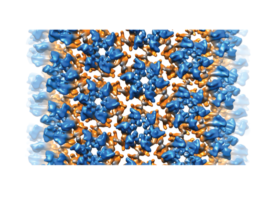 emd_8582.png emd_8582.png | 257 KB | ||
| Archive directory |  http://ftp.pdbj.org/pub/emdb/structures/EMD-8582ftp://ftp.pdbj.org/pub/emdb/structures/EMD-8582 http://ftp.pdbj.org/pub/emdb/structures/EMD-8582ftp://ftp.pdbj.org/pub/emdb/structures/EMD-8582 | HTTPS FTP |
-Related structure data
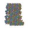
| Related structure data |  5up4MC M: atomic model generated by this map C: citing same article ( |
|---|---|
| Similar structure data |
-Links
| EMDB pages | EMDB (EBI/PDBe) / EMDataResource |
|---|---|
| Related items in Molecule of the Month |

-Map
| File | Download / File: emd_8582.map.gz / Format: CCP4 / Size: 465.5 MB / Type: IMAGE STORED AS FLOATING POINT NUMBER (4 BYTES) | ||||||||||||||||||||||||||||||||||||||||||||||||||||||||||||
|---|---|---|---|---|---|---|---|---|---|---|---|---|---|---|---|---|---|---|---|---|---|---|---|---|---|---|---|---|---|---|---|---|---|---|---|---|---|---|---|---|---|---|---|---|---|---|---|---|---|---|---|---|---|---|---|---|---|---|---|---|---|
| Annotation | CryoEM structure of HIV-1 capsid protein and spacer peptide-1 (CA-SP1) | ||||||||||||||||||||||||||||||||||||||||||||||||||||||||||||
| Voxel size | X=Y=Z: 1.1 Å | ||||||||||||||||||||||||||||||||||||||||||||||||||||||||||||
| Density |
| ||||||||||||||||||||||||||||||||||||||||||||||||||||||||||||
| Symmetry | Space group: 1 | ||||||||||||||||||||||||||||||||||||||||||||||||||||||||||||
| Details | EMDB XML:
CCP4 map header:
| ||||||||||||||||||||||||||||||||||||||||||||||||||||||||||||
-Supplemental data
- Sample components
-Entire : HIV-1 CA-SP1
| Entire | Name: HIV-1 CA-SP1 |
|---|---|
| Components |
|
-Supramolecule #1: HIV-1 CA-SP1
| Supramolecule | Name: HIV-1 CA-SP1 / type: complex / ID: 1 / Parent: 0 / Macromolecule list: all |
|---|---|
| Source (natural) | Organism: Human immunodeficiency virus 1 |
| Recombinant expression | Organism:  Escherichia coli (E. coli) Escherichia coli (E. coli) |
-Macromolecule #1: HIV-1 Capsid Protein and spacer peptide 1
| Macromolecule | Name: HIV-1 Capsid Protein and spacer peptide 1 / type: protein_or_peptide / ID: 1 / Number of copies: 18 / Enantiomer: LEVO |
|---|---|
| Source (natural) | Organism: Human immunodeficiency virus 1 |
| Molecular weight | Theoretical: 24.654268 KDa |
| Recombinant expression | Organism: Escherichia coli (E. coli) |
| Sequence | String: PIVQNLQGQM VHQAISPRTL NAWVKVVEEK AFSPEVIPMF SALSEGATPQ DLNTMLNTVG GHQAAMQMLK ETINEEAAEW DRLHPVHAG PIAPGQMREP RGSDIAGTTS TLQEQIGWMT HNPPIPVGEI YKRWIILGLN KIVRMYSPTS ILDIRQGPKE P FRDYVDRF ...String: PIVQNLQGQM VHQAISPRTL NAWVKVVEEK AFSPEVIPMF SALSEGATPQ DLNTMLNTVG GHQAAMQMLK ETINEEAAEW DRLHPVHAG PIAPGQMREP RGSDIAGTTS TLQEQIGWMT HNPPIPVGEI YKRWIILGLN KIVRMYSPTS ILDIRQGPKE P FRDYVDRF YKTLRAEQAS QEVKNWMTET LLVQNANPDC KTILKALGPG ATLEEMMTAC QGV |
-Experimental details
-Structure determination
| Method | cryo EM |
|---|---|
 Processing Processing | helical reconstruction |
| Aggregation state | helical array |
-Sample preparation
| Concentration | 2.2 mg/mL | |||||||||
|---|---|---|---|---|---|---|---|---|---|---|
| Buffer | pH: 8 Component:
| |||||||||
| Grid | Model: Quantifoil / Material: COPPER / Mesh: 300 / Pretreatment - Type: GLOW DISCHARGE | |||||||||
| Vitrification | Cryogen name: ETHANE / Chamber humidity: 90 % / Chamber temperature: 298 K / Instrument: HOMEMADE PLUNGER |
- Electron microscopy
| Microscope | FEI TITAN KRIOS |
|---|---|
| Electron beam | Acceleration voltage: 300 kV / Electron source: FIELD EMISSION GUN |
| Electron optics | Illumination mode: FLOOD BEAM / Imaging mode: BRIGHT FIELDBright-field microscopy / Nominal defocus max: 3.5 µm / Nominal defocus min: 1.0 µm / Nominal magnification: 59000 |
| Image recording | Film or detector model: KODAK SO-163 FILM / Number real images: 200 / Average electron dose: 23.0 e/Å2 |
| Experimental equipment |  Model: Titan Krios / Image courtesy: FEI Company |
-Image processing
| CTF correction | Software - Name: CTFFIND (ver. 3) |
|---|---|
| Final angle assignment | Type: NOT APPLICABLE |
| Final reconstruction | Applied symmetry - Helical parameters - Δz: 13.46 Å Applied symmetry - Helical parameters - Δ&Phi: 128.88 ° Applied symmetry - Helical parameters - Axial symmetry: C1 (asymmetric) Resolution.type: BY AUTHOR / Resolution: 9.0 Å / Resolution method: FSC 0.143 CUT-OFF / Software - Name: FREALIGN / Number images used: 14000 |
| FSC plot (resolution estimation) | 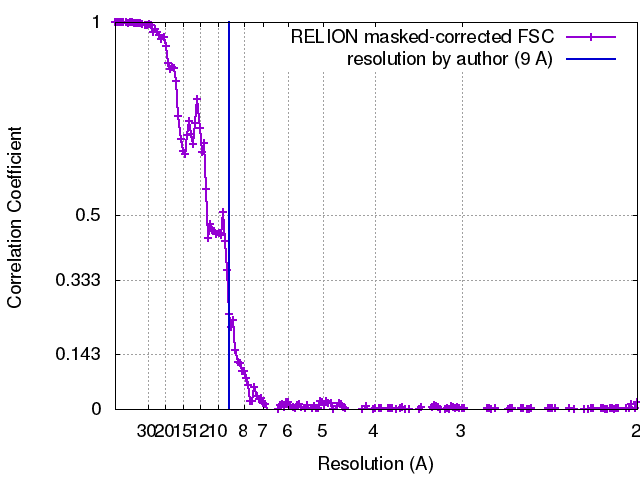 |
-Atomic model buiding 1
| Initial model |
| ||||||||||||||||
|---|---|---|---|---|---|---|---|---|---|---|---|---|---|---|---|---|---|
| Output model | PDB-5up4: |