 Movie
Movie Controller


+ Open data

- Basic information
| Entry | Database: EMDB / ID: EMD-8481 | ||||||||||||
|---|---|---|---|---|---|---|---|---|---|---|---|---|---|
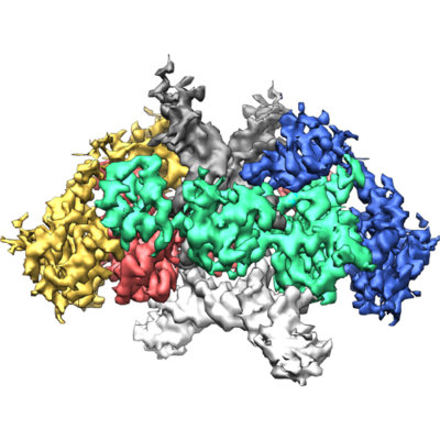
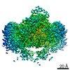
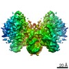
| Title | Structure of tetrameric HIV-1 Strand Transfer Complex Intasome | ||||||||||||
 Map data Map data | cryoEM reconstruction of a tetrameric HIV-1 strand transfer complex intasome | ||||||||||||
 Sample Sample |
| ||||||||||||
 Keywords Keywords |  integrase / integration / transposase / transesterification / VIRAL PROTEIN integrase / integration / transposase / transesterification / VIRAL PROTEIN | ||||||||||||
| Function / homology |  Function and homology information Function and homology informationRNA endonuclease activity / nucleotidyltransferase activity / HIV-1 retropepsin / : / retroviral ribonuclease H / exoribonuclease H / : / exoribonuclease H activity / host multivesicular body / DNA integration ...RNA endonuclease activity / nucleotidyltransferase activity / HIV-1 retropepsin / : / retroviral ribonuclease H / exoribonuclease H / : / exoribonuclease H activity / host multivesicular body / DNA integration / RNA-directed DNA polymerase / viral genome integration into host DNA / viral penetration into host nucleus / establishment of integrated proviral latency / RNA-directed DNA polymerase activity / Transferases; Transferring phosphorus-containing groups; Nucleotidyltransferases / RNA-DNA hybrid ribonuclease activity / viral nucleocapsid / endonuclease activity / DNA recombination / Hydrolases; Acting on ester bonds / nucleic acid binding / DNA-directed DNA polymerase / aspartic-type endopeptidase activity / DNA-directed DNA polymerase activity / symbiont entry into host cell / symbiont-mediated suppression of host gene expression / lipid binding / host cell nucleus / structural molecule activity / host cell plasma membrane / virion membrane / proteolysis / DNA binding / RNA binding / zinc ion binding / membraneSimilarity search - Function | ||||||||||||
| Biological species |   Human immunodeficiency virus 1 / Human immunodeficiency virus 1 /  Homo sapiens (human) Homo sapiens (human) | ||||||||||||
| Method | single particle reconstruction / cryo EM / Resolution: 3.9 Å | ||||||||||||
 Authors Authors | Lyumkis D / Passos D | ||||||||||||
| Funding support |  United States, 3 items United States, 3 items
| ||||||||||||
 Citation Citation | Journal: Science / Year: 2017 Title: Cryo-EM structures and atomic model of the HIV-1 strand transfer complex intasome. Authors: Dario Oliveira Passos / Min Li / Renbin Yang / Stephanie V Rebensburg / Rodolfo Ghirlando / Youngmin Jeon / Nikoloz Shkriabai / Mamuka Kvaratskhelia / Robert Craigie / Dmitry Lyumkis / Abstract: Like all retroviruses, HIV-1 irreversibly inserts a viral DNA (vDNA) copy of its RNA genome into host target DNA (tDNA). The intasome, a higher-order nucleoprotein complex composed of viral integrase ...Like all retroviruses, HIV-1 irreversibly inserts a viral DNA (vDNA) copy of its RNA genome into host target DNA (tDNA). The intasome, a higher-order nucleoprotein complex composed of viral integrase (IN) and the ends of linear vDNA, mediates integration. Productive integration into host chromatin results in the formation of the strand transfer complex (STC) containing catalytically joined vDNA and tDNA. HIV-1 intasomes have been refractory to high-resolution structural studies. We used a soluble IN fusion protein to facilitate structural studies, through which we present a high-resolution cryo-electron microscopy (cryo-EM) structure of the core tetrameric HIV-1 STC and a higher-order form that adopts carboxyl-terminal domain rearrangements. The distinct STC structures highlight how HIV-1 can use the common retroviral intasome core architecture to accommodate different IN domain modules for assembly. | ||||||||||||
| History |
|
- Structure visualization
| Movie |
Movie viewer |
|---|---|
| Structure viewer | EM map: SurfViewMolmilJmol/JSmol |
| Supplemental images |
- Downloads & links
-EMDB archive
| Map data | emd_8481.map.gz | 23.4 MB | EMDB map data format | |
|---|---|---|---|---|
| Header (meta data) | emd-8481-v30.xmlemd-8481.xml | 25.1 KB 25.1 KB | Display Display | EMDB header |
| FSC (resolution estimation) | emd_8481_fsc.xml | 6.8 KB | Display | FSC data file |
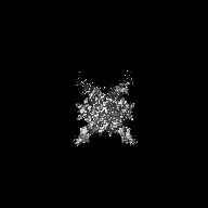
| Images |  emd_8481.png emd_8481.png | 172.6 KB | ||
| Filedesc metadata | emd-8481.cif.gz | 7.5 KB | ||
| Others | emd_8481_half_map_1.map.gzemd_8481_half_map_2.map.gz | 2.8 MB 2.8 MB | ||
| Archive directory |  http://ftp.pdbj.org/pub/emdb/structures/EMD-8481ftp://ftp.pdbj.org/pub/emdb/structures/EMD-8481 http://ftp.pdbj.org/pub/emdb/structures/EMD-8481ftp://ftp.pdbj.org/pub/emdb/structures/EMD-8481 | HTTPS FTP |
-Related structure data
| Related structure data |  5u1cMC  8483C C: citing same article ( M: atomic model generated by this map |
|---|---|
| Similar structure data |


-Links
| EMDB pages | EMDB (EBI/PDBe) / EMDataResource |
|---|---|
| Related items in Molecule of the Month |

-Map
| File | Download / File: emd_8481.map.gz / Format: CCP4 / Size: 27 MB / Type: IMAGE STORED AS FLOATING POINT NUMBER (4 BYTES) | ||||||||||||||||||||||||||||||||||||||||||||||||||||||||||||||||||||
|---|---|---|---|---|---|---|---|---|---|---|---|---|---|---|---|---|---|---|---|---|---|---|---|---|---|---|---|---|---|---|---|---|---|---|---|---|---|---|---|---|---|---|---|---|---|---|---|---|---|---|---|---|---|---|---|---|---|---|---|---|---|---|---|---|---|---|---|---|---|
| Annotation | cryoEM reconstruction of a tetrameric HIV-1 strand transfer complex intasome | ||||||||||||||||||||||||||||||||||||||||||||||||||||||||||||||||||||
| Projections & slices | Image control
Images are generated by Spider. | ||||||||||||||||||||||||||||||||||||||||||||||||||||||||||||||||||||
| Voxel size | X=Y=Z: 1.31 Å | ||||||||||||||||||||||||||||||||||||||||||||||||||||||||||||||||||||
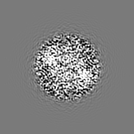
| Density |
| ||||||||||||||||||||||||||||||||||||||||||||||||||||||||||||||||||||
| Symmetry | Space group: 1 | ||||||||||||||||||||||||||||||||||||||||||||||||||||||||||||||||||||
| Details | EMDB XML:
CCP4 map header:
| ||||||||||||||||||||||||||||||||||||||||||||||||||||||||||||||||||||
 Z (Sec.)
 Y (Row.)
 X (Col.)


-Supplemental data
-Half map: cryoEM reconstruction of a tetrameric HIV-1 strand transfer...
| File | emd_8481_half_map_1.map | ||||||||||||
|---|---|---|---|---|---|---|---|---|---|---|---|---|---|
| Annotation | cryoEM reconstruction of a tetrameric HIV-1 strand transfer complex intasome, map 1 | ||||||||||||
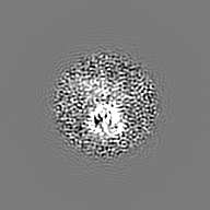
| Projections & Slices |
| ||||||||||||
| Density Histograms |


-Half map: cryoEM reconstruction of a tetrameric HIV-1 strand transfer...
| File | emd_8481_half_map_2.map | ||||||||||||
|---|---|---|---|---|---|---|---|---|---|---|---|---|---|
| Annotation | cryoEM reconstruction of a tetrameric HIV-1 strand transfer complex intasome, map 2 | ||||||||||||
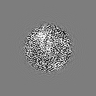
| Projections & Slices |
| ||||||||||||
| Density Histograms |


- Sample components
-Entire : complex formed by a tetrameric assembly of Sso7d-fusion HIV-1 Int...
| Entire | Name: complex formed by a tetrameric assembly of Sso7d-fusion HIV-1 Integrase with the product of DNA strand transfer |
|---|---|
| Components |
|
-Supramolecule #1: complex formed by a tetrameric assembly of Sso7d-fusion HIV-1 Int...
| Supramolecule | Name: complex formed by a tetrameric assembly of Sso7d-fusion HIV-1 Integrase with the product of DNA strand transfer type: complex / ID: 1 / Parent: 0 / Macromolecule list: #1-#4 |
|---|---|
| Source (natural) | Organism: Human immunodeficiency virus 1 |
| Molecular weight | Theoretical: 228 KDa |
-Macromolecule #1: HIV-1 Integrase, Sso7d chimera
| Macromolecule | Name: HIV-1 Integrase, Sso7d chimera / type: protein_or_peptide / ID: 1 / Number of copies: 4 / Enantiomer: LEVO |
|---|---|
| Source (natural) | Organism: Human immunodeficiency virus 1 |
| Molecular weight | Theoretical: 42.320273 KDa |
| Recombinant expression | Organism:  Escherichia coli (E. coli) Escherichia coli (E. coli) |
| Sequence | String: MGSSHHHHHH SSGLVPRGSH MATVKFKYKG EEKEVDISKI KKVWRVGKMI SFTYDEGGGK TGRGAVSEKD APKELLQMLE KQKKGGGGG GGGGGGFLDG IDKAQEEHEK YHSNWRAMAS DFNLPPVVAK EIVASCDKCQ LKGEAMHGQV DCSPGIWQLD C THLEGKVI ...String: MGSSHHHHHH SSGLVPRGSH MATVKFKYKG EEKEVDISKI KKVWRVGKMI SFTYDEGGGK TGRGAVSEKD APKELLQMLE KQKKGGGGG GGGGGGFLDG IDKAQEEHEK YHSNWRAMAS DFNLPPVVAK EIVASCDKCQ LKGEAMHGQV DCSPGIWQLD C THLEGKVI LVAVHVASGY IEAEVIPAET GQETAYFLLK LAGRWPVKTV HTDNGSNFTS TTVKAACWWA GIKQEFGIPY NP QSQGVIQ SMNKELKKII GQVRDQAEHL KTAVQMAVFI HNFKRKGGIG GYSAGERIVD IIATDIQTKE LQKQITKIQN FRV YYRDSR DPVWKGPAKL LWKGEGAVVI QDNSDIKVVP RRKAKIIRDY GKQMAGDDCV ASRQDED UniProtKB: DNA-binding protein, Integrase |
-Macromolecule #2: DNA (11-MER)
| Macromolecule | Name: DNA (11-MER) / type: dna / ID: 2 / Number of copies: 2 / Classification: DNA |
|---|---|
| Source (natural) | Organism: Homo sapiens (human) |
| Molecular weight | Theoretical: 3.349197 KDa |
| Sequence | String: (DG)(DT)(DA)(DC)(DG)(DC)(DT)(DG)(DA)(DC) (DT) |
-Macromolecule #3: DNA (23-MER)
| Macromolecule | Name: DNA (23-MER) / type: dna / ID: 3 / Number of copies: 2 / Classification: DNA |
|---|---|
| Source (natural) | Organism: Human immunodeficiency virus 1 |
| Molecular weight | Theoretical: 7.015546 KDa |
| Sequence | String: (DA)(DC)(DT)(DG)(DC)(DT)(DA)(DG)(DA)(DG) (DA)(DT)(DT)(DT)(DT)(DC)(DC)(DA)(DC)(DA) (DC)(DT)(DG) |
-Macromolecule #4: DNA (37-MER)
| Macromolecule | Name: DNA (37-MER) / type: dna / ID: 4 / Number of copies: 2 / Classification: DNA |
|---|---|
| Source (natural) | Organism: Homo sapiens (human) |
| Molecular weight | Theoretical: 11.414358 KDa |
| Sequence | String: (DC)(DA)(DG)(DT)(DG)(DT)(DG)(DG)(DA)(DA) (DA)(DA)(DT)(DC)(DT)(DC)(DT)(DA)(DG)(DC) (DA)(DG)(DT)(DT)(DA)(DC)(DA)(DG)(DT) (DC)(DA)(DG)(DC)(DG)(DT)(DA)(DC) |
-Macromolecule #5: ZINC ION
| Macromolecule | Name: ZINC ION / type: ligand / ID: 5 / Number of copies: 2 / Formula: ZN |
|---|---|
| Molecular weight | Theoretical: 65.409 Da |
-Macromolecule #6: MAGNESIUM ION
| Macromolecule | Name: MAGNESIUM ION / type: ligand / ID: 6 / Number of copies: 2 / Formula: MG |
|---|---|
| Molecular weight | Theoretical: 24.305 Da |
-Experimental details
-Structure determination
| Method | cryo EM |
|---|---|
 Processing Processing | single particle reconstruction |
| Aggregation state | particle |
-Sample preparation
| Concentration | 0.5 mg/mL | |||||||||||||||
|---|---|---|---|---|---|---|---|---|---|---|---|---|---|---|---|---|
| Buffer | pH: 8 Component:
| |||||||||||||||
| Grid | Model: Quantifoil / Material: GOLD / Mesh: 400 / Pretreatment - Type: PLASMA CLEANING / Pretreatment - Time: 6 sec. / Pretreatment - Atmosphere: OTHER | |||||||||||||||
| Vitrification | Cryogen name: ETHANE / Chamber humidity: 50 % / Chamber temperature: 277 K / Instrument: HOMEMADE PLUNGER Details: Sample containing HIV STC intasomes in SEC buffer was applied onto freshly plasma-treated (6 seconds, Gatan Solarus plasma cleaner) holey gold UltrAuFoil grids (Quantifoil), adsorbed for 30 ...Details: Sample containing HIV STC intasomes in SEC buffer was applied onto freshly plasma-treated (6 seconds, Gatan Solarus plasma cleaner) holey gold UltrAuFoil grids (Quantifoil), adsorbed for 30 seconds, then plunged into liquid ethane using a manual cryo-plunger in an ambient environment of 4 degrees C.. |
- Electron microscopy
| Microscope | FEI TITAN KRIOS |
|---|---|
| Electron beam | Acceleration voltage: 300 kV / Electron source: FIELD EMISSION GUN |
| Electron optics | C2 aperture diameter: 100.0 µm / Calibrated defocus max: 4.0 µm / Calibrated defocus min: 1.5 µm / Calibrated magnification: 38167 / Illumination mode: FLOOD BEAM / Imaging mode: BRIGHT FIELDBright-field microscopy / Cs: 2.7 mm / Nominal magnification: 22500 |
| Sample stage | Specimen holder model: FEI TITAN KRIOS AUTOGRID HOLDER / Cooling holder cryogen: NITROGEN |
| Temperature | Min: 90.0 K / Max: 90.0 K |
| Image recording | Film or detector model: GATAN K2 SUMMIT (4k x 4k) / Detector mode: COUNTING / Digitization - Dimensions - Width: 3838 pixel / Digitization - Dimensions - Height: 3710 pixel / Digitization - Frames/image: 1-100 / Number grids imaged: 1 / Number real images: 1225 / Average exposure time: 20.0 sec. / Average electron dose: 95.0 e/Å2 Details: Individual frames were gain-corrected, aligned, and summed with the application of an exposure filter using MotionCor2, according to the nominal dose rate. |
| Experimental equipment |  Model: Titan Krios / Image courtesy: FEI Company |
-Image processing
| Particle selection | Number selected: 274764 |
|---|---|
| Startup model | Type of model: INSILICO MODEL / In silico model: common lines model using OptiMod Details: An initial model was generated directly from the class averages using OptiMod. |
| Initial angle assignment | Type: PROJECTION MATCHING Projection matching processing - Angular sampling: 7.5 degrees Software - Name: RELION (ver. 1.3) / Details: Relion 3D classification, auto mode |
| Final 3D classification | Software - Name: FREALIGN (ver. 3.11) |
| Final angle assignment | Type: PROJECTION MATCHING / Software - Name: FREALIGN (ver. 9.11) / Details: Frealign 3D classification and refinement |
| Final reconstruction | Applied symmetry - Point group: C2 (2 fold cyclic) / Algorithm: FOURIER SPACE / Resolution.type: BY AUTHOR / Resolution: 3.9 Å / Resolution method: FSC 0.143 CUT-OFF / Software - Name: FREALIGN (ver. 9.11) / Details: Resolution-limited refinement used throughout / Number images used: 83766 |
| FSC plot (resolution estimation) | 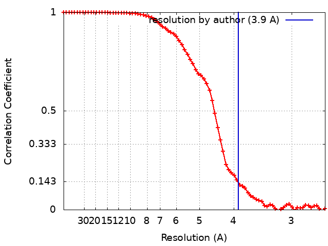 |
-Atomic model buiding 1
| Details | To generate ensemble models, the complete intasome model was iteratively relaxed - using two-fold symmetry and a combination of Rosetta and Phenix - against one half map (the working map) and inspected for consistency with the second half map (the free map). The model was then adjusted manually using Coot. Final ensemble modeling used half maps for all aspects of refinement and evaluation: 500 models were generated as described using Rosetta. From the 100 top-scoring models (scored by Rosetta energy), the ten models with the best map-to-model FSC were selected and refined in real space using secondary-structure restraints in Phenix. Molprobity was used throughout the refinement process. |
|---|---|
| Refinement | Space: REAL / Protocol: FLEXIBLE FIT / Overall B value: 180 / Target criteria: FSC 0.5 |
| Output model | PDB-5u1c: |
