 Movie
Movie Controller

[English] 日本語
 Yorodumi
- EMDB-3247: Subtomogram average of a piliated type IVa pilus machine in wild-... -
+ Open data

- Basic information
| Entry | Database: EMDB / ID: EMD-3247 | |||||||||
|---|---|---|---|---|---|---|---|---|---|---|
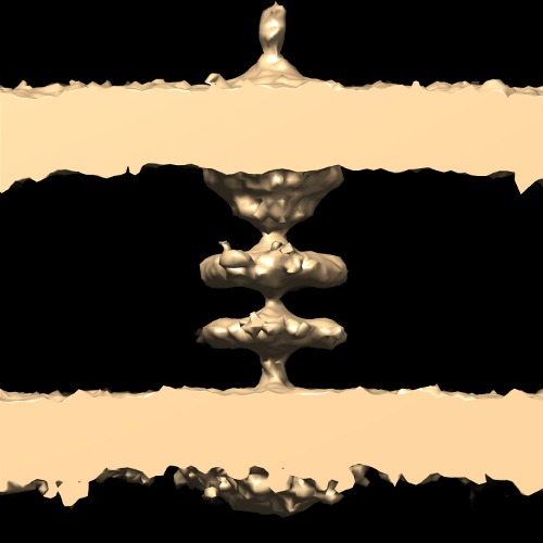
| Title | Subtomogram average of a piliated type IVa pilus machine in wild-type Myxococcus xanthus cells | |||||||||
 Map data Map data | Subtomogram average of a piliated type IVa pilus machine in wild-type Myxococcus xanthus cells | |||||||||
 Sample Sample |
| |||||||||
 Keywords Keywords |  type IV pilus / motor / motility / adhesion type IV pilus / motor / motility / adhesion | |||||||||
| Function / homology |  Function and homology informationtype IV pilus assembly / type IV pilus-dependent motility / pilus assembly / protein secretion / cell outer membrane / cell division / ATP hydrolysis activity / ATP binding / membrane / metal ion binding ...type IV pilus assembly / type IV pilus-dependent motility / pilus assembly / protein secretion / cell outer membrane / cell division / ATP hydrolysis activity / ATP binding / membrane / metal ion binding / plasma membrane / cytoplasm Function and homology informationtype IV pilus assembly / type IV pilus-dependent motility / pilus assembly / protein secretion / cell outer membrane / cell division / ATP hydrolysis activity / ATP binding / membrane / metal ion binding ...type IV pilus assembly / type IV pilus-dependent motility / pilus assembly / protein secretion / cell outer membrane / cell division / ATP hydrolysis activity / ATP binding / membrane / metal ion binding / plasma membrane / cytoplasmSimilarity search - Function | |||||||||
| Biological species |  Myxococcus xanthus (bacteria) Myxococcus xanthus (bacteria) | |||||||||
| Method | subtomogram averaging / cryo EM | |||||||||
 Authors Authors | Chang YW / Rettberg LA / Treuner-Lange A / Iwasa J / Sogaard-Andersen L / Jensen GJ | |||||||||
 Citation Citation | Journal: Science / Year: 2016 Title: Architecture of the type IVa pilus machine. Authors: Yi-Wei Chang / Lee A Rettberg / Anke Treuner-Lange / Janet Iwasa / Lotte Søgaard-Andersen / Grant J Jensen /   Abstract: Type IVa pili are filamentous cell surface structures observed in many bacteria. They pull cells forward by extending, adhering to surfaces, and then retracting. We used cryo-electron tomography of ...Type IVa pili are filamentous cell surface structures observed in many bacteria. They pull cells forward by extending, adhering to surfaces, and then retracting. We used cryo-electron tomography of intact Myxococcus xanthus cells to visualize type IVa pili and the protein machine that assembles and retracts them (the type IVa pilus machine, or T4PM) in situ, in both the piliated and nonpiliated states, at a resolution of 3 to 4 nanometers. We found that T4PM comprises an outer membrane pore, four interconnected ring structures in the periplasm and cytoplasm, a cytoplasmic disc and dome, and a periplasmic stem. By systematically imaging mutants lacking defined T4PM proteins or with individual proteins fused to tags, we mapped the locations of all 10 T4PM core components and the minor pilins, thereby providing insights into pilus assembly, structure, and function. | |||||||||
| History |
|
- Structure visualization
| Movie |
Movie viewer |
|---|---|
| Structure viewer | EM map: SurfViewMolmilJmol/JSmol |
| Supplemental images |

- Downloads & links
-EMDB archive
| Map data | emd_3247.map.gz | 3 MB | EMDB map data format | |
|---|---|---|---|---|
| Header (meta data) | emd-3247-v30.xmlemd-3247.xml | 7.7 KB 7.7 KB | Display Display | EMDB header |
| Images | emd_3247.tifemd_3247_1.tif | 97.9 KB 60 KB | ||
| Archive directory |  http://ftp.pdbj.org/pub/emdb/structures/EMD-3247ftp://ftp.pdbj.org/pub/emdb/structures/EMD-3247 http://ftp.pdbj.org/pub/emdb/structures/EMD-3247ftp://ftp.pdbj.org/pub/emdb/structures/EMD-3247 | HTTPS FTP |
-Related structure data
| Related structure data |  3jc8MC  3248C  3249C  3250C  3251C 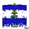 3252C 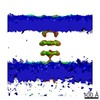 3253C 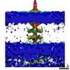 3254C 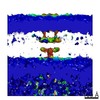 3255C  3256C 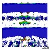 3257C 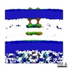 3258C 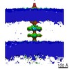 3259C 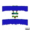 3260C 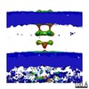 3261C 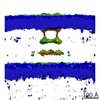 3262C 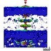 3263C 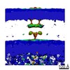 3264C  3jc9C M: atomic model generated by this map C: citing same article ( |
|---|---|
| Similar structure data |
-Links
| EMDB pages | EMDB (EBI/PDBe) / EMDataResource |
|---|---|
| Related items in Molecule of the Month |

-Map
| File | Download / File: emd_3247.map.gz / Format: CCP4 / Size: 3.3 MB / Type: IMAGE STORED AS FLOATING POINT NUMBER (4 BYTES) | ||||||||||||||||||||||||||||||||||||||||||||||||||||||||||||
|---|---|---|---|---|---|---|---|---|---|---|---|---|---|---|---|---|---|---|---|---|---|---|---|---|---|---|---|---|---|---|---|---|---|---|---|---|---|---|---|---|---|---|---|---|---|---|---|---|---|---|---|---|---|---|---|---|---|---|---|---|---|
| Annotation | Subtomogram average of a piliated type IVa pilus machine in wild-type Myxococcus xanthus cells | ||||||||||||||||||||||||||||||||||||||||||||||||||||||||||||
| Voxel size | X=Y=Z: 7.8 Å | ||||||||||||||||||||||||||||||||||||||||||||||||||||||||||||
| Density |
| ||||||||||||||||||||||||||||||||||||||||||||||||||||||||||||
| Symmetry | Space group: 1 | ||||||||||||||||||||||||||||||||||||||||||||||||||||||||||||
| Details | EMDB XML:
CCP4 map header:
| ||||||||||||||||||||||||||||||||||||||||||||||||||||||||||||
-Supplemental data
- Sample components
-Entire : Wild-type Myxococcus xanthus DK1622 cells
| Entire | Name: Wild-type Myxococcus xanthus DK1622 cells |
|---|---|
| Components |
|
-Supramolecule #1000: Wild-type Myxococcus xanthus DK1622 cells
| Supramolecule | Name: Wild-type Myxococcus xanthus DK1622 cells / type: sample / ID: 1000 / Number unique components: 1 |
|---|
-Supramolecule #1: Type IVa pilus
| Supramolecule | Name: Type IVa pilus / type: organelle_or_cellular_component / ID: 1 / Recombinant expression: No |
|---|---|
| Source (natural) | Organism: Myxococcus xanthus (bacteria) / Strain: DK1622 |
-Experimental details
-Structure determination
| Method | cryo EM |
|---|---|
 Processing Processing | subtomogram averaging |
| Aggregation state | cell |
-Sample preparation
| Vitrification | Cryogen name: ETHANE-PROPANE MIXTURE / Instrument: FEI VITROBOT MARK III |
|---|
- Electron microscopy
| Microscope | FEI POLARA 300 |
|---|---|
| Electron beam | Acceleration voltage: 300 kV / Electron source: FIELD EMISSION GUN |
| Electron optics | Illumination mode: FLOOD BEAM / Imaging mode: BRIGHT FIELDBright-field microscopy / Nominal magnification: 27500 |
| Specialist optics | Energy filter - Name: GATAN / Energy filter - Lower energy threshold: 0.0 eV / Energy filter - Upper energy threshold: 20.0 eV |
| Sample stage | Specimen holder model: GATAN LIQUID NITROGEN / Tilt series - Axis1 - Min angle: -60 ° / Tilt series - Axis1 - Max angle: 60 ° |
| Date | Aug 26, 2013 |
| Image recording | Category: CCD / Film or detector model: GATAN K2 SUMMIT (4k x 4k) / Average electron dose: 150 e/Å2 |
| Experimental equipment |  Model: Tecnai Polara / Image courtesy: FEI Company |
-Image processing
| Final reconstruction | Applied symmetry - Point group: C2 (2 fold cyclic) / Algorithm: OTHER / Software - Name: IMOD, TOMO3D, PEET / Number subtomograms used: 134 |
|---|
