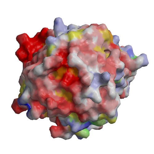
Functional site
| 1) | chain | B |
| residue | 344 | |
| type | ||
| sequence | R | |
| description | binding site for residue CL A 503 | |
| source | : AC3 | |
| 2) | chain | B |
| residue | 226 | |
| type | ||
| sequence | H | |
| description | binding site for residue EDO B 502 | |
| source | : AC4 | |
| 3) | chain | B |
| residue | 299 | |
| type | ||
| sequence | K | |
| description | binding site for residue EDO B 502 | |
| source | : AC4 | |
| 4) | chain | B |
| residue | 23 | |
| type | ||
| sequence | K | |
| description | binding site for residue CIT B 503 | |
| source | : AC5 | |
| 5) | chain | B |
| residue | 24 | |
| type | ||
| sequence | N | |
| description | binding site for residue CIT B 503 | |
| source | : AC5 | |
| 6) | chain | B |
| residue | 122 | |
| type | ||
| sequence | I | |
| description | binding site for residue CIT B 503 | |
| source | : AC5 | |
| 7) | chain | B |
| residue | 125 | |
| type | ||
| sequence | R | |
| description | binding site for residue CIT B 503 | |
| source | : AC5 | |
| 8) | chain | B |
| residue | 309 | |
| type | ||
| sequence | D | |
| description | binding site for residue CIT B 503 | |
| source | : AC5 | |
| 9) | chain | B |
| residue | 332 | |
| type | ||
| sequence | F | |
| description | binding site for residue CIT B 503 | |
| source | : AC5 | |
| 10) | chain | B |
| residue | 335 | |
| type | ||
| sequence | R | |
| description | binding site for residue CIT B 503 | |
| source | : AC5 | |
| 11) | chain | B |
| residue | 374 | |
| type | ||
| sequence | L | |
| description | binding site for residue CIT B 503 | |
| source | : AC5 | |
| 12) | chain | B |
| residue | 375 | |
| type | ||
| sequence | R | |
| description | binding site for residue CIT B 503 | |
| source | : AC5 | |
| 13) | chain | B |
| residue | 401 | |
| type | ||
| sequence | R | |
| description | binding site for residue CIT B 503 | |
| source | : AC5 | |
| 14) | chain | B |
| residue | 203 | |
| type | ||
| sequence | N | |
| description | binding site for residue CL B 504 | |
| source | : AC6 | |
| 15) | chain | B |
| residue | 96 | |
| type | ||
| sequence | R | |
| description | binding site for Di-peptide 0V5 B 501 and CYS B 120 | |
| source | : AC7 | |
| 16) | chain | B |
| residue | 119 | |
| type | ||
| sequence | G | |
| description | binding site for Di-peptide 0V5 B 501 and CYS B 120 | |
| source | : AC7 | |
| 17) | chain | B |
| residue | 121 | |
| type | ||
| sequence | T | |
| description | binding site for Di-peptide 0V5 B 501 and CYS B 120 | |
| source | : AC7 | |
| 18) | chain | B |
| residue | 122 | |
| type | ||
| sequence | I | |
| description | binding site for Di-peptide 0V5 B 501 and CYS B 120 | |
| source | : AC7 | |
| 19) | chain | B |
| residue | 123 | |
| type | ||
| sequence | G | |
| description | binding site for Di-peptide 0V5 B 501 and CYS B 120 | |
| source | : AC7 | |
| 20) | chain | B |
| residue | 125 | |
| type | ||
| sequence | R | |
| description | binding site for Di-peptide 0V5 B 501 and CYS B 120 | |
| source | : AC7 | |
| 21) | chain | B |
| residue | 401 | |
| type | ||
| sequence | R | |
| description | binding site for Di-peptide 0V5 B 501 and CYS B 120 | |
| source | : AC7 | |
| 22) | chain | B |
| residue | 120 | |
| type | ACT_SITE | |
| sequence | C | |
| description | Proton donor => ECO:0000255|HAMAP-Rule:MF_00111 | |
| source | Swiss-Prot : SWS_FT_FI1 | |
| 23) | chain | B |
| residue | 331 | |
| type | BINDING | |
| sequence | V | |
| description | BINDING => ECO:0000255|HAMAP-Rule:MF_00111 | |
| source | Swiss-Prot : SWS_FT_FI2 | |
| 24) | chain | B |
| residue | 23 | |
| type | BINDING | |
| sequence | K | |
| description | BINDING => ECO:0000255|HAMAP-Rule:MF_00111 | |
| source | Swiss-Prot : SWS_FT_FI2 | |
| 25) | chain | B |
| residue | 96 | |
| type | BINDING | |
| sequence | R | |
| description | BINDING => ECO:0000255|HAMAP-Rule:MF_00111 | |
| source | Swiss-Prot : SWS_FT_FI2 | |
| 26) | chain | B |
| residue | 125 | |
| type | BINDING | |
| sequence | R | |
| description | BINDING => ECO:0000255|HAMAP-Rule:MF_00111 | |
| source | Swiss-Prot : SWS_FT_FI2 | |
| 27) | chain | B |
| residue | 309 | |
| type | BINDING | |
| sequence | D | |
| description | BINDING => ECO:0000255|HAMAP-Rule:MF_00111 | |
| source | Swiss-Prot : SWS_FT_FI2 | |
| 28) | chain | B |
| residue | 120 | |
| type | MOD_RES | |
| sequence | C | |
| description | 2-(S-cysteinyl)pyruvic acid O-phosphothioketal => ECO:0000255|HAMAP-Rule:MF_00111 | |
| source | Swiss-Prot : SWS_FT_FI3 | |
