|
|
|
1)
|
chain |
B |
| residue |
219 |
| type |
|
| sequence |
Q
|
| description |
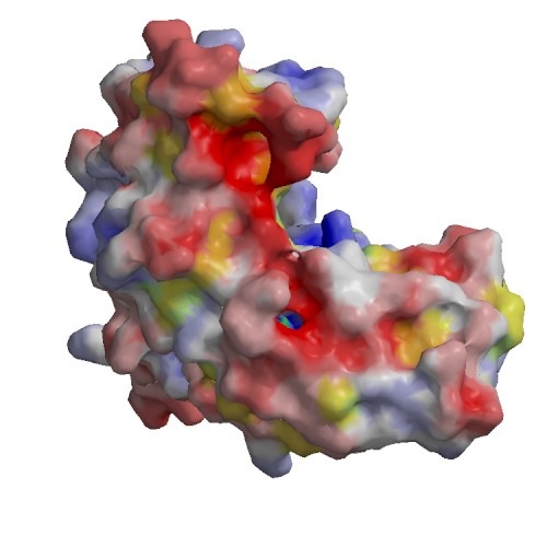
binding site for residue BEZ B 301
|
| source |
: AC1
|
|
|
2)
|
chain |
B |
| residue |
222 |
| type |
|
| sequence |
R
|
| description |
binding site for residue BEZ B 301
|
| source |
: AC1
|
|
|
3)
|
chain |
B |
| residue |
41 |
| type |
|
| sequence |
R
|
| description |
binding site for Di-peptide ASP D 424 and ZS8 D 425
|
| source |
: AC4
|
|
|
4)
|
chain |
B |
| residue |
42 |
| type |
|
| sequence |
N
|
| description |
binding site for Di-peptide ASP D 424 and ZS8 D 425
|
| source |
: AC4
|
|
|
5)
|
chain |
B |
| residue |
49 |
| type |
|
| sequence |
K
|
| description |
binding site for Di-peptide ASP D 424 and ZS8 D 425
|
| source |
: AC4
|
|
|
6)
|
chain |
B |
| residue |
117 |
| type |
|
| sequence |
F
|
| description |
binding site for Di-peptide ASP D 424 and ZS8 D 425
|
| source |
: AC4
|
|
|
7)
|
chain |
B |
| residue |
41 |
| type |
|
| sequence |
R
|
| description |
binding site for Di-peptide ZS8 D 425 and LEU D 426
|
| source |
: AC5
|
|
|
8)
|
chain |
B |
| residue |
42 |
| type |
|
| sequence |
N
|
| description |
binding site for Di-peptide ZS8 D 425 and LEU D 426
|
| source |
: AC5
|
|
|
9)
|
chain |
B |
| residue |
117 |
| type |
|
| sequence |
F
|
| description |
binding site for Di-peptide ZS8 D 425 and LEU D 426
|
| source |
: AC5
|
|
|
10)
|
chain |
B |
| residue |
120 |
| type |
|
| sequence |
K
|
| description |
binding site for Di-peptide ZS8 D 425 and LEU D 426
|
| source |
: AC5
|
|
|
11)
|
chain |
B |
| residue |
3 |
| type |
MOD_RES |
| sequence |
K
|
| description |
N6-acetyllysine => ECO:0007744|PubMed:19608861
|
| source |
Swiss-Prot : SWS_FT_FI3
|
|
|
12)
|
chain |
B |
| residue |
68 |
| type |
MOD_RES |
| sequence |
K
|
| description |
N6-acetyllysine => ECO:0007744|PubMed:19608861
|
| source |
Swiss-Prot : SWS_FT_FI3
|
|
|
13)
|
chain |
B |
| residue |
56 |
| type |
SITE |
| sequence |
R
|
| description |
Interaction with phosphoserine on interacting protein => ECO:0000250|UniProtKB:P63103
|
| source |
Swiss-Prot : SWS_FT_FI1
|
|
|
14)
|
chain |
B |
| residue |
127 |
| type |
SITE |
| sequence |
R
|
| description |
Interaction with phosphoserine on interacting protein => ECO:0000250|UniProtKB:P63103
|
| source |
Swiss-Prot : SWS_FT_FI1
|
|
|
15)
|
chain |
B |
| residue |
210 |
| type |
MOD_RES |
| sequence |
S
|
| description |
Phosphoserine => ECO:0000250|UniProtKB:P63102
|
| source |
Swiss-Prot : SWS_FT_FI7
|
|
|
16)
|
chain |
B |
| residue |
1 |
| type |
MOD_RES |
| sequence |
M
|
| description |
N-acetylmethionine => ECO:0000269|Ref.8, ECO:0007744|PubMed:22223895, ECO:0007744|PubMed:22814378, ECO:0007744|PubMed:25944712
|
| source |
Swiss-Prot : SWS_FT_FI2
|
|
|
17)
|
chain |
B |
| residue |
58 |
| type |
MOD_RES |
| sequence |
S
|
| description |
Phosphoserine; by PKA and PKB/AKT1 => ECO:0000269|PubMed:11956222, ECO:0000269|PubMed:12865427, ECO:0000269|PubMed:15883165, ECO:0000269|PubMed:16376338
|
| source |
Swiss-Prot : SWS_FT_FI4
|
|
|
18)
|
chain |
B |
| residue |
184 |
| type |
MOD_RES |
| sequence |
S
|
| description |
Phosphoserine; by MAPK8 => ECO:0000269|PubMed:15071501, ECO:0000269|PubMed:15696159
|
| source |
Swiss-Prot : SWS_FT_FI5
|
|