|
|
|
1)
|
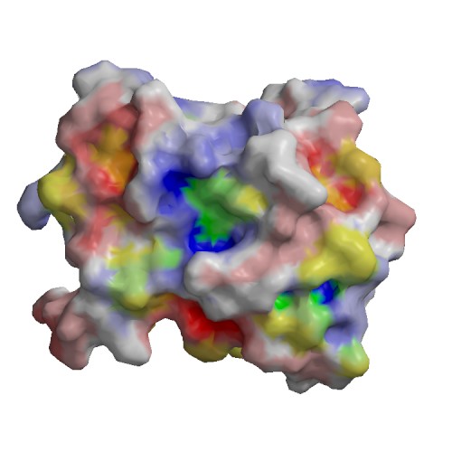
chain |
A |
| residue |
191 |
| type |
SITE |
| sequence |
F
|
| description |
Involved in dimerization of the capsid protein => ECO:0000269|PubMed:25100849
|
| source |
Swiss-Prot : SWS_FT_FI2
|
|
|
2)
|
chain |
A |
| residue |
192 |
| type |
SITE |
| sequence |
Y
|
| description |
Involved in dimerization of the capsid protein => ECO:0000269|PubMed:25100849
|
| source |
Swiss-Prot : SWS_FT_FI2
|
|
|
3)
|
chain |
A |
| residue |
225 |
| type |
SITE |
| sequence |
N
|
| description |
Involved in dimerization of the capsid protein => ECO:0000269|PubMed:25100849
|
| source |
Swiss-Prot : SWS_FT_FI2
|
|
|
4)
|
chain |
A |
| residue |
267 |
| type |
SITE |
| sequence |
W
|
| description |
Cleavage; by autolysis => ECO:0000250|UniProtKB:P03315
|
| source |
Swiss-Prot : SWS_FT_FI3
|
|
|
5)
|
chain |
A |
| residue |
144 |
| type |
ACT_SITE |
| sequence |
H
|
| description |
Charge relay system => ECO:0000255|PROSITE-ProRule:PRU01027
|
| source |
Swiss-Prot : SWS_FT_FI1
|
|
|
6)
|
chain |
A |
| residue |
166 |
| type |
ACT_SITE |
| sequence |
D
|
| description |
Charge relay system => ECO:0000255|PROSITE-ProRule:PRU01027
|
| source |
Swiss-Prot : SWS_FT_FI1
|
|
|
7)
|
chain |
A |
| residue |
218 |
| type |
ACT_SITE |
| sequence |
S
|
| description |
Charge relay system => ECO:0000255|PROSITE-ProRule:PRU01027
|
| source |
Swiss-Prot : SWS_FT_FI1
|
|