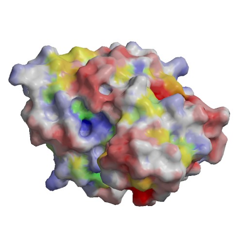
Functional site
| 1) | chain | A |
| residue | 429 | |
| type | ||
| sequence | D | |
| description | BINDING SITE FOR RESIDUE CA A 497 | |
| source | : AC1 | |
| 2) | chain | A |
| residue | 431 | |
| type | ||
| sequence | D | |
| description | BINDING SITE FOR RESIDUE CA A 497 | |
| source | : AC1 | |
| 3) | chain | A |
| residue | 433 | |
| type | ||
| sequence | C | |
| description | BINDING SITE FOR RESIDUE CA A 497 | |
| source | : AC1 | |
| 4) | chain | A |
| residue | 435 | |
| type | ||
| sequence | C | |
| description | BINDING SITE FOR RESIDUE CA A 497 | |
| source | : AC1 | |
| 5) | chain | A |
| residue | 445-457 | |
| type | prosite | |
| sequence | WWFDACGPSNLNG | |
| description | FIBRINOGEN_C_1 Fibrinogen C-terminal domain signature. WWFdaCgpSnlNG | |
| source | prosite : PS00514 | |
| 6) | chain | A |
| residue | 429 | |
| type | BINDING | |
| sequence | D | |
| description | BINDING => ECO:0000269|PubMed:15893672, ECO:0000269|PubMed:16732286, ECO:0007744|PDB:1Z3U | |
| source | Swiss-Prot : SWS_FT_FI1 | |
| 7) | chain | A |
| residue | 431 | |
| type | BINDING | |
| sequence | D | |
| description | BINDING => ECO:0000269|PubMed:15893672, ECO:0000269|PubMed:16732286, ECO:0007744|PDB:1Z3U | |
| source | Swiss-Prot : SWS_FT_FI1 | |
| 8) | chain | A |
| residue | 433 | |
| type | BINDING | |
| sequence | C | |
| description | BINDING => ECO:0000269|PubMed:15893672, ECO:0000269|PubMed:16732286, ECO:0007744|PDB:1Z3U | |
| source | Swiss-Prot : SWS_FT_FI1 | |
| 9) | chain | A |
| residue | 435 | |
| type | BINDING | |
| sequence | C | |
| description | BINDING => ECO:0000269|PubMed:15893672, ECO:0000269|PubMed:16732286, ECO:0007744|PDB:1Z3U | |
| source | Swiss-Prot : SWS_FT_FI1 | |
| 10) | chain | A |
| residue | 304 | |
| type | CARBOHYD | |
| sequence | N | |
| description | N-linked (GlcNAc...) asparagine => ECO:0000255 | |
| source | Swiss-Prot : SWS_FT_FI2 | |
