Loading
PDBj
MenuPDBj@FacebookPDBj@TwitterPDBj@YouTubewwPDB FoundationwwPDB
RCSB PDBPDBeBMRBAdv. SearchSearch help
Search by PDB author
2JU6
DownloadVisualize
BU of 2ju6 by Molmil
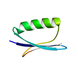
Solid-State Protein Structure Determination with Proton-Detected Triple Resonance 3D Magic-Angle Spinning NMR Spectroscopy
Descriptor: Immunoglobulin G-binding protein G
Authors:Zhou, D.H, Shea, J.J, Nieuwkoop, A.J, Franks, W, Wylie, B.J, Mullen, C, Sandoz, D, Rienstra, C.M.
Deposit date:2007-08-15
Release date:2007-12-04
Last modified:2021-10-20
Method:SOLID-STATE NMR
Cite:Solid-State Protein-Structure Determination with Proton-Detected Triple-Resonance 3D Magic-Angle-Spinning NMR Spectroscopy.
Angew.Chem.Int.Ed.Engl., 46, 2007
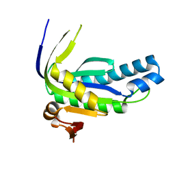
2MH2
DownloadVisualize
BU of 2mh2 by Molmil
Structural insights into the DNA recognition and protein interaction domains reveal fundamental homologous DNA pairing properties of HOP2
Descriptor: Homologous-pairing protein 2 homolog
Authors:Moktan, H, Guiraldelli, M.F, Eyter, C.A, Zhao, W, Camerini-Otero, R.D, Sung, P, Zhou, D.H, Pezza, R.J.
Deposit date:2013-11-13
Release date:2014-04-16
Last modified:2014-08-06
Method:SOLUTION NMR
Cite:Solution Structure and DNA-binding Properties of the Winged Helix Domain of the Meiotic Recombination HOP2 Protein.
J.Biol.Chem., 289, 2014
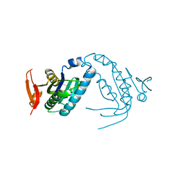
2K0P
DownloadVisualize
BU of 2k0p by Molmil
Determination of a Protein Structure in the Solid State from NMR Chemical Shifts
Descriptor: Immunoglobulin G-binding protein G
Authors:Robustelli, P, Cavalli, A, Salvatella, X, Vendruscolo, M.
Deposit date:2008-02-11
Release date:2009-03-03
Last modified:2021-10-20
Method:SOLID-STATE NMR
Cite:Determination of protein structures in the solid state from NMR chemical shifts.
Structure, 16, 2008
6AO9
DownloadVisualize
BU of 6ao9 by Molmil
Crystal structure of monomeric guanylyl cyclase domain of RhoGC fusion protein from the aquatic fungus Blastocladiella emersonii
Descriptor: Bacterio-rhodopsin/guanylyl cyclase 1 fusion protein
Authors:Prem Kumar, R, Oprian, D.D.
Deposit date:2017-08-15
Release date:2017-11-22
Last modified:2023-10-04
Method:X-RAY DIFFRACTION (1.13 Å)
Cite:Structure and monomer/dimer equilibrium for the guanylyl cyclase domain of the optogenetics protein RhoGC.
J. Biol. Chem., 292, 2017
6AOA
DownloadVisualize
BU of 6aoa by Molmil
Monomeric crystal structure of the E497/C566D double mutant of the guanylyl cyclase domain of the RhoGC fusion protein from the aquatic fungus Blastocladiella emersonii
Descriptor: Bacterio-rhodopsin/guanylyl cyclase 1 fusion protein
Authors:Prem Kumar, R, Oprian, D.D.
Deposit date:2017-08-15
Release date:2017-11-22
Last modified:2023-10-04
Method:X-RAY DIFFRACTION (1.4 Å)
Cite:Structure and monomer/dimer equilibrium for the guanylyl cyclase domain of the optogenetics protein RhoGC.
J. Biol. Chem., 292, 2017
6AOB
DownloadVisualize
BU of 6aob by Molmil
Crystal structure of non-canonical dimeric guanylyl cyclase domain of RhoGC fusion protein from the aquatic fungus Blastocladiella emersonii
Descriptor: Bacterio-rhodopsin/guanylyl cyclase 1 fusion protein, L(+)-TARTARIC ACID, MANGANESE (II) ION
Authors:Prem Kumar, R, Oprian, D.D.
Deposit date:2017-08-15
Release date:2017-11-22
Last modified:2023-10-04
Method:X-RAY DIFFRACTION (1.7 Å)
Cite:Structure and monomer/dimer equilibrium for the guanylyl cyclase domain of the optogenetics protein RhoGC.
J. Biol. Chem., 292, 2017

218853

PDB entries from 2024-04-24

PDB statisticsPDBj update infoContact PDBjnumon