Loading
PDBj
MenuPDBj@FacebookPDBj@TwitterPDBj@YouTubewwPDB FoundationwwPDB
RCSB PDBPDBeBMRBAdv. SearchSearch help
Search by PDB author
4EF0
DownloadVisualize
BU of 4ef0 by Molmil
Crystal Structure of the first catalytic domain of protein disulfide isomerase P5
Descriptor: Protein disulfide-isomerase A6
Authors:Vinaik, R, Kozlov, G, Gehring, K.
Deposit date:2012-03-28
Release date:2013-04-03
Last modified:2023-09-13
Method:X-RAY DIFFRACTION (1.5 Å)
Cite:Crystal Structure of the first catalytic domain of protein disulfide isomerase P5
To be Published
4GWR
DownloadVisualize
BU of 4gwr by Molmil
Crystal Structure of the second catalytic domain of protein disulfide isomerase P5
Descriptor: Protein disulfide-isomerase A6
Authors:Vinaik, R, Kozlov, G, Gehring, K.
Deposit date:2012-09-03
Release date:2013-09-04
Last modified:2023-11-08
Method:X-RAY DIFFRACTION (1.81 Å)
Cite:To be published
To be Published, 2013
4I6X
DownloadVisualize
BU of 4i6x by Molmil
Crystal Structure of Non-catalyic Domain of Protein Disulfide Isomerase-related (PDIr) Protein
Descriptor: Protein disulfide-isomerase A5
Authors:Kozlov, G, Vinaik, R, Gehring, K.
Deposit date:2012-11-30
Release date:2013-04-03
Last modified:2013-05-08
Method:X-RAY DIFFRACTION (1.5 Å)
Cite:Structure of the Non-Catalytic Domain of the Protein Disulfide Isomerase-Related Protein (PDIR) Reveals Function in Protein Binding.
Plos One, 8, 2013
4IOT
DownloadVisualize
BU of 4iot by Molmil
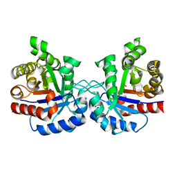
High-resolution Structure of Triosephosphate isomerase from E. coli
Descriptor: SULFATE ION, Triosephosphate isomerase
Authors:Vinaik, R, Kozlov, G, Gehring, K, Montreal-Kingston Bacterial Structural Genomics Initiative (BSGI)
Deposit date:2013-01-08
Release date:2013-01-23
Last modified:2023-09-20
Method:X-RAY DIFFRACTION (1.85 Å)
Cite:Triosephosphate isomerase is a common crystallization contaminant of soluble His-tagged proteins produced in Escherichia coli.
Acta Crystallogr.,Sect.F, 69, 2013
4EEW
DownloadVisualize
BU of 4eew by Molmil
Crystal structure of the Ubl domain of BAG6
Descriptor: Large proline-rich protein BAG6
Authors:Kozlov, G, LePage, K, Vinaik, R, Gehring, K.
Deposit date:2012-03-28
Release date:2012-04-11
Last modified:2023-09-13
Method:X-RAY DIFFRACTION (1.3 Å)
Cite:Crystal structure of the Ubl domain of BAG6
To be Published

218500

PDB entries from 2024-04-17

PDB statisticsPDBj update infoContact PDBjnumon