Loading
PDBj
MenuPDBj@FacebookPDBj@TwitterPDBj@YouTubewwPDB FoundationwwPDB
RCSB PDBPDBeBMRBAdv. SearchSearch help
Search by PDB author
5KYH
DownloadVisualize
BU of 5kyh by Molmil
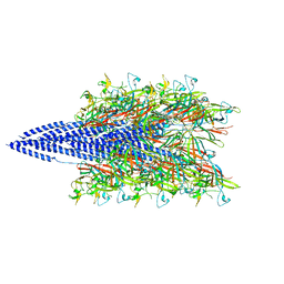
Structure of Iho670 Flagellar-like Filament
Descriptor: Iho670
Authors:Braun, T, Vos, M, Kalisman, N, Sherman, N.E, Rachel, R, Wirth, R, Schroeder, G.F, Egelman, E.H.
Deposit date:2016-07-21
Release date:2016-09-07
Last modified:2019-12-11
Method:ELECTRON MICROSCOPY (4 Å)
Cite:Archaeal flagellin combines a bacterial type IV pilin domain with an Ig-like domain.
Proc.Natl.Acad.Sci.USA, 113, 2016
3ILY
DownloadVisualize
BU of 3ily by Molmil
Apo crystal structure of protein tyrosine phosphatase from Entamoeba histolytica featuring a disordered active site
Descriptor: Protein tyrosine phosphatase, putative
Authors:Seattle Structural Genomics Center for Infectious Disease (SSGCID)
Deposit date:2009-08-07
Release date:2009-08-25
Last modified:2023-09-06
Method:X-RAY DIFFRACTION (2.2 Å)
Cite:Crystal structure and putative substrate identification for the Entamoeba histolytica low molecular weight tyrosine phosphatase.
Mol.Biochem.Parasitol., 193, 2014
3IDO
DownloadVisualize
BU of 3ido by Molmil
Crystal structure of protein tyrosine phosphatase from Entamoeba histolytica with a phosphotyrosine crude mimic HEPES molecule in the active site
Descriptor: 4-(2-HYDROXYETHYL)-1-PIPERAZINE ETHANESULFONIC ACID, Protein tyrosine phosphatase
Authors:Seattle Structural Genomics Center for Infectious Disease (SSGCID)
Deposit date:2009-07-21
Release date:2009-07-28
Last modified:2023-09-06
Method:X-RAY DIFFRACTION (2.2 Å)
Cite:Crystal structure and putative substrate identification for the Entamoeba histolytica low molecular weight tyrosine phosphatase.
Mol.Biochem.Parasitol., 193, 2014
3JVI
DownloadVisualize
BU of 3jvi by Molmil
Product state mimic crystal structure of protein tyrosine phosphatase from Entamoeba histolytica
Descriptor: Protein tyrosine phosphatase, SULFATE ION
Authors:Seattle Structural Genomics Center for Infectious Disease (SSGCID)
Deposit date:2009-09-16
Release date:2009-09-22
Last modified:2023-09-06
Method:X-RAY DIFFRACTION (1.8 Å)
Cite:Crystal structure and putative substrate identification for the Entamoeba histolytica low molecular weight tyrosine phosphatase.
Mol.Biochem.Parasitol., 193, 2014
3JS5
DownloadVisualize
BU of 3js5 by Molmil
Crystal structure of protein tyrosine phosphatase from Entamoeba histolytica with Hepes in the active site. High resolution, alternative crystal form with 1 molecule in asymmetric unit
Descriptor: 4-(2-HYDROXYETHYL)-1-PIPERAZINE ETHANESULFONIC ACID, Protein tyrosine phosphatase, SODIUM ION
Authors:Seattle Structural Genomics Center for Infectious Disease (SSGCID)
Deposit date:2009-09-09
Release date:2009-09-22
Last modified:2023-09-06
Method:X-RAY DIFFRACTION (1.94 Å)
Cite:Crystal structure and putative substrate identification for the Entamoeba histolytica low molecular weight tyrosine phosphatase.
Mol.Biochem.Parasitol., 193, 2014

218500

PDB entries from 2024-04-17

PDB statisticsPDBj update infoContact PDBjnumon