Loading
PDBj
MenuPDBj@FacebookPDBj@TwitterPDBj@YouTubewwPDB FoundationwwPDB
RCSB PDBPDBeBMRBAdv. SearchSearch help
Search by PDB author
6CXB
DownloadVisualize
BU of 6cxb by Molmil
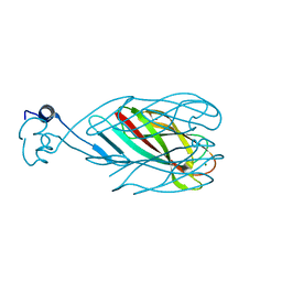
Structure of N-truncated R1-type pyocin tail fiber at 1.7 angstrom resolution
Descriptor: GLYCEROL, MAGNESIUM ION, R1-type pyocin tail fiber protein
Authors:Salazar, A.J, Sherekar, M, Saccettini, J.C.
Deposit date:2018-04-02
Release date:2019-02-20
Last modified:2023-10-04
Method:X-RAY DIFFRACTION (1.701 Å)
Cite:R pyocin tail fiber structure reveals a receptor-binding domain with a lectin fold.
PLoS ONE, 14, 2019
4FB8
DownloadVisualize
BU of 4fb8 by Molmil
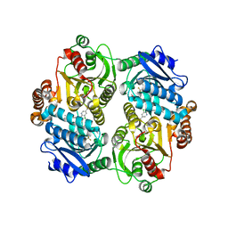
Crystal Structure of apo Acyl-CoA Carboxylase
Descriptor: Probable propionyl-CoA carboxylase beta chain 6
Authors:Reddy, M.C.M, Bruning, J.B, Sherekar, M, Valluru, S, Ehrenfeld, H, Sacchettini, J.C, TB Structural Genomics Consortium (TBSGC)
Deposit date:2012-05-22
Release date:2014-02-19
Last modified:2023-09-13
Method:X-RAY DIFFRACTION (3 Å)
Cite:Structure, Activity, and Inhibition of the Carboxyltransferase beta-Subunit of Acetyl Coenzyme A Carboxylase (AccD6) from Mycobacterium tuberculosis.
Antimicrob.Agents Chemother., 58, 2014
4G2R
DownloadVisualize
BU of 4g2r by Molmil
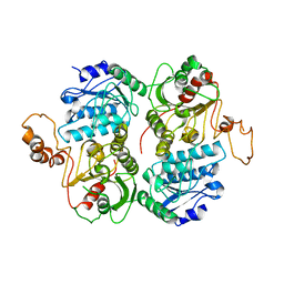
Crystal Structure of the carboxyltransferase subunit of ACC (AccD6) in complex with inhibitor haloxyfop from Mycobacterium tuberculosis
Descriptor: (2R)-2-(4-{[3-chloro-5-(trifluoromethyl)pyridin-2-yl]oxy}phenoxy)propanoic acid, AccD6, Carboxyltransferase beta-subunit of Acyl-CoA Carboxylase
Authors:Reddy, M.C.M, Bruning, J.B, Thurman, C, Sherekar, M, Valluru, S, Ehrenfeld, H, Sacchettini, J.C, TB Structural Genomics Consortium (TBSGC)
Deposit date:2012-07-12
Release date:2014-02-19
Last modified:2023-09-13
Method:X-RAY DIFFRACTION (2.28 Å)
Cite:Structure, Activity, and Inhibition of the Carboxyltransferase beta-Subunit of Acetyl Coenzyme A Carboxylase (AccD6) from Mycobacterium tuberculosis.
Antimicrob.Agents Chemother., 58, 2014
6CU2
DownloadVisualize
BU of 6cu2 by Molmil
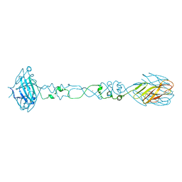
Structure of N-truncated R2-type pyocin tail fiber at 2.6 angstrom resolution
Descriptor: MAGNESIUM ION, NICKEL (II) ION, R2-type pyocin
Authors:Salazar, A.J, Sherekar, M, Saccettini, J.C.
Deposit date:2018-03-23
Release date:2019-02-20
Last modified:2023-10-04
Method:X-RAY DIFFRACTION (2.58 Å)
Cite:R pyocin tail fiber structure reveals a receptor-binding domain with a lectin fold.
PLoS ONE, 14, 2019
6CT8
DownloadVisualize
BU of 6ct8 by Molmil
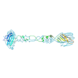
Selenomethionine structure of N-truncated R2-type pyocin tail fiber at 2.6 angstrom resolution
Descriptor: NICKEL (II) ION, R2-type pyocin
Authors:Salazar, A.J, Sherekar, M, Saccettini, J.C.
Deposit date:2018-03-22
Release date:2019-02-20
Method:X-RAY DIFFRACTION (2.62 Å)
Cite:R pyocin tail fiber structure reveals a receptor-binding domain with a lectin fold.
PLoS ONE, 14, 2019

218853

PDB entries from 2024-04-24

PDB statisticsPDBj update infoContact PDBjnumon