Loading
PDBj
MenuPDBj@FacebookPDBj@TwitterPDBj@YouTubewwPDB FoundationwwPDB
RCSB PDBPDBeBMRBAdv. SearchSearch help
Search by PDB author
1C3X
DownloadVisualize
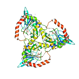
BU of 1c3x by Molmil
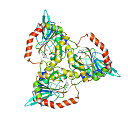
PURINE NUCLEOSIDE PHOSPHORYLASE FROM CELLULOMONAS SP. IN COMPLEX WITH 8-IODO-GUANINE
Descriptor: 8-IODO-GUANINE, CALCIUM ION, PENTOSYLTRANSFERASE, ...
Authors:Tebbe, J, Bzowska, A, Wielgus-Kutrowska, B, Schroeder, W, Kazimierczuk, Z, Shugar, D, Saenger, W, Koellner, G.
Deposit date:1999-07-29
Release date:1999-12-22
Last modified:2024-02-07
Method:X-RAY DIFFRACTION (2.4 Å)
Cite:Crystal structure of the purine nucleoside phosphorylase (PNP) from Cellulomonas sp. and its implication for the mechanism of trimeric PNPs.
J.Mol.Biol., 294, 1999
1QE5
DownloadVisualize
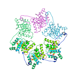
BU of 1qe5 by Molmil
PURINE NUCLEOSIDE PHOSPHORYLASE FROM CELLULOMONAS SP. IN COMPLEX WITH PHOSPHATE
Descriptor: CALCIUM ION, PENTOSYLTRANSFERASE, PHOSPHATE ION
Authors:Tebbe, J, Bzowska, A, Wielgus-Kutrowska, B, Schroeder, W, Kazimierczuk, Z, Shugar, D, Saenger, W, Koellner, G.
Deposit date:1999-07-13
Release date:1999-12-12
Last modified:2023-08-16
Method:X-RAY DIFFRACTION (2.2 Å)
Cite:Crystal structure of the purine nucleoside phosphorylase (PNP) from Cellulomonas sp. and its implication for the mechanism of trimeric PNPs.
J.Mol.Biol., 294, 1999
1NLF
DownloadVisualize
BU of 1nlf by Molmil
Crystal Structure of DNA Helicase RepA in complex with sulfate at 1.95 A resolution
Descriptor: Regulatory protein repA, SULFATE ION
Authors:Xu, H, Strater, N, Schroeder, W, Bottcher, C, Ludwig, K, Saenger, W.
Deposit date:2003-01-07
Release date:2003-04-29
Last modified:2024-02-14
Method:X-RAY DIFFRACTION (1.95 Å)
Cite:Structure of DNA helicase RepA in complex with sulfate at 1.95 A resolution implicates structural changes to an "open" form.
Acta Crystallogr.,Sect.D, 59, 2003
4JXS
DownloadVisualize
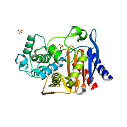
BU of 4jxs by Molmil
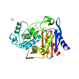
X-ray crystal structure of AmpC beta-lactamase from E. coli in complex with a non-covalent inhibitor 3-[(4-CARBOXYBENZYL)SULFAMOYL]THIOPHENE-2-CARBOXYLIC ACID (compound 4)
Descriptor: 3-[(4-carboxybenzyl)sulfamoyl]thiophene-2-carboxylic acid, Beta-lactamase, PHOSPHATE ION
Authors:Powers, R.A, Hendershot, J.M.
Deposit date:2013-03-28
Release date:2014-05-07
Last modified:2023-09-20
Method:X-RAY DIFFRACTION (1.9 Å)
Cite:Structure-based efforts to optimize a non-beta-lactam inhibitor of AmpC beta-lactamase.
Bioorg.Med.Chem., 22, 2014
4JXW
DownloadVisualize
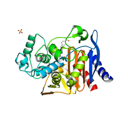
BU of 4jxw by Molmil
X-ray crystal structure of AmpC beta-lactamase from E. coli in complex with a non-covalent inhibitor 3-{[3-(4-CARBOXYPHENYL)PROPYL]SULFAMOYL}THIOPHENE-2-CARBOXYLIC ACID (compound 6)
Descriptor: 3-{[3-(4-carboxyphenyl)propyl]sulfamoyl}thiophene-2-carboxylic acid, Beta-lactamase, PHOSPHATE ION
Authors:Powers, R.A, Hendershot, J.M.
Deposit date:2013-03-28
Release date:2014-05-07
Last modified:2024-02-28
Method:X-RAY DIFFRACTION (2.3 Å)
Cite:Structure-based efforts to optimize a non-beta-lactam inhibitor of AmpC beta-lactamase.
Bioorg.Med.Chem., 22, 2014
4JXV
DownloadVisualize
BU of 4jxv by Molmil
X-ray crystal structure of AmpC beta-lactamase from E. coli in complex with a non-covalent inhibitor 3-{[2-(4-CARBOXYPHENYL)ETHYL]SULFAMOYL}THIOPHENE-2-CARBOXYLIC ACID (compound 5)
Descriptor: 3-{[2-(4-carboxyphenyl)ethyl]sulfamoyl}thiophene-2-carboxylic acid, Beta-lactamase, PHOSPHATE ION
Authors:Powers, R.A, Hendershot, J.M.
Deposit date:2013-03-28
Release date:2014-04-30
Last modified:2024-02-28
Method:X-RAY DIFFRACTION (1.76 Å)
Cite:Structure-based efforts to optimize a non-beta-lactam inhibitor of AmpC beta-lactamase.
Bioorg.Med.Chem., 22, 2014

218500

PDB entries from 2024-04-17

PDB statisticsPDBj update infoContact PDBjnumon