Loading
PDBj
MenuPDBj@FacebookPDBj@TwitterPDBj@YouTubewwPDB FoundationwwPDB
RCSB PDBPDBeBMRBAdv. SearchSearch help
Search by PDB author
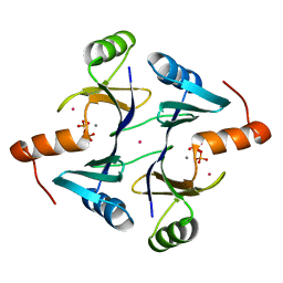
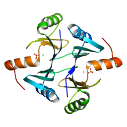
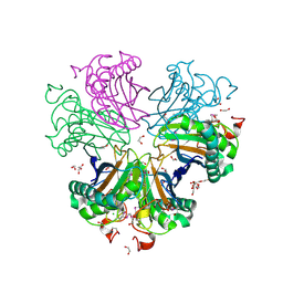
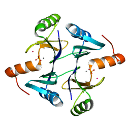
1LQK
DownloadVisualize
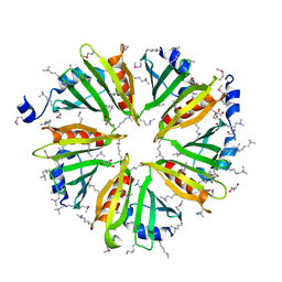
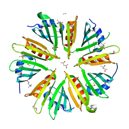
BU of 1lqk by Molmil
High Resolution Structure of Fosfomycin Resistance Protein A (FosA)
Descriptor: MANGANESE (II) ION, PHOSPHATE ION, POTASSIUM ION, ...
Authors:Rife, C.L, Pharris, R.E, Newcomer, M.E, Armstrong, R.N.
Deposit date:2002-05-10
Release date:2002-09-11
Last modified:2024-02-14
Method:X-RAY DIFFRACTION (1.35 Å)
Cite:Crystal structure of a genomically encoded fosfomycin resistance protein (FosA) at 1.19 A resolution by MAD phasing off the L-III edge of Tl(+)
J.Am.Chem.Soc., 124, 2002
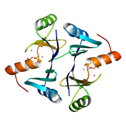
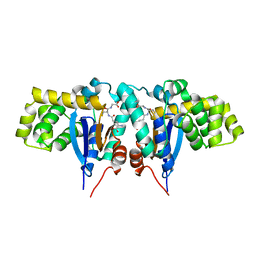
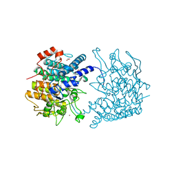
1LQO
DownloadVisualize
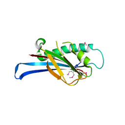
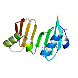
BU of 1lqo by Molmil
Crystal Strutcure of the Fosfomycin Resistance Protein A (FosA) Containing Bound Thallium Cations
Descriptor: MANGANESE (II) ION, PHOSPHATE ION, PROBABLE Fosfomycin Resistance Protein, ...
Authors:Rife, C.L, Pharris, R.E, Newcomer, M.E, Armstrong, R.N.
Deposit date:2002-05-11
Release date:2002-09-11
Last modified:2024-02-14
Method:X-RAY DIFFRACTION (2 Å)
Cite:Crystal structure of a genomically encoded fosfomycin resistance protein (FosA) at 1.19 A resolution by MAD phasing off the L-III edge of Tl(+)
J.Am.Chem.Soc., 124, 2002
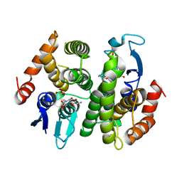
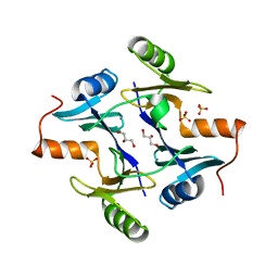
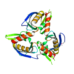
1LQP
DownloadVisualize
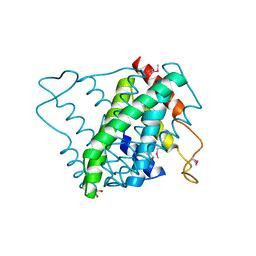
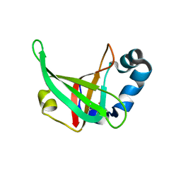
BU of 1lqp by Molmil
CRYSTAL STRUCTURE OF THE FOSFOMYCIN RESISTANCE PROTEIN (FOSA) CONTAINING BOUND SUBSTRATE
Descriptor: FOSFOMYCIN, MANGANESE (II) ION, POTASSIUM ION, ...
Authors:Rife, C.L, Pharris, R.E, Newcomer, M.E, Armstrong, R.N.
Deposit date:2002-05-11
Release date:2002-09-11
Last modified:2024-02-14
Method:X-RAY DIFFRACTION (1.19 Å)
Cite:Crystal structure of a genomically encoded fosfomycin resistance protein (FosA) at 1.19 A resolution by MAD phasing off the L-III edge of Tl(+)
J.Am.Chem.Soc., 124, 2002
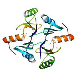
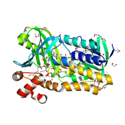
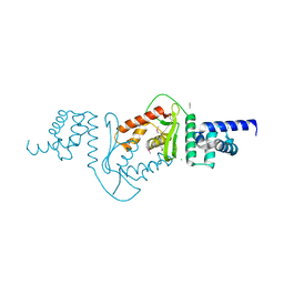
1N2A
DownloadVisualize
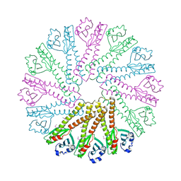
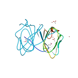
BU of 1n2a by Molmil
Crystal Structure of a Bacterial Glutathione Transferase from Escherichia coli with Glutathione Sulfonate in the Active Site
Descriptor: GLUTATHIONE SULFONIC ACID, Glutathione S-transferase
Authors:Rife, C.L, Parsons, J.F, Xiao, G, Gilliland, G.L, Armstrong, R.N.
Deposit date:2002-10-22
Release date:2003-11-04
Last modified:2024-02-14
Method:X-RAY DIFFRACTION (1.9 Å)
Cite:Conserved structural elements in glutathione transferase homologues encoded in the genome of Escherichia coli
Proteins, 53, 2003
1NKI
DownloadVisualize
BU of 1nki by Molmil
CRYSTAL STRUCTURE OF THE FOSFOMYCIN RESISTANCE PROTEIN A (FOSA) CONTAINING BOUND PHOSPHONOFORMATE
Descriptor: MANGANESE (II) ION, PHOSPHONOFORMIC ACID, POTASSIUM ION, ...
Authors:Rife, C.L, Pharris, R.E, Newcomer, M.E, Armstrong, R.N.
Deposit date:2003-01-03
Release date:2004-01-13
Last modified:2023-08-16
Method:X-RAY DIFFRACTION (0.95 Å)
Cite:Phosphonoformate: a minimal transition state analogue inhibitor of the fosfomycin resistance protein, FosA.
Biochemistry, 43, 2004
1NNR
DownloadVisualize
BU of 1nnr by Molmil
Crystal structure of a probable fosfomycin resistance protein (PA1129) from Pseudomonas aeruginosa with sulfate present in the active site
Descriptor: MANGANESE (II) ION, SULFATE ION, probable fosfomycin resistance protein
Authors:Rife, C.L, Pharris, R.E, Newcomer, M.E, Armstrong, R.N.
Deposit date:2003-01-14
Release date:2004-01-27
Last modified:2023-08-16
Method:X-RAY DIFFRACTION (2.25 Å)
Cite:Phosphonoformate: a minimal transition state analogue inhibitor of the fosfomycin resistance protein, FosA.
Biochemistry, 43, 2004
1R4W
DownloadVisualize
BU of 1r4w by Molmil
Crystal structure of Mitochondrial class kappa glutathione transferase
Descriptor: GLUTATHIONE, Glutathione S-transferase, mitochondrial
Authors:Ladner, J.E, Parsons, J.F, Rife, C.L, Gilliland, G.L, Armstrong, R.N.
Deposit date:2003-10-08
Release date:2004-02-03
Last modified:2024-02-14
Method:X-RAY DIFFRACTION (2.5 Å)
Cite:Parallel Evolutionary Pathways for Glutathione Transferases: Structure and Mechanism of the Mitochondrial Class Kappa Enzyme rGSTK1-1
Biochemistry, 43, 2004
1NPB
DownloadVisualize
BU of 1npb by Molmil
Crystal structure of the fosfomycin resistance protein from transposon Tn2921
Descriptor: GLYCEROL, SULFATE ION, fosfomycin-resistance protein
Authors:Pakhomova, S, Rife, C.L, Armstrong, R.N, Newcomer, M.E.
Deposit date:2003-01-17
Release date:2004-03-02
Last modified:2023-08-16
Method:X-RAY DIFFRACTION (2.5 Å)
Cite:Structure of fosfomycin resistance protein FosA from transposon Tn2921.
Protein Sci., 13, 2004
3OZ2
DownloadVisualize
BU of 3oz2 by Molmil
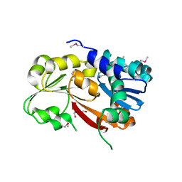
Crystal structure of a geranylgeranyl bacteriochlorophyll reductase-like (Ta0516) from Thermoplasma acidophilum at 1.60 A resolution
Descriptor: (2R)-3-{[(R)-{[(2S)-2,3-dihydroxypropyl]oxy}(hydroxy)phosphoryl]oxy}-2-[(6Z)-tridec-6-enoyloxy]propyl (9Z)-octadec-9-enoate, 1,2-ETHANEDIOL, Digeranylgeranylglycerophospholipid reductase, ...
Authors:Joint Center for Structural Genomics (JCSG)
Deposit date:2010-09-24
Release date:2010-10-27
Last modified:2023-02-01
Method:X-RAY DIFFRACTION (1.6 Å)
Cite:Insights into substrate specificity of geranylgeranyl reductases revealed by the structure of digeranylgeranylglycerophospholipid reductase, an essential enzyme in the biosynthesis of archaeal membrane lipids.
J.Mol.Biol., 404, 2010
3HSA
DownloadVisualize
BU of 3hsa by Molmil
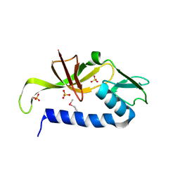
Crystal structure of pleckstrin homology domain (YP_926556.1) from SHEWANELLA AMAZONENSIS SB2B at 1.99 A resolution
Descriptor: DI(HYDROXYETHYL)ETHER, GLYCEROL, pleckstrin homology domain
Authors:Joint Center for Structural Genomics (JCSG)
Deposit date:2009-06-10
Release date:2009-06-23
Last modified:2023-02-01
Method:X-RAY DIFFRACTION (1.99 Å)
Cite:Bacterial pleckstrin homology domains: a prokaryotic origin for the PH domain.
J.Mol.Biol., 396, 2010
2QTP
DownloadVisualize
BU of 2qtp by Molmil
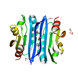
Crystal structure of a duf1185 family protein (spo0826) from silicibacter pomeroyi dss-3 at 2.10 A resolution
Descriptor: Uncharacterized protein
Authors:Joint Center for Structural Genomics (JCSG)
Deposit date:2007-08-02
Release date:2007-08-21
Last modified:2023-01-25
Method:X-RAY DIFFRACTION (2.1 Å)
Cite:Structures of the first representatives of Pfam family PF06684 (DUF1185) reveal a novel variant of the Bacillus chorismate mutase fold and suggest a role in amino-acid metabolism.
Acta Crystallogr.,Sect.F, 66, 2010
3NL9
DownloadVisualize
BU of 3nl9 by Molmil
Crystal structure of a putative NTP pyrophosphohydrolase (Exig_1061) from EXIGUOBACTERIUM SP. 255-15 at 1.78 A resolution
Descriptor: 1,2-ETHANEDIOL, putative NTP pyrophosphohydrolase
Authors:Joint Center for Structural Genomics (JCSG)
Deposit date:2010-06-21
Release date:2010-07-21
Last modified:2023-02-01
Method:X-RAY DIFFRACTION (1.78 Å)
Cite:Structure of a putative NTP pyrophosphohydrolase: YP_001813558.1 from Exiguobacterium sibiricum 255-15.
Acta Crystallogr.,Sect.F, 66, 2010
3B77
DownloadVisualize
BU of 3b77 by Molmil
Crystal structure of a ph domain containing bacterial protein (exig_2160) from exiguobacterium sibiricum 255-15 at 2.42 A resolution
Descriptor: Uncharacterized protein
Authors:Joint Center for Structural Genomics (JCSG)
Deposit date:2007-10-30
Release date:2007-11-13
Last modified:2023-01-25
Method:X-RAY DIFFRACTION (2.42 Å)
Cite:Bacterial pleckstrin homology domains: a prokaryotic origin for the PH domain.
J.Mol.Biol., 396, 2010
3BYQ
DownloadVisualize
BU of 3byq by Molmil
Crystal structure of a duf1185 family protein (bb2672) from bordetella bronchiseptica rb50 at 1.70 A resolution
Descriptor: 1,2-ETHANEDIOL, CHLORIDE ION, TETRAETHYLENE GLYCOL, ...
Authors:Joint Center for Structural Genomics (JCSG)
Deposit date:2008-01-16
Release date:2008-01-29
Last modified:2023-02-01
Method:X-RAY DIFFRACTION (1.7 Å)
Cite:Structures of the first representatives of Pfam family PF06684 (DUF1185) reveal a novel variant of the Bacillus chorismate mutase fold and suggest a role in amino-acid metabolism.
Acta Crystallogr.,Sect.F, 66, 2010
3CGH
DownloadVisualize
BU of 3cgh by Molmil
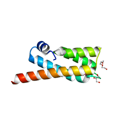
Crystal structure of a susd homolog (bt_3984) from bacteroides thetaiotaomicron vpi-5482 at 1.70 A resolution
Descriptor: ACETATE ION, SusD homolog, ZINC ION
Authors:Joint Center for Structural Genomics (JCSG)
Deposit date:2008-03-05
Release date:2008-03-18
Last modified:2023-02-01
Method:X-RAY DIFFRACTION (1.7 Å)
Cite:Structure of BT_3984, a member of the SusD/RagB family of nutrient-binding molecules.
Acta Crystallogr.,Sect.F, 66, 2010
3CM1
DownloadVisualize
BU of 3cm1 by Molmil
Crystal structure of SsgA-like sporulation-specific cell division protein (YP_290167.1) from Thermobifida fusca YX-ER1 at 2.60 A resolution
Descriptor: SsgA-like sporulation-specific cell division protein
Authors:Joint Center for Structural Genomics (JCSG), Chruszcz, M, Minor, W, Wang, S.
Deposit date:2008-03-20
Release date:2008-04-01
Last modified:2023-02-01
Method:X-RAY DIFFRACTION (2.6 Å)
Cite:Structural and functional characterizations of SsgB, a conserved activator of developmental cell division in morphologically complex actinomycetes.
J.Biol.Chem., 284, 2009
3DEE
DownloadVisualize
BU of 3dee by Molmil
CRYSTAL STRUCTURE OF A PUTATIVE REGULATORY PROTEIN INVOLVED IN TRANSCRIPTION (NGO1945) FROM NEISSERIA GONORRHOEAE FA 1090 AT 2.25 A RESOLUTION
Descriptor: CHLORIDE ION, IMIDAZOLE, Putative regulatory protein
Authors:Joint Center for Structural Genomics (JCSG)
Deposit date:2008-06-09
Release date:2008-08-05
Last modified:2023-02-01
Method:X-RAY DIFFRACTION (2.1 Å)
Cite:The structure of the first representative of Pfam family PF09836 reveals a two-domain organization and suggests involvement in transcriptional regulation.
Acta Crystallogr.,Sect.F, 66, 2010
3DCX
DownloadVisualize
BU of 3dcx by Molmil
Crystal structure of a duf1696 family protein with a pleckstrin-homology domain (shew_0819) from shewanella loihica pv-4 at 2.00 A resolution
Descriptor: (4S)-2-METHYL-2,4-PENTANEDIOL, CHLORIDE ION, Protein of Unknown Function (DUF1696) with Pleckstrin-homology Domains
Authors:Joint Center for Structural Genomics (JCSG)
Deposit date:2008-06-04
Release date:2008-07-29
Last modified:2023-02-01
Method:X-RAY DIFFRACTION (2 Å)
Cite:Bacterial pleckstrin homology domains: a prokaryotic origin for the PH domain.
J.Mol.Biol., 396, 2010
3DUE
DownloadVisualize
BU of 3due by Molmil
CRYSTAL STRUCTURE OF A PUTATIVE PERIPLASMIC PROTEIN FROM DUF2874 FAMILY (BVU_2987) FROM BACTEROIDES VULGATUS ATCC 8482 AT 1.85 A RESOLUTION
Descriptor: CACODYLATE ION, Putative periplasmic protein
Authors:Joint Center for Structural Genomics (JCSG)
Deposit date:2008-07-17
Release date:2008-08-05
Last modified:2023-02-01
Method:X-RAY DIFFRACTION (1.85 Å)
Cite:The structure of BVU2987 from Bacteroides vulgatus reveals a superfamily of bacterial periplasmic proteins with possible inhibitory function.
Acta Crystallogr.,Sect.F, 66, 2010
3F1Z
DownloadVisualize
BU of 3f1z by Molmil
Crystal structure of putative nucleic acid-binding lipoprotein (YP_001337197.1) from Klebsiella pneumoniae subsp. pneumoniae MGH 78578 at 2.46 A resolution
Descriptor: DI(HYDROXYETHYL)ETHER, putative nucleic acid-binding lipoprotein
Authors:Joint Center for Structural Genomics (JCSG)
Deposit date:2008-10-28
Release date:2008-11-18
Last modified:2023-02-01
Method:X-RAY DIFFRACTION (2.46 Å)
Cite:The structure of KPN03535 (gi|152972051), a novel putative lipoprotein from Klebsiella pneumoniae, reveals an OB-fold.
Acta Crystallogr.,Sect.F, 66, 2010
3H50
DownloadVisualize
BU of 3h50 by Molmil
CRYSTAL STRUCTURE OF A TETRACENOMYCIN POLYKETIDE SYNTHESIS PROTEIN (TCMJ) FROM XANTHOMONAS CAMPESTRIS PV. CAMPESTRIS AT 1.60 A RESOLUTION
Descriptor: ACETATE ION, Tetracenomycin polyketide synthesis protein, ZINC ION
Authors:Joint Center for Structural Genomics (JCSG)
Deposit date:2009-04-21
Release date:2009-05-05
Last modified:2023-02-01
Method:X-RAY DIFFRACTION (1.6 Å)
Cite:Conformational changes associated with the binding of zinc acetate at the putative active site of XcTcmJ, a cupin from Xanthomonas campestris pv. campestris.
Acta Crystallogr.,Sect.F, 66, 2010
3IRB
DownloadVisualize
BU of 3irb by Molmil
Crystal structure of protein with unknown function from DUF35 family (13815350) from SULFOLOBUS SOLFATARICUS at 1.80 A resolution
Descriptor: ACETIC ACID, SULFATE ION, ZINC ION, ...
Authors:Joint Center for Structural Genomics (JCSG)
Deposit date:2009-08-21
Release date:2009-09-01
Last modified:2023-02-01
Method:X-RAY DIFFRACTION (1.8 Å)
Cite:The structure of SSO2064, the first representative of Pfam family PF01796, reveals a novel two-domain zinc-ribbon OB-fold architecture with a potential acyl-CoA-binding role.
Acta Crystallogr.,Sect.F, 66, 2010
3L5O
DownloadVisualize
BU of 3l5o by Molmil
Crystal structure of protein with unknown function from DUF364 family (ZP_00559375.1) from Desulfitobacterium hafniense DCB-2 at 2.01 A resolution
Descriptor: 1,2-ETHANEDIOL, CHLORIDE ION, IMIDAZOLE, ...
Authors:Joint Center for Structural Genomics, Joint Center for Structural Genomics (JCSG)
Deposit date:2009-12-22
Release date:2010-02-02
Last modified:2023-02-01
Method:X-RAY DIFFRACTION (2.01 Å)
Cite:Structure of the first representative of Pfam family PF04016 (DUF364) reveals enolase and Rossmann-like folds that combine to form a unique active site with a possible role in heavy-metal chelation.
Acta Crystallogr.,Sect.F, 66, 2010
2OOK
DownloadVisualize
BU of 2ook by Molmil
Crystal structure of a protein with unknown function (YP_749275.1) from Shewanella Frigidimarina NCIMB 400 at 1.80 A resolution
Descriptor: 1,2-ETHANEDIOL, Hypothetical protein
Authors:Joint Center for Structural Genomics (JCSG)
Deposit date:2007-01-25
Release date:2007-02-06
Last modified:2023-01-25
Method:X-RAY DIFFRACTION (1.8 Å)
Cite:Open and closed conformations of two SpoIIAA-like proteins (YP_749275.1 and YP_001095227.1) provide insights into membrane association and ligand binding.
Acta Crystallogr.,Sect.F, 66, 2010
2OOC
DownloadVisualize
BU of 2ooc by Molmil
Crystal structure of Histidine Phosphotransferase ShpA (NP_419930.1) from Caulobacter crescentus at 1.52 A resolution
Descriptor: GLYCEROL, Histidine phosphotransferase, TETRAETHYLENE GLYCOL
Authors:Joint Center for Structural Genomics (JCSG)
Deposit date:2007-01-25
Release date:2007-02-06
Last modified:2023-01-25
Method:X-RAY DIFFRACTION (1.52 Å)
Cite:Crystal structure of histidine phosphotransfer protein ShpA, an essential regulator of stalk biogenesis in Caulobacter crescentus.
J.Mol.Biol., 390, 2009


123>

218500

PDB entries from 2024-04-17

PDB statisticsPDBj update infoContact PDBjnumon