Loading
PDBj
MenuPDBj@FacebookPDBj@TwitterPDBj@YouTubewwPDB FoundationwwPDB
RCSB PDBPDBeBMRBAdv. SearchSearch help
Search by PDB author
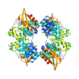
5V8T
DownloadVisualize
BU of 5v8t by Molmil
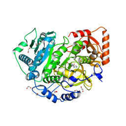
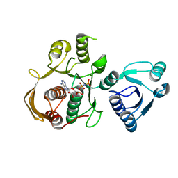
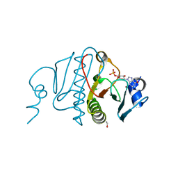
Crystal structure of SMT fusion Peptidyl-prolyl cis-trans isomerase from Burkholderia pseudomallei complexed with SF354
Descriptor: 2-{[3,5-bis(2-methoxyethoxy)benzene-1-carbonyl]amino}ethyl (2S)-1-(benzylsulfonyl)piperidine-2-carboxylate, FORMIC ACID, MAGNESIUM ION, ...
Authors:Lorimer, D.D, Dranow, D.M, Seufert, F, Abendroth, J, Holzgrabe, U.
Deposit date:2017-03-22
Release date:2018-03-14
Last modified:2023-10-04
Method:X-RAY DIFFRACTION (2.1 Å)
Cite:Crystal structure of SMT fusion Peptidyl-prolyl cis-trans isomerase from Burkholderia pseudomallei complexed with SF354
to be published
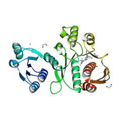
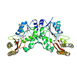
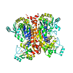
6X9N
DownloadVisualize
BU of 6x9n by Molmil
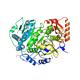
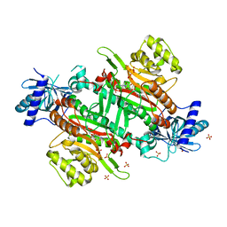
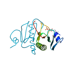
Pseudomonas aeruginosa MurC with AZ5595
Descriptor: (2R)-2-({4-[(5-tert-butyl-1-methyl-1H-pyrazol-3-yl)amino]-1H-pyrazolo[3,4-d]pyrimidin-6-yl}amino)-2-phenylethan-1-ol, 1,2-ETHANEDIOL, CHLORIDE ION, ...
Authors:Horanyi, P.S, Mayclin, S.J, Durand-Reville, T.F, Lorimer, D.D, Edwards, T.E, Seattle Structural Genomics Center for Infectious Disease (SSGCID)
Deposit date:2020-06-03
Release date:2020-09-02
Last modified:2023-10-18
Method:X-RAY DIFFRACTION (2.25 Å)
Cite:Pseudomonas aeruginosa MurC with AZ5595
To Be Published
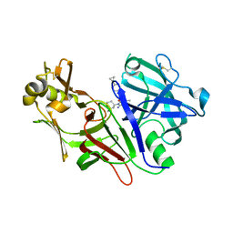
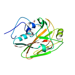
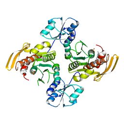
8DSR
DownloadVisualize
BU of 8dsr by Molmil
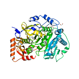
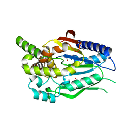
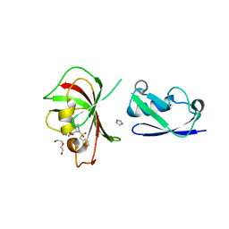
Structure of Plasmepsin X (PM10, PMX) from Plasmodium falciparum 3D7 in complex with UCB7362
Descriptor: (2E,6S)-6-{2-chloro-3-[(2-cyclopropylpyrimidin-5-yl)amino]phenyl}-2-imino-6-methyl-3-[(2S,4S)-2-methyloxan-4-yl]-1,3-diazinan-4-one, Plasmepsin X
Authors:Abendroth, J, Lorimer, D.D.
Deposit date:2022-07-22
Release date:2022-10-19
Last modified:2023-10-25
Method:X-RAY DIFFRACTION (2.85 Å)
Cite:Discovery and Characterization of Potent, Efficacious, Orally Available Antimalarial Plasmepsin X Inhibitors and Preclinical Safety Assessment of UCB7362 .
J.Med.Chem., 65, 2022
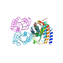
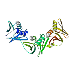
8DGD
DownloadVisualize
BU of 8dgd by Molmil
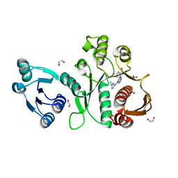
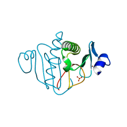
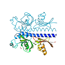
Crystal Structure of GAF domain-containing protein, from Klebsiella pneumoniae
Descriptor: 2-(N-MORPHOLINO)-ETHANESULFONIC ACID, GAF domain-containing protein, ZINC ION
Authors:Seattle Structural Genomics Center for Infectious Disease, Delker, S.L, Weiss, M.J, Lorimer, D.D, Edwards, T.E, Seattle Structural Genomics Center for Infectious Disease (SSGCID)
Deposit date:2022-06-23
Release date:2023-03-01
Last modified:2024-03-13
Method:X-RAY DIFFRACTION (1.9 Å)
Cite:Crystal Structure of GAF domain-containing protein, from Klebsiella pneumoniae
to be published
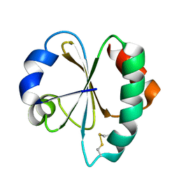
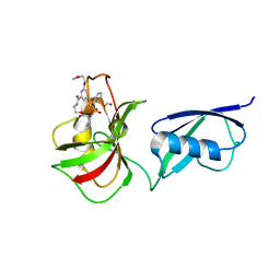
3VFI
DownloadVisualize
BU of 3vfi by Molmil
Crystal Structure of a Metagenomic Thioredoxin
Descriptor: thioredoxin
Authors:Craig, T.K, Gardberg, A, Lorimer, D.D, Burgin Jr, A.B, Segall, A, Rohwer, F.
Deposit date:2012-01-09
Release date:2013-01-23
Method:X-RAY DIFFRACTION (1.75 Å)
Cite:Structure of a Metagenomic Thioredoxin
To be Published
5IFI
DownloadVisualize
BU of 5ifi by Molmil
CRYSTAL STRUCTURE OF ACETYL-COA SYNTHETASE IN COMPLEX WITH ADENOSINE-5'-PROPYLPHOSPHATE FROM CRYPTOCOCCUS NEOFORMANS H99
Descriptor: 1,2-ETHANEDIOL, ADENOSINE-5'-MONOPHOSPHATE-PROPYL ESTER, Acetyl-coenzyme A synthetase, ...
Authors:Seattle Structural Genomics Center for Infectious Disease (SSGCID), SSGCID, Fox III, D, Edwards, T.E, Lorimer, D.D, Mutz, M.W.
Deposit date:2016-02-26
Release date:2016-03-16
Last modified:2023-09-27
Method:X-RAY DIFFRACTION (1.95 Å)
Cite:Crystal structure of Acetyl-CoA Synthetase in complex with Adenosine-5'-propylphosphate from Cryptococcus neoformans H99
To Be Published
5K85
DownloadVisualize
BU of 5k85 by Molmil
Crystal Structure of Acetyl-CoA Synthetase in Complex with Adenosine-5'-propylphosphate and Coenzyme A from Cryptococcus neoformans H99
Descriptor: 1,2-ETHANEDIOL, ADENOSINE-5'-MONOPHOSPHATE-PROPYL ESTER, Acetyl-coenzyme A synthetase, ...
Authors:Seattle Structural Genomics Center for Infectious Disease (SSGCID), Fox III, D, Delker, S.L, Potts, K.T, Lorimer, D.D, Edwards, T.E, Mutz, M.W, SSGCID
Deposit date:2016-05-27
Release date:2016-08-17
Last modified:2023-09-27
Method:X-RAY DIFFRACTION (2.3 Å)
Cite:CRYSTAL STRUCTURE OF ACETYL-COA SYNTHETASE IN COMPLEX WITH ADENOSINE-5'-PROPYLPHOSPHATE AND COENZYME A FROM CRYPTOCOCCUS NEOFORMANS H99
To Be Published
5K8F
DownloadVisualize
BU of 5k8f by Molmil
Crystal structure of Acetyl-CoA Synthetase in complex with ATP and Acetyl-AMP from Cryptococcus neoformans H99
Descriptor: 1,2-ETHANEDIOL, ADENOSINE-5'-TRIPHOSPHATE, Acetyl-coenzyme A synthetase, ...
Authors:Seattle Structural Genomics Center for Infectious Disease (SSGCID), Fox III, D, Delker, S.L, Potts, K.T, Lorimer, D.D, Edwards, T.E, Mutz, M.W.
Deposit date:2016-05-30
Release date:2016-07-13
Last modified:2023-09-27
Method:X-RAY DIFFRACTION (2.45 Å)
Cite:Crystal structure of Acetyl-CoA Synthetase in complex with ATP and Acetyl-AMP from Cryptococcus neoformans H99
To Be Published
6X9F
DownloadVisualize
BU of 6x9f by Molmil
Pseudomonas aeruginosa MurC with AZ8074
Descriptor: 1,2-ETHANEDIOL, CHLORIDE ION, DIMETHYL SULFOXIDE, ...
Authors:Horanyi, P.S, Mayclin, S.J, Durand-Reville, T.F, Lorimer, D.D, Edwards, T.E, Seattle Structural Genomics Center for Infectious Disease (SSGCID)
Deposit date:2020-06-02
Release date:2020-09-02
Last modified:2023-10-18
Method:X-RAY DIFFRACTION (2.35 Å)
Cite:Pseudomonas aeruginosa MurC with AZ8074
To Be Published
6CAU
DownloadVisualize
BU of 6cau by Molmil
UDP-N-acetylmuramate--alanine ligase from Acinetobacter baumannii AB5075-UW with AMPPNP
Descriptor: MAGNESIUM ION, PHOSPHOAMINOPHOSPHONIC ACID-ADENYLATE ESTER, UDP-N-acetylmuramate--L-alanine ligase
Authors:Horanyi, P.S, Abendroth, J, Lorimer, D.D, Edwards, T.E, Seattle Structural Genomics Center for Infectious Disease (SSGCID)
Deposit date:2018-01-31
Release date:2018-03-07
Last modified:2024-03-13
Method:X-RAY DIFFRACTION (2.5 Å)
Cite:UDP-N-acetylmuramate--alanine ligase from Acinetobacter baumannii AB5075-UW with AMPPNP
To be Published
6CHD
DownloadVisualize
BU of 6chd by Molmil
Crystal Structure of Human Lysyl-tRNA Synthetase complexed with L-Lysylsulfamoyl Adenosine
Descriptor: 1,2-ETHANEDIOL, 5'-O-[(L-LYSYLAMINO)SULFONYL]ADENOSINE, GLYCEROL, ...
Authors:Dranow, D.M, Abendroth, J, Lorimer, D.D.
Deposit date:2018-02-22
Release date:2018-03-07
Last modified:2023-10-04
Method:X-RAY DIFFRACTION (2.5 Å)
Cite:Crystal Structure of Human Lysyl-tRNA Synthetase complexed with L-Lysylsulfamoyl Adenosine
to be published
6CKG
DownloadVisualize
BU of 6ckg by Molmil
D-glycerate 3-kinase from Cryptococcus neoformans var. grubii serotype A (H99 / ATCC 208821 / CBS 10515 / FGSC 9487)
Descriptor: 1,2-ETHANEDIOL, D-glycerate 3-kinase
Authors:Horanyi, P.S, Abendroth, J, Lorimer, D.D, Edwards, T.E, Seattle Structural Genomics Center for Infectious Disease (SSGCID)
Deposit date:2018-02-28
Release date:2018-04-04
Last modified:2024-03-13
Method:X-RAY DIFFRACTION (2 Å)
Cite:D-glycerate 3-kinase from Cryptococcus neoformans var. grubii serotype A (H99 / ATCC 208821 / CBS 10515 / FGSC 9487)
To be Published
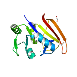
6DAV
DownloadVisualize
BU of 6dav by Molmil
Crystal Structure of Human DHFR complexed with NADP and N10formyltetrahydrofolate
Descriptor: 1,2-ETHANEDIOL, Dihydrofolate reductase, N-(4-{[(2,4-diaminopteridin-6-yl)methyl](hydroxymethyl)amino}benzene-1-carbonyl)-L-glutamic acid, ...
Authors:Mayclin, S.J, Dranow, D.M, Lorimer, D.D.
Deposit date:2018-05-02
Release date:2018-05-16
Last modified:2024-03-13
Method:X-RAY DIFFRACTION (1.55 Å)
Cite:Crystal Structure of Human DHFR complexed with NADP and N10formyltetrahydrofolate
to be published
3OJ7
DownloadVisualize
BU of 3oj7 by Molmil
Crystal structure of a histidine triad family protein from entamoeba histolytica, bound to sulfate
Descriptor: Putative histidine triad family protein, SULFATE ION, ZINC ION
Authors:Seattle Structural Genomics Center for Infectious Disease (SSGCID)
Deposit date:2010-08-20
Release date:2010-09-08
Last modified:2023-09-06
Method:X-RAY DIFFRACTION (1.4 Å)
Cite:Structures of a histidine triad family protein from Entamoeba histolytica bound to sulfate, AMP and GMP.
Acta Crystallogr F Struct Biol Commun, 71, 2015
3OXK
DownloadVisualize
BU of 3oxk by Molmil
Crystal structure of a histidine triad family protein from Entamoeba histolytica, bound to GMP
Descriptor: ETHANOL, GUANOSINE-5'-MONOPHOSPHATE, Putative histidine triad family protein, ...
Authors:Seattle Structural Genomics Center for Infectious Disease (SSGCID)
Deposit date:2010-09-21
Release date:2010-10-06
Last modified:2023-09-06
Method:X-RAY DIFFRACTION (1.55 Å)
Cite:Structures of a histidine triad family protein from Entamoeba histolytica bound to sulfate, AMP and GMP.
Acta Crystallogr F Struct Biol Commun, 71, 2015
3OMF
DownloadVisualize
BU of 3omf by Molmil
Crystal structure of a histidine triad family protein from Entamoeba histolytica, bound to AMP
Descriptor: ADENOSINE MONOPHOSPHATE, Putative histidine triad family protein, ZINC ION
Authors:SSGCID, Seattle Structural Genomics Center for Infectious Disease (SSGCID)
Deposit date:2010-08-26
Release date:2010-09-08
Last modified:2023-09-06
Method:X-RAY DIFFRACTION (1.8 Å)
Cite:Structures of a histidine triad family protein from Entamoeba histolytica bound to sulfate, AMP and GMP.
Acta Crystallogr F Struct Biol Commun, 71, 2015
5KLX
DownloadVisualize
BU of 5klx by Molmil
Crystal Structure of SMT Fusion Peptidyl-Prolyl Cis-Trans Isomerase from Burkholderia Pseudomallei Complexed with SF110
Descriptor: 2-[(2~{S})-1-(phenylmethyl)sulfonylpiperidin-2-yl]carbonyloxyethyl pyridine-3-carboxylate, Chimera protein of Ubiquitin-like protein SMT3 and Peptidyl-prolyl cis-trans isomerase, DI(HYDROXYETHYL)ETHER, ...
Authors:Fox III, D, Edwards, T.E.
Deposit date:2016-06-26
Release date:2017-05-31
Last modified:2023-09-27
Method:X-RAY DIFFRACTION (2.45 Å)
Cite:Crystal Structure of SMT Fusion Peptidyl-Prolyl Cis-Trans Isomerase from Burkholderia Pseudomallei Complexed with SF110
To Be Published
6PZJ
DownloadVisualize
BU of 6pzj by Molmil
Structure of the N-terminal domain (residues 43-304) of Methyl-accepting chemotaxis protein from Leptospira interrogans serogroup Icterohaemorrhagiae serovar Copenhageni (strain Fiocruz L1-130)
Descriptor: 1,2-ETHANEDIOL, CHLORIDE ION, Methyl-accepting chemotaxis protein
Authors:Seattle Structural Genomics Center for Infectious Disease (SSGCID)
Deposit date:2019-07-31
Release date:2019-08-14
Last modified:2024-03-13
Method:X-RAY DIFFRACTION (1.75 Å)
Cite:Structure of the N-terminal domain (residues 43-304) of Methyl-accepting chemotaxis protein from Leptospira interrogans serogroup Icterohaemorrhagiae serovar Copenhageni (strain Fiocruz L1-130)
to be published
5KWV
DownloadVisualize
BU of 5kwv by Molmil
Crystal Structure of a Pantoate-beta-alanine Ligase from Neisseria gonorrhoeae with bound AMPPNP
Descriptor: PHOSPHOAMINOPHOSPHONIC ACID-ADENYLATE ESTER, Pantothenate synthetase
Authors:Seattle Structural Genomics Center for Infectious Disease (SSGCID)
Deposit date:2016-07-19
Release date:2016-07-27
Last modified:2023-10-04
Method:X-RAY DIFFRACTION (2.25 Å)
Cite:Crystal Structure of a Pantoate-beta-alanine Ligase from Neisseria gonorrhoeae with bound AMPPNP
to be published
7RCR
DownloadVisualize
BU of 7rcr by Molmil
Crystal Structure of the refolded Hemagglutinin head domain of Influenza A virus A/Ohio/09/2015
Descriptor: 1,2-ETHANEDIOL, Hemagglutinin
Authors:Seattle Structural Genomics Center for Infectious Disease, Seattle Structural Genomics Center for Infectious Disease (SSGCID)
Deposit date:2021-07-08
Release date:2021-08-04
Last modified:2024-04-03
Method:X-RAY DIFFRACTION (1.6 Å)
Cite:Crystal Structure of the refolded Hemagglutinin head domain of Influenzavirus A Influenza A virus A/Ohio/09/2015
to be published
7RZO
DownloadVisualize
BU of 7rzo by Molmil
Crystal structure of a dihydrofolate reductase (folA) from Stenotrophomonas maltophilia
Descriptor: 1,2-ETHANEDIOL, CHLORIDE ION, Dihydrofolate reductase
Authors:Seattle Structural Genomics Center for Infectious Disease (SSGCID)
Deposit date:2021-08-27
Release date:2021-10-20
Last modified:2023-10-18
Method:X-RAY DIFFRACTION (1.8 Å)
Cite:Crystal Structure of dihydrofolate reductase from Stenotrophomonas maltophilia K279a
to be published
7RZM
DownloadVisualize
BU of 7rzm by Molmil
Crystal Structure of dnaN DNA polymerase III beta subunit from Stenotrophomonas maltophilia K279a
Descriptor: DNA polymerase III subunit beta
Authors:Seattle Structural Genomics Center for Infectious Disease (SSGCID)
Deposit date:2021-08-27
Release date:2021-10-20
Last modified:2023-10-18
Method:X-RAY DIFFRACTION (2.15 Å)
Cite:Crystal Structure of dnaN DNA polymerase III beta subunit from Stenotrophomonas maltophilia K279a
To Be Published
8EGL
DownloadVisualize
BU of 8egl by Molmil
Crystal Structure of Guanylate kinase from Pseudomonas aeruginosa PAO1 in complex with GMP and ADP
Descriptor: ADENOSINE-5'-DIPHOSPHATE, GUANOSINE-5'-MONOPHOSPHATE, Guanylate kinase
Authors:Seattle Structural Genomics Center for Infectious Disease (SSGCID)
Deposit date:2022-09-12
Release date:2022-09-28
Last modified:2023-10-18
Method:X-RAY DIFFRACTION (2.35 Å)
Cite:Crystal Structure of Guanylate kinase from Pseudomonas aeruginosa PAO1 in complex with GMP and ADP
to be published
8EGN
DownloadVisualize
BU of 8egn by Molmil
Crystal Structure of UDP-N-acetylmuramate-L-alanine ligase (UDP-N-acetylmuramoyl-L-alanine synthetase, MurC) Pseudomonas aeruginosa in complex with ligand AZ-13643701
Descriptor: (1R,2S)-1-({4-[(5-tert-butyl-1-methyl-1H-pyrazol-3-yl)amino]-1H-pyrazolo[3,4-d]pyrimidin-6-yl}amino)-2,3-dihydro-1H-inden-2-ol, SULFATE ION, UDP-N-acetylmuramate--L-alanine ligase
Authors:Seattle Structural Genomics Center for Infectious Disease (SSGCID)
Deposit date:2022-09-12
Release date:2022-09-28
Last modified:2023-10-18
Method:X-RAY DIFFRACTION (1.95 Å)
Cite:Crystal Structure of UDP-N-acetylmuramate-L-alanine ligase (UDP-N-acetylmuramoyl-L-alanine synthetase, MurC) Pseudomonas aeruginosa in complex with ligand AZ-13643701
to be published
8EGM
DownloadVisualize
BU of 8egm by Molmil
Crystal Structure of UDP-N-acetylmuramate-L-alanine ligase (UDP-N-acetylmuramoyl-L-alanine synthetase, MurC) Pseudomonas aeruginosa in complex with compound AZ13644908
Descriptor: (2R)-2-({4-[(5-tert-butyl-1-methyl-1H-pyrazol-3-yl)amino]-1H-pyrazolo[3,4-d]pyrimidin-6-yl}amino)-2-phenylethyl (2-aminoethyl)carbamate, SULFATE ION, UDP-N-acetylmuramate--L-alanine ligase
Authors:Seattle Structural Genomics Center for Infectious Disease (SSGCID)
Deposit date:2022-09-12
Release date:2022-10-05
Last modified:2023-10-18
Method:X-RAY DIFFRACTION (2.2 Å)
Cite:Crystal Structure of UDP-N-acetylmuramate-L-alanine ligase (UDP-N-acetylmuramoyl-L-alanine synthetase, MurC) Pseudomonas aeruginosa
to be published

218500

PDB entries from 2024-04-17

PDB statisticsPDBj update infoContact PDBjnumon