Loading
PDBj
MenuPDBj@FacebookPDBj@TwitterPDBj@YouTubewwPDB FoundationwwPDB
RCSB PDBPDBeBMRBAdv. SearchSearch help
Search by PDB author
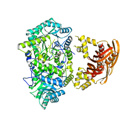
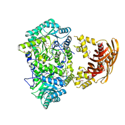
1SR9
DownloadVisualize
BU of 1sr9 by Molmil
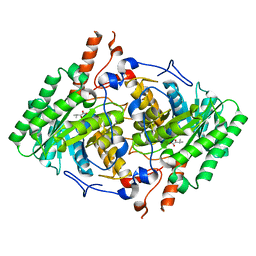
Crystal Structure of LeuA from Mycobacterium tuberculosis
Descriptor: 2-isopropylmalate synthase, 3-METHYL-2-OXOBUTANOIC ACID, CHLORIDE ION, ...
Authors:Koon, N, Squire, C.J, Baker, E.N.
Deposit date:2004-03-22
Release date:2004-05-18
Last modified:2017-10-11
Method:X-RAY DIFFRACTION (2 Å)
Cite:Crystal structure of LeuA from Mycobacterium tuberculosis, a key enzyme in leucine biosynthesis
Proc.Natl.Acad.Sci.USA, 101, 2004
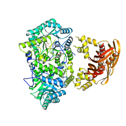
3FIG
DownloadVisualize
BU of 3fig by Molmil
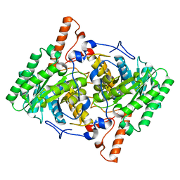
Crystal Structure of Leucine-bound LeuA from Mycobacterium tuberculosis
Descriptor: 2-isopropylmalate synthase, GLYCEROL, LEUCINE, ...
Authors:Koon, N, Squire, C.J, Baker, E.N.
Deposit date:2008-12-11
Release date:2008-12-23
Last modified:2023-11-01
Method:X-RAY DIFFRACTION (2.3 Å)
Cite:Crystal structure of LeuA from Mycobacterium tuberculosis, a key enzyme in leucine biosynthesis.
Proc.Natl.Acad.Sci.USA, 101, 2004
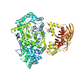
3HPS
DownloadVisualize
BU of 3hps by Molmil
Crystal structure of Mycobacterium tuberculosis LeuA complexed with ketoisocaproate (KIC)
Descriptor: 2-OXO-4-METHYLPENTANOIC ACID, 2-isopropylmalate synthase, GLYCEROL, ...
Authors:Koon, N, Squire, C.J, Baker, E.N.
Deposit date:2009-06-04
Release date:2010-05-19
Last modified:2023-09-06
Method:X-RAY DIFFRACTION (1.8 Å)
Cite:Probing the active site of M. tuberculosis LeuA
To be Published
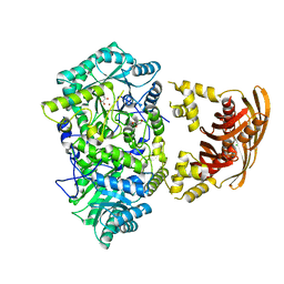
3HQ1
DownloadVisualize
BU of 3hq1 by Molmil
Crystal structure of Mycobacterium tuberculosis LeuA complexed with citrate and Mn2+
Descriptor: 2-isopropylmalate synthase, CHLORIDE ION, CITRATE ANION, ...
Authors:Koon, N, Squire, C.J, Baker, E.N.
Deposit date:2009-06-05
Release date:2010-05-19
Last modified:2023-09-06
Method:X-RAY DIFFRACTION (1.7 Å)
Cite:Probing the active site of M. tuberculosis LeuA
To be Published
3HPZ
DownloadVisualize
BU of 3hpz by Molmil
Crystal structure of Mycobacterium tuberculosis LeuA complexed with bromopyruvate
Descriptor: 2-isopropylmalate synthase, Bromopyruvate, CHLORIDE ION, ...
Authors:Koon, N, Squire, C.J, Baker, E.N.
Deposit date:2009-06-05
Release date:2010-05-19
Last modified:2023-09-06
Method:X-RAY DIFFRACTION (2.2 Å)
Cite:Probing the active site of M. tuberculosis LeuA
To be Published
3U6W
DownloadVisualize
BU of 3u6w by Molmil
Truncated M. tuberculosis LeuA (1-425) complexed with KIV
Descriptor: 2-isopropylmalate synthase, 3-METHYL-2-OXOBUTANOIC ACID, GLYCEROL, ...
Authors:Koon, N, Baker, E.N, Squire, C.J.
Deposit date:2011-10-13
Release date:2012-03-14
Last modified:2023-09-13
Method:X-RAY DIFFRACTION (2.21 Å)
Cite:Removal of the C-terminal regulatory domain of alpha-isopropylmalate synthase disrupts functional substrate binding.
Biochemistry, 51, 2012
3HPX
DownloadVisualize
BU of 3hpx by Molmil
Crystal structure of Mycobacterium tuberculosis LeuA active site domain 1-425 (truncation mutant delta:426-644)
Descriptor: 2-isopropylmalate synthase, GLYCEROL, NICKEL (II) ION
Authors:Koon, N, Squire, C.J, Baker, E.N.
Deposit date:2009-06-05
Release date:2010-05-19
Last modified:2023-09-06
Method:X-RAY DIFFRACTION (2.03 Å)
Cite:Removal of the C-terminal regulatory domain of alpha-isopropylmalate synthase disrupts functional substrate binding
Biochemistry, 51, 2012
3RMJ
DownloadVisualize
BU of 3rmj by Molmil
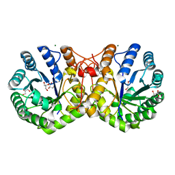
Crystal structure of truncated alpha-Isopropylmalate Synthase from Neisseria meningitidis
Descriptor: 2-isopropylmalate synthase, GLYCEROL, MAGNESIUM ION, ...
Authors:Huisman, F.H.A, Baker, H.M, Koon, N, Baker, E.N, Parker, E.J.
Deposit date:2011-04-20
Release date:2012-03-14
Last modified:2023-11-01
Method:X-RAY DIFFRACTION (1.95 Å)
Cite:Removal of the C-terminal regulatory domain of alpha-isopropylmalate synthase disrupts functional substrate binding
Biochemistry, 51, 2012
3IR8
DownloadVisualize
BU of 3ir8 by Molmil
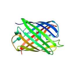
Red fluorescent protein mKeima at pH 7.0
Descriptor: Large stokes shift fluorescent protein
Authors:Henderson, J.N, Osborn, M.F, Koon, N, Gepshtein, R, Huppert, D, Remington, S.J.
Deposit date:2009-08-21
Release date:2009-09-08
Last modified:2023-11-15
Method:X-RAY DIFFRACTION (1.63 Å)
Cite:Excited state proton transfer in the red fluorescent protein mKeima.
J.Am.Chem.Soc., 131, 2009
2IYJ
DownloadVisualize
BU of 2iyj by Molmil
Crystal structure of the N-terminal dimer domain of E.coli DsbC
Descriptor: SULFATE ION, THIOL DISULFIDE INTERCHANGE PROTEIN DSBC
Authors:Yeh, S.-M, Metcalf, P.
Deposit date:2006-07-18
Release date:2007-07-24
Last modified:2023-12-13
Method:X-RAY DIFFRACTION (2 Å)
Cite:Structures of Dimerization Domains of the Escherichia Coli Disulfide-Bond Isomerase Enzymes Dsbc and Dsbg.
Acta Crystallogr.,Sect.D, 63, 2007
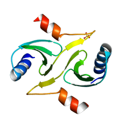
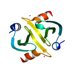
2IY2
DownloadVisualize
BU of 2iy2 by Molmil
Crystal structure of the N-terminal dimer domain of E.coli DsbG
Descriptor: CADMIUM ION, THIOL DISULFIDE INTERCHANGE PROTEIN DSBG
Authors:Yeh, S.-M, Metcalf, P.
Deposit date:2006-07-12
Release date:2007-07-17
Last modified:2023-12-13
Method:X-RAY DIFFRACTION (1.9 Å)
Cite:Structures of the Dimerization Domains of the Escherichia Coli Disulfide-Bond Isomerase Enzymes Dsbc and Dsbg
Acta Crystallogr.,Sect.D, 63, 2007

218853

PDB entries from 2024-04-24

PDB statisticsPDBj update infoContact PDBjnumon