Loading
PDBj
MenuPDBj@FacebookPDBj@TwitterPDBj@YouTubewwPDB FoundationwwPDB
RCSB PDBPDBeBMRBAdv. SearchSearch help
Search by PDB author
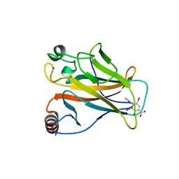
1UOL
DownloadVisualize
BU of 1uol by Molmil
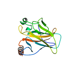
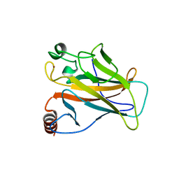
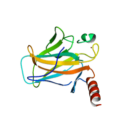
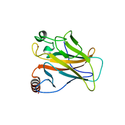
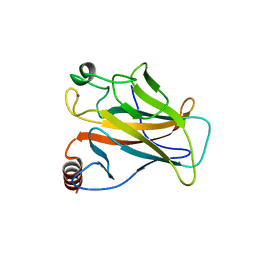
Crystal structure of the human p53 core domain mutant M133L/V203A/N239Y/N268D at 1.9 A resolution.
Descriptor: CELLULAR TUMOR ANTIGEN P53, ZINC ION
Authors:Joerger, A.C, Allen, M.D, Fersht, A.R.
Deposit date:2003-09-19
Release date:2003-10-16
Last modified:2011-07-13
Method:X-RAY DIFFRACTION (1.9 Å)
Cite:Crystal Structure of a Superstable Mutant of Human P53 Core Domain. Insights Into the Mechanism of Rescuing Oncogenic Mutations
J.Biol.Chem., 279, 2004
1HL2
DownloadVisualize
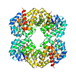
BU of 1hl2 by Molmil
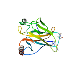
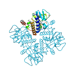
Crystal structure of N-acetylneuraminate lyase from E. coli mutant L142R in complex with b-hydroxypyruvate
Descriptor: 3-HYDROXYPYRUVIC ACID, N-ACETYLNEURAMINATE LYASE SUBUNIT
Authors:Joerger, A.C, Fersht, A.R.
Deposit date:2003-03-12
Release date:2003-05-09
Last modified:2023-12-13
Method:X-RAY DIFFRACTION (1.8 Å)
Cite:Mimicking Natural Evolution in Vitro: An N-Acetylneuraminate Lyase Mutant with an Increased Dihydrodipicolinate Synthase Activity
Proc.Natl.Acad.Sci.USA, 100, 2003
2J1Z
DownloadVisualize
BU of 2j1z by Molmil
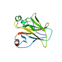
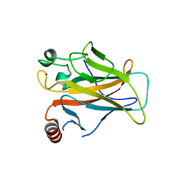
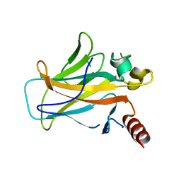
Human p53 core domain mutant M133L-V203A-N239Y-N268D-F270L
Descriptor: CELLULAR TUMOR ANTIGEN P53, ZINC ION
Authors:Joerger, A.C, Fersht, A.R.
Deposit date:2006-08-15
Release date:2006-09-20
Last modified:2023-12-13
Method:X-RAY DIFFRACTION (1.8 Å)
Cite:Structural Basis for Understanding Oncogenic P53 Mutations and Designing Rescue Drugs.
Proc.Natl.Acad.Sci.USA, 103, 2006
2J1Y
DownloadVisualize
BU of 2j1y by Molmil
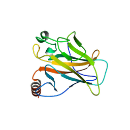
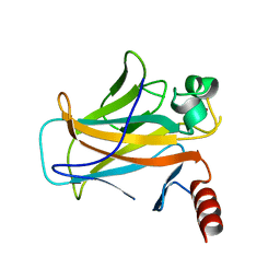
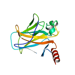
Human p53 core domain mutant M133L-V203A-N239Y-G245S-N268D
Descriptor: CALCIUM ION, CELLULAR TUMOR ANTIGEN P53, ZINC ION
Authors:Joerger, A.C, Fersht, A.R.
Deposit date:2006-08-15
Release date:2006-09-20
Last modified:2023-12-13
Method:X-RAY DIFFRACTION (1.69 Å)
Cite:Structural Basis for Understanding Oncogenic P53 Mutations and Designing Rescue Drugs.
Proc.Natl.Acad.Sci.USA, 103, 2006
2J20
DownloadVisualize
BU of 2j20 by Molmil
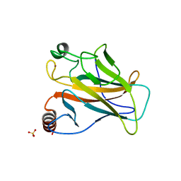
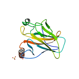
Human p53 core domain mutant M133L-V203A-N239Y-N268D-R273C
Descriptor: CELLULAR TUMOR ANTIGEN P53, SULFATE ION, ZINC ION
Authors:Joerger, A.C, Ang, H.C, Fersht, A.R.
Deposit date:2006-08-15
Release date:2006-09-20
Last modified:2023-12-13
Method:X-RAY DIFFRACTION (1.8 Å)
Cite:Structural Basis for Understanding Oncogenic P53 Mutations and Designing Rescue Drugs.
Proc.Natl.Acad.Sci.USA, 103, 2006
2J1X
DownloadVisualize
BU of 2j1x by Molmil
Human p53 core domain mutant M133L-V203A-Y220C-N239Y-N268D
Descriptor: CELLULAR TUMOR ANTIGEN P53, ZINC ION
Authors:Joerger, A.C, Fersht, A.R.
Deposit date:2006-08-15
Release date:2006-09-20
Last modified:2023-12-13
Method:X-RAY DIFFRACTION (1.65 Å)
Cite:Structural Basis for Understanding Oncogenic P53 Mutations and Designing Rescue Drugs.
Proc.Natl.Acad.Sci.USA, 103, 2006
2J1W
DownloadVisualize
BU of 2j1w by Molmil
Human p53 core domain mutant M133L-V143A-V203A-N239Y-N268D
Descriptor: CELLULAR TUMOR ANTIGEN P53, ZINC ION
Authors:Joerger, A.C, Fersht, A.R.
Deposit date:2006-08-15
Release date:2006-09-20
Last modified:2023-12-13
Method:X-RAY DIFFRACTION (1.8 Å)
Cite:Structural Basis for Understanding Oncogenic P53 Mutations and Designing Rescue Drugs.
Proc.Natl.Acad.Sci.USA, 103, 2006
2J21
DownloadVisualize
BU of 2j21 by Molmil
Human p53 core domain mutant M133L-V203A-N239Y-N268D-R282W
Descriptor: CELLULAR TUMOR ANTIGEN P53, ZINC ION
Authors:Joerger, A.C, Fersht, A.R.
Deposit date:2006-08-15
Release date:2006-09-20
Last modified:2023-12-13
Method:X-RAY DIFFRACTION (1.6 Å)
Cite:Structural Basis for Understanding Oncogenic P53 Mutations and Designing Rescue Drugs.
Proc.Natl.Acad.Sci.USA, 103, 2006
2BIM
DownloadVisualize
BU of 2bim by Molmil
human p53 core domain mutant M133L-V203A-N239Y-N268D-R273H
Descriptor: CELLULAR TUMOR ANTIGEN P53, SULFATE ION, ZINC ION
Authors:Joerger, A.C, Fersht, A.R.
Deposit date:2005-01-25
Release date:2005-01-26
Last modified:2023-12-13
Method:X-RAY DIFFRACTION (1.98 Å)
Cite:Structures of P53 Cancer Mutants and Mechanism of Rescue by Second-Site Suppressor Mutations
J.Biol.Chem., 280, 2005
2BIQ
DownloadVisualize
BU of 2biq by Molmil
human p53 core domain mutant T123A-M133L-H168R-V203A-N239Y-R249S- N268D
Descriptor: CELLULAR TUMOR ANTIGEN P53, ZINC ION
Authors:Joerger, A.C, Fersht, A.R.
Deposit date:2005-01-25
Release date:2005-01-26
Last modified:2023-12-13
Method:X-RAY DIFFRACTION (1.8 Å)
Cite:Structures of P53 Cancer Mutants and Mechanism of Rescue by Second-Site Suppressor Mutations
J.Biol.Chem., 280, 2005
2BIN
DownloadVisualize
BU of 2bin by Molmil
human p53 core domain mutant M133L-H168R-V203A-N239Y-N268D
Descriptor: CELLULAR TUMOR ANTIGEN P53, ZINC ION
Authors:Joerger, A.C, Fersht, A.R.
Deposit date:2005-01-25
Release date:2005-01-26
Last modified:2023-12-13
Method:X-RAY DIFFRACTION (1.9 Å)
Cite:Structures of P53 Cancer Mutants and Mechanism of Rescue by Second-Site Suppressor Mutations
J.Biol.Chem., 280, 2005
2BIO
DownloadVisualize
BU of 2bio by Molmil
human p53 core domain mutant M133L-V203A-N239Y-R249S-N268D
Descriptor: CELLULAR TUMOR ANTIGEN P53, ZINC ION
Authors:Joerger, A.C, Fersht, A.R.
Deposit date:2005-01-25
Release date:2005-01-26
Last modified:2023-12-13
Method:X-RAY DIFFRACTION (1.9 Å)
Cite:Structures of P53 Cancer Mutants and Mechanism of Rescue by Second-Site Suppressor Mutations
J.Biol.Chem., 280, 2005
2BIP
DownloadVisualize
BU of 2bip by Molmil
human p53 core domain mutant M133L-H168R-V203A-N239Y-R249S-N268D
Descriptor: CELLULAR TUMOR ANTIGEN P53, ZINC ION
Authors:Joerger, A.C, Fersht, A.R.
Deposit date:2005-01-25
Release date:2005-01-26
Last modified:2023-12-13
Method:X-RAY DIFFRACTION (1.8 Å)
Cite:Structures of P53 Cancer Mutants and Mechanism of Rescue by Second-Site Suppressor Mutations
J.Biol.Chem., 280, 2005
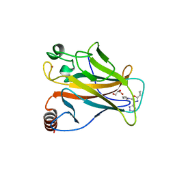
5AB9
DownloadVisualize
BU of 5ab9 by Molmil
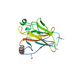
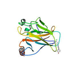
Structure of the p53 cancer mutant Y220C with bound small molecule 7- ethyl-3-(piperidin-4-yl)-1H-indole
Descriptor: 7-ethyl-3-(piperidin-4-yl)-1H-indole, CELLULAR TUMOR ANTIGEN P53, DI(HYDROXYETHYL)ETHER, ...
Authors:Joerger, A.C.
Deposit date:2015-08-04
Release date:2015-12-16
Last modified:2024-01-10
Method:X-RAY DIFFRACTION (1.36 Å)
Cite:Exploiting Transient Protein States for the Design of Small-Molecule Stabilizers of Mutant P53.
Structure, 23, 2015
5ABA
DownloadVisualize
BU of 5aba by Molmil
Structure of the p53 cancer mutant Y220C with bound small-molecule stabilizer PhiKan5149
Descriptor: 1-{1-[(3-bromo-5-chloro-2-hydroxyphenyl)methyl piperidin-4-yl}piperidin-4-ol], CELLULAR TUMOR ANTIGEN P53, ZINC ION
Authors:Joerger, A.C.
Deposit date:2015-08-04
Release date:2015-12-16
Last modified:2024-01-10
Method:X-RAY DIFFRACTION (1.62 Å)
Cite:Exploiting Transient Protein States for the Design of Small-Molecule Stabilizers of Mutant P53.
Structure, 23, 2015
5AOI
DownloadVisualize
BU of 5aoi by Molmil
Structure of the p53 cancer mutant Y220C in complex with an indole- based small molecule
Descriptor: 2-(5-BROMO-7-ETHYL-2-METHYL-1H-INDOLE-3-YL)ETHAN-1-AMIN, CELLULAR TUMOR ANTIGEN P53, DI(HYDROXYETHYL)ETHER, ...
Authors:Joerger, A.C, Wilcken, R.
Deposit date:2015-09-10
Release date:2015-12-16
Last modified:2024-01-10
Method:X-RAY DIFFRACTION (1.78 Å)
Cite:Exploiting Transient Protein States for the Design of Small-Molecule Stabilizers of Mutant P53.
Structure, 23, 2015
5AOM
DownloadVisualize
BU of 5aom by Molmil
Structure of the p53 cancer mutant Y220C with bound small molecule PhiKan883
Descriptor: CELLULAR TUMOR ANTIGEN P53, GLYCEROL, N-(5-chloranyl-2-oxidanyl-phenyl)piperidine-4-carboxamide, ...
Authors:Joerger, A.C, Boeckler, F.M, Wilcken, R.
Deposit date:2015-09-10
Release date:2015-12-16
Last modified:2024-01-10
Method:X-RAY DIFFRACTION (1.74 Å)
Cite:Exploiting Transient Protein States for the Design of Small-Molecule Stabilizers of Mutant P53.
Structure, 23, 2015
5AOL
DownloadVisualize
BU of 5aol by Molmil
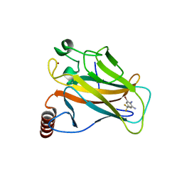
Structure of the p53 cancer mutant Y220C with bound 3-bromo-5-(trifluoromethyl)benzene-1,2-diamine
Descriptor: 3-BROMO-5-(TRIFLUOROMETHYL)BENZENE-1,2-DIAMINE, CELLULAR TUMOR ANTIGEN P53, ZINC ION
Authors:Joerger, A.C, Harbrecht, H.
Deposit date:2015-09-10
Release date:2015-12-16
Last modified:2024-01-10
Method:X-RAY DIFFRACTION (1.5 Å)
Cite:Exploiting Transient Protein States for the Design of Small-Molecule Stabilizers of Mutant P53.
Structure, 23, 2015
5AOJ
DownloadVisualize
BU of 5aoj by Molmil
Structure of the p53 cancer mutant Y220C in complex with 2-hydroxy-3, 5-diiodo-4-(1H-pyrrol-1-yl)benzoic acid
Descriptor: 2-hydroxy-3,5-diiodo-4-(1H-pyrrol-1-yl)benzoic acid, CELLULAR TUMOR ANTIGEN P53, DI(HYDROXYETHYL)ETHER, ...
Authors:Joerger, A.C, Baud, M.G, Bauer, M.R, Fersht, A.R.
Deposit date:2015-09-10
Release date:2015-12-16
Last modified:2024-01-10
Method:X-RAY DIFFRACTION (1.47 Å)
Cite:Exploiting Transient Protein States for the Design of Small-Molecule Stabilizers of Mutant P53.
Structure, 23, 2015
5AOK
DownloadVisualize
BU of 5aok by Molmil
Structure of the p53 cancer mutant Y220C with bound small molecule PhiKan7099
Descriptor: 5-[2-cyclopropyl-5-(1H-pyrrol-1-yl)-1,3-oxazol-4-yl]-1H-1,2,3,4-tetrazole, CELLULAR TUMOR ANTIGEN P53, DI(HYDROXYETHYL)ETHER, ...
Authors:Joerger, A.C.
Deposit date:2015-09-10
Release date:2015-12-16
Last modified:2024-01-10
Method:X-RAY DIFFRACTION (1.35 Å)
Cite:Exploiting Transient Protein States for the Design of Small-Molecule Stabilizers of Mutant P53.
Structure, 23, 2015
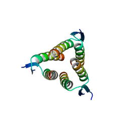
2WQJ
DownloadVisualize
BU of 2wqj by Molmil
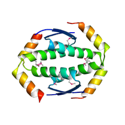
Crystal structure of a truncated variant of the human p73 tetramerization domain
Descriptor: TUMOR PROTEIN P73
Authors:Joerger, A.C.
Deposit date:2009-08-21
Release date:2009-10-06
Last modified:2019-05-08
Method:X-RAY DIFFRACTION (2 Å)
Cite:Structural Evolution of P53, P63, and P73: Implication for Heterotetramer Formation.
Proc.Natl.Acad.Sci.USA, 106, 2009
2WQI
DownloadVisualize
BU of 2wqi by Molmil
Crystal structure of the human p73 tetramerization domain
Descriptor: TUMOR PROTEIN P73
Authors:Joerger, A.C.
Deposit date:2009-08-21
Release date:2009-10-06
Last modified:2019-05-08
Method:X-RAY DIFFRACTION (1.7 Å)
Cite:Structural Evolution of P53, P63, and P73: Implication for Heterotetramer Formation.
Proc.Natl.Acad.Sci.USA, 106, 2009
2WTT
DownloadVisualize
BU of 2wtt by Molmil
Structure of the human p73 tetramerization domain (crystal form II)
Descriptor: TUMOR PROTEIN P73
Authors:Joerger, A.C.
Deposit date:2009-09-21
Release date:2009-10-13
Last modified:2023-12-20
Method:X-RAY DIFFRACTION (2.3 Å)
Cite:Structural Evolution of P53, P63, and P73: Implication for Heterotetramer Formation.
Proc.Natl.Acad.Sci.USA, 106, 2009
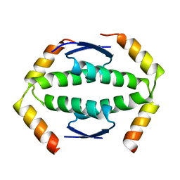
1E4A
DownloadVisualize
BU of 1e4a by Molmil
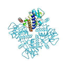
L-Fuculose 1-Phosphate Aldolase from Escherichia coli Mutant Del(27)
Descriptor: BETA-MERCAPTOETHANOL, L-FUCULOSE 1-PHOSPHATE ALDOLASE, SULFATE ION, ...
Authors:Joerger, A.C, Schulz, G.E.
Deposit date:2000-06-30
Release date:2000-11-06
Last modified:2019-01-30
Method:X-RAY DIFFRACTION (2.15 Å)
Cite:Structures of L-Fuculose-1-Phosphate Aldolase Mutants Outlining Motions During Catalysis
J.Mol.Biol., 303, 2000
1E47
DownloadVisualize
BU of 1e47 by Molmil
L-Fuculose 1-Phosphate Aldolase from Escherichia coli Mutant E73Q
Descriptor: 1,3-DIHYDROXYACETONEPHOSPHATE, BETA-MERCAPTOETHANOL, L-FUCULOSE 1-PHOSPHATE ALDOLASE, ...
Authors:Joerger, A.C, Schulz, G.E.
Deposit date:2000-06-30
Release date:2000-11-06
Last modified:2019-01-30
Method:X-RAY DIFFRACTION (2.15 Å)
Cite:Structures of l-fuculose-1-phosphate aldolase mutants outlining motions during catalysis.
J. Mol. Biol., 303, 2000

218500

PDB entries from 2024-04-17

PDB statisticsPDBj update infoContact PDBjnumon