Loading
PDBj
MenuPDBj@FacebookPDBj@TwitterPDBj@YouTubewwPDB FoundationwwPDB
RCSB PDBPDBeBMRBAdv. SearchSearch help
Search by PDB author
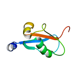
1KLV
DownloadVisualize
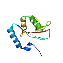
BU of 1klv by Molmil
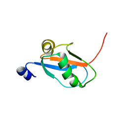
Solution Structure and Backbone Dynamics of GABARAP, GABAA Receptor associated protein
Descriptor: GABA(A) Receptor associated protein
Authors:Kouno, T, Miura, K, Tada, M, Kanematsu, T, Tate, S, Shirakawa, M, Hirata, M, Kawano, K.
Deposit date:2001-12-13
Release date:2003-10-07
Last modified:2022-02-23
Method:SOLUTION NMR
Cite:1H, 13C and '5N resonance assignments of GABARAP, GABAA receptor associated protein.
J.Biomol.Nmr, 22, 2002
1KM7
DownloadVisualize
BU of 1km7 by Molmil
Solution Structure and Backbone Dynamics of GABARAP, GABAA Receptor Associated Protein
Descriptor: GABA(A) Receptor Associated Protein
Authors:Kouno, T, Miura, K, Tada, M, Kanematsu, T, Tate, S, Shirakawa, M, Hirata, M, Kawano, K.
Deposit date:2001-12-14
Release date:2003-10-07
Last modified:2022-02-23
Method:SOLUTION NMR
Cite:1H, 13C and '5N resonance assignments of GABARAP, GABAA receptor associated protein.
J.Biomol.Nmr, 22, 2002
8I75
DownloadVisualize
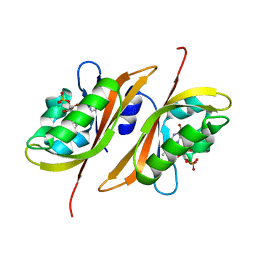
BU of 8i75 by Molmil
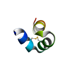
Crystal structure of decarboxylated osteocalcin at pH 2.0
Descriptor: Osteocalcin
Authors:Yokoyama, T, Nabeshima, Y, Obita, T, Mizuguchi, M.
Deposit date:2023-01-31
Release date:2024-01-31
Method:X-RAY DIFFRACTION (1.33 Å)
Cite:Crystal structures of decarboxylated osteocalcin and implications for the interaction with its receptor
To Be Published
8I76
DownloadVisualize
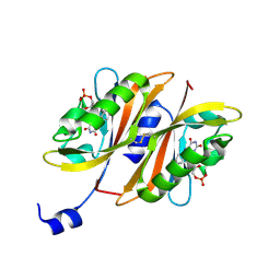
BU of 8i76 by Molmil
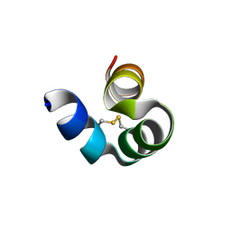
Crystal structure of decarboxylated osteocalcin at pH 2.0 without glycerol
Descriptor: Osteocalcin
Authors:Yokoyama, T, Nabeshima, Y, Obita, T, Mizuguchi, M.
Deposit date:2023-01-31
Release date:2024-01-31
Method:X-RAY DIFFRACTION (1.381 Å)
Cite:Crystal structures of decarboxylated osteocalcin and implications for the interaction with its receptor
To Be Published
8I74
DownloadVisualize
BU of 8i74 by Molmil
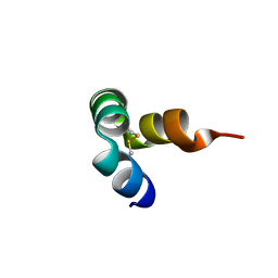
Crystal structure of decarboxylated osteocalcin at pH 8.5
Descriptor: Osteocalcin
Authors:Yokoyama, T, Nabeshima, Y, Obita, T, Mizuguchi, M.
Deposit date:2023-01-31
Release date:2024-01-31
Method:X-RAY DIFFRACTION (1.36 Å)
Cite:Crystal structures of decarboxylated osteocalcin and implications for the interaction with its receptor
To Be Published
1V49
DownloadVisualize
BU of 1v49 by Molmil
Solution structure of microtubule-associated protein light chain-3
Descriptor: Microtubule-associated proteins 1A/1B light chain 3B
Authors:Kouno, T, Mizuguchi, M, Tanida, I, Ueno, T, Kominami, E, Kawano, K.
Deposit date:2003-11-11
Release date:2004-12-28
Last modified:2023-12-27
Method:SOLUTION NMR
Cite:Solution structure of microtubule-associated protein light chain 3 and identification of its functional subdomains.
J.Biol.Chem., 280, 2005
2Z6D
DownloadVisualize
BU of 2z6d by Molmil
Crystal structure of LOV1 domain of phototropin2 from Arabidopsis thaliana
Descriptor: FLAVIN MONONUCLEOTIDE, Phototropin-2
Authors:Nakasako, M, Matsuoka, D, Tokutomi, S.
Deposit date:2007-07-29
Release date:2008-07-29
Last modified:2023-11-01
Method:X-RAY DIFFRACTION (2 Å)
Cite:Structural basis of the LOV1 dimerization of Arabidopsis phototropins 1 and 2
J.Mol.Biol., 381, 2008
2Z6C
DownloadVisualize
BU of 2z6c by Molmil
Crystal structure of LOV1 domain of phototropin1 from Arabidopsis thaliana
Descriptor: FLAVIN MONONUCLEOTIDE, Phototropin-1
Authors:Nakasako, M, Matsuoka, D, Tokutomi, S.
Deposit date:2007-07-29
Release date:2008-07-29
Last modified:2023-11-01
Method:X-RAY DIFFRACTION (2.1 Å)
Cite:Structural basis of the LOV1 dimerization of Arabidopsis phototropins 1 and 2
J.Mol.Biol., 381, 2008
1HAK
DownloadVisualize
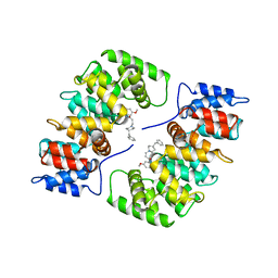
BU of 1hak by Molmil
CRYSTAL STRUCTURE OF RECOMBINANT HUMAN PLACENTAL ANNEXIN V COMPLEXED WITH K-201 AS A CALCIUM CHANNEL ACTIVITY INHIBITOR
Descriptor: 4-[3-{1-(4-BENZYL)PIPERODINYL}PROPIONYL]-7-METHOXY-2,3,4,5-TERTRAHYDRO-1,4-BENZOTHIAZEPINE, ANNEXIN V
Authors:Ago, H, Inagaki, E, Miyano, M.
Deposit date:1997-12-10
Release date:1999-02-16
Last modified:2023-08-09
Method:X-RAY DIFFRACTION (3 Å)
Cite:Crystal structure of annexin V with its ligand K-201 as a calcium channel activity inhibitor.
J.Mol.Biol., 274, 1997
1LYP
DownloadVisualize
BU of 1lyp by Molmil
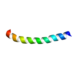
THE SOLUTION STRUCTURE OF THE ACTIVE DOMAIN OF CAP18: A LIPOPOLYSACCHARIDE BINDING PROTEIN FROM RABBIT LEUKOCYTES
Descriptor: CAP18
Authors:Chen, C, Huang, T.-H.
Deposit date:1995-01-12
Release date:1995-03-31
Last modified:2022-02-23
Method:SOLUTION NMR
Cite:The solution structure of the active domain of CAP18--a lipopolysaccharide binding protein from rabbit leukocytes.
FEBS Lett., 370, 1995
2DCW
DownloadVisualize
BU of 2dcw by Molmil
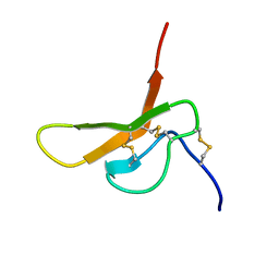
The solution structure of horseshoe crab antimicrobial peptide tachystatin b with the inhibitory cystine-knot motif
Descriptor: Tachystatin-B2
Authors:Fujitani, N, Kawano, K.
Deposit date:2006-01-16
Release date:2007-01-23
Last modified:2022-03-09
Method:SOLUTION NMR
Cite:The solution structure of horseshoe crab antimicrobial peptide tachystatin B with an inhibitory cystine-knot motif
J.Pept.Sci., 13, 2007
2DCV
DownloadVisualize
BU of 2dcv by Molmil
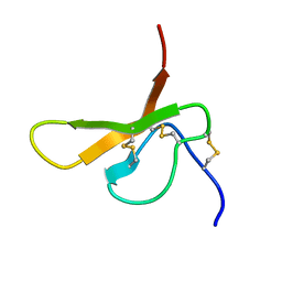
The solution structure of horseshoe crab antimicrobial peptide tachystatin b with the inhibitory cystine-knot motif
Descriptor: Tachystatin-B1
Authors:Fujitani, N, Kohno, T.
Deposit date:2006-01-16
Release date:2007-01-23
Last modified:2022-03-09
Method:SOLUTION NMR
Cite:The solution structure of horseshoe crab antimicrobial peptide tachystatin B with an inhibitory cystine-knot motif
J.Pept.Sci., 13, 2007

218853

PDB entries from 2024-04-24

PDB statisticsPDBj update infoContact PDBjnumon