Loading
PDBj
MenuPDBj@FacebookPDBj@TwitterPDBj@YouTubewwPDB FoundationwwPDB
RCSB PDBPDBeBMRBAdv. SearchSearch help
Search by PDB author
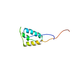
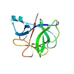
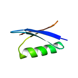
1GB1
DownloadVisualize
BU of 1gb1 by Molmil
A NOVEL, HIGHLY STABLE FOLD OF THE IMMUNOGLOBULIN BINDING DOMAIN OF STREPTOCOCCAL PROTEIN G
Descriptor: PROTEIN G
Authors:Gronenborn, A.M, Clore, G.M.
Deposit date:1991-05-15
Release date:1993-04-15
Last modified:2022-02-23
Method:SOLUTION NMR
Cite:A novel, highly stable fold of the immunoglobulin binding domain of streptococcal protein G.
Science, 253, 1991
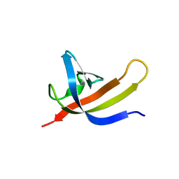
2GB1
DownloadVisualize
BU of 2gb1 by Molmil
A NOVEL, HIGHLY STABLE FOLD OF THE IMMUNOGLOBULIN BINDING DOMAIN OF STREPTOCOCCAL PROTEIN G
Descriptor: PROTEIN G
Authors:Gronenborn, A.M, Clore, G.M.
Deposit date:1991-05-15
Release date:1993-04-15
Last modified:2022-03-09
Method:SOLUTION NMR
Cite:A novel, highly stable fold of the immunoglobulin binding domain of streptococcal protein G.
Science, 253, 1991
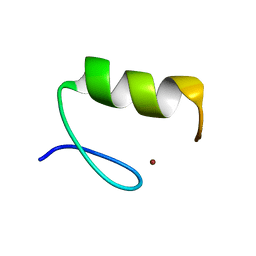
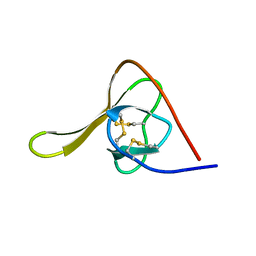
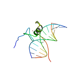
4ZNF
DownloadVisualize
BU of 4znf by Molmil
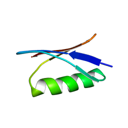
HIGH-RESOLUTION THREE-DIMENSIONAL STRUCTURE OF A SINGLE ZINC FINGER FROM A HUMAN ENHANCER BINDING PROTEIN IN SOLUTION
Descriptor: ZINC FINGER, ZINC ION
Authors:Gronenborn, A.M, Clore, G.M, Omichinski, J.G.
Deposit date:1990-07-09
Release date:1992-01-15
Last modified:2022-03-16
Method:SOLUTION NMR
Cite:High-resolution three-dimensional structure of a single zinc finger from a human enhancer binding protein in solution.
Biochemistry, 29, 1990
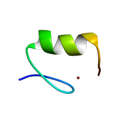
3ZNF
DownloadVisualize
BU of 3znf by Molmil
HIGH-RESOLUTION THREE-DIMENSIONAL STRUCTURE OF A SINGLE ZINC FINGER FROM A HUMAN ENHANCER BINDING PROTEIN IN SOLUTION
Descriptor: ZINC FINGER, ZINC ION
Authors:Gronenborn, A.M, Clore, G.M, Omichinski, J.G.
Deposit date:1990-07-09
Release date:1992-01-15
Last modified:2022-03-16
Method:SOLUTION NMR
Cite:High-resolution three-dimensional structure of a single zinc finger from a human enhancer binding protein in solution.
Biochemistry, 29, 1990
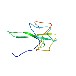
5T82
DownloadVisualize
BU of 5t82 by Molmil
HIV-1 reverse transcriptase thumb subdomain
Descriptor: Reverse transcriptase
Authors:Gronenborn, A.M, Sharaf, N.G, Byeon, I.-J.L.
Deposit date:2016-09-06
Release date:2017-01-18
Last modified:2023-06-14
Method:SOLUTION NMR
Cite:NMR structure of the HIV-1 reverse transcriptase thumb subdomain.
J. Biomol. NMR, 66, 2016
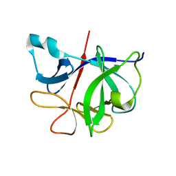
6I1B
DownloadVisualize
BU of 6i1b by Molmil
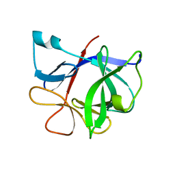
HIGH-RESOLUTION THREE-DIMENSIONAL STRUCTURE OF INTERLEUKIN-1 BETA IN SOLUTION BY THREE-AND FOUR-DIMENSIONAL NUCLEAR MAGNETIC RESONANCE SPECTROSCOPY
Descriptor: INTERLEUKIN-1 BETA
Authors:Clore, G.M, Gronenborn, A.M.
Deposit date:1991-01-22
Release date:1992-10-15
Last modified:2022-03-16
Method:SOLUTION NMR
Cite:High-resolution three-dimensional structure of interleukin 1 beta in solution by three- and four-dimensional nuclear magnetic resonance spectroscopy.
Biochemistry, 30, 1991
7I1B
DownloadVisualize
BU of 7i1b by Molmil
HIGH-RESOLUTION THREE-DIMENSIONAL STRUCTURE OF INTERLEUKIN-1 BETA IN SOLUTION BY THREE-AND FOUR-DIMENSIONAL NUCLEAR MAGNETIC RESONANCE SPECTROSCOPY
Descriptor: INTERLEUKIN-1 BETA
Authors:Clore, G.M, Gronenborn, A.M.
Deposit date:1991-01-22
Release date:1992-10-15
Last modified:2022-03-23
Method:SOLUTION NMR
Cite:High-resolution three-dimensional structure of interleukin 1 beta in solution by three- and four-dimensional nuclear magnetic resonance spectroscopy.
Biochemistry, 30, 1991
8EZ6
DownloadVisualize
BU of 8ez6 by Molmil
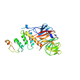
The DBC1/SIRT1 Interaction is Choreographed by Post-translational Modification
Descriptor: Cell cycle and apoptosis regulator protein 2
Authors:Krzysiak, T.C, Gronenborn, A.M.
Deposit date:2022-10-31
Release date:2024-03-27
Last modified:2024-04-03
Method:X-RAY DIFFRACTION (2 Å)
Cite:Inhibitory Protein-Protein Interactions of the SIRT1 Deacetylase are Choreographed by Post-translational Modification
Protein Sci., 2024
9ILB
DownloadVisualize
BU of 9ilb by Molmil
HUMAN INTERLEUKIN-1 BETA
Descriptor: PROTEIN (HUMAN INTERLEUKIN-1 BETA)
Authors:Yu, B, Blaber, M, Gronenborn, A.M, Clore, G.M, Caspar, D.L.D.
Deposit date:1998-10-22
Release date:1999-01-06
Last modified:2023-09-20
Method:X-RAY DIFFRACTION (2.28 Å)
Cite:Disordered water within a hydrophobic protein cavity visualized by x-ray crystallography.
Proc.Natl.Acad.Sci.USA, 96, 1999
6HIR
DownloadVisualize
BU of 6hir by Molmil
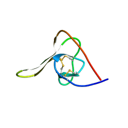
SOLUTION STRUCTURE OF RECOMBINANT HIRUDIN AND THE LYS-47 (RIGHT ARROW) GLU MUTANT. A NUCLEAR MAGNETIC RESONANCE AND HYBRID DISTANCE GEOMETRY-DYNAMICAL SIMULATED ANNEALING STUDY
Descriptor: HIRUDIN VARIANT-1
Authors:Clore, G.M, Gronenborn, A.M.
Deposit date:1990-01-09
Release date:1990-01-15
Last modified:2017-11-29
Method:SOLUTION NMR
Cite:Solution structure of recombinant hirudin and the Lys-47----Glu mutant: a nuclear magnetic resonance and hybrid distance geometry-dynamical simulated annealing study.
Biochemistry, 28, 1989
7TA8
DownloadVisualize
BU of 7ta8 by Molmil
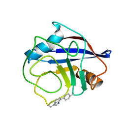
NMR structure of crosslinked cyclophilin A
Descriptor: Peptidyl-prolyl cis-trans isomerase A
Authors:Lu, M, Toptygin, D, Xiang, Y, Shi, Y, Schwieters, C.D, Lipinski, E.C, Ahn, J, Byeon, I.-J.L, Gronenborn, A.M.
Deposit date:2021-12-20
Release date:2022-06-01
Last modified:2023-06-14
Method:SOLUTION NMR
Cite:The Magic of Linking Rings: Discovery of a Unique Photoinduced Fluorescent Protein Crosslink.
J.Am.Chem.Soc., 144, 2022
3TRX
DownloadVisualize
BU of 3trx by Molmil
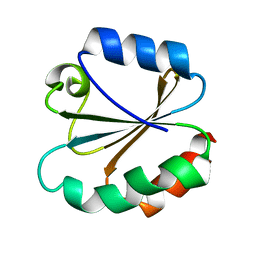
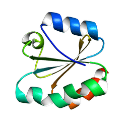
HIGH-RESOLUTION THREE-DIMENSIONAL STRUCTURE OF REDUCED RECOMBINANT HUMAN THIOREDOXIN IN SOLUTION
Descriptor: THIOREDOXIN
Authors:Forman-Kay, J.D, Clore, G.M, Gronenborn, A.M.
Deposit date:1990-12-17
Release date:1992-01-15
Last modified:2022-03-16
Method:SOLUTION NMR
Cite:High-resolution three-dimensional structure of reduced recombinant human thioredoxin in solution.
Biochemistry, 30, 1991
7R7P
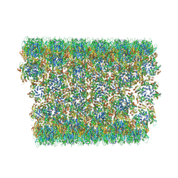
DownloadVisualize
BU of 7r7p by Molmil
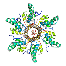
Immature HIV-1 CACTD-SP1 lattice with Bevirimat (BVM) and Inositol hexakisphosphate (IP6)
Descriptor: 3alpha-[(3-carboxy-3-methylbutanoyl)oxy]-8alpha,9beta,10alpha,13alpha,17alpha,19beta-lup-20(29)-en-28-oic acid, Gag polyprotein, INOSITOL HEXAKISPHOSPHATE
Authors:Sarkar, S, Zadrozny, K.K, Zadorozhnyi, R, Russell, R.W, Quinn, C.M, Kleinpeter, A, Ablan, S, Meshkin, H, Perilla, J.R, Ganser-Pornillos, B.K, Pornillos, O, Freed, E.O, Gronenborn, A.M, Polenova, T.
Deposit date:2021-06-25
Release date:2023-02-15
Last modified:2023-03-22
Method:SOLID-STATE NMR
Cite:Structural basis of HIV-1 maturation inhibitor binding and activity.
Nat Commun, 14, 2023
7R7Q
DownloadVisualize
BU of 7r7q by Molmil
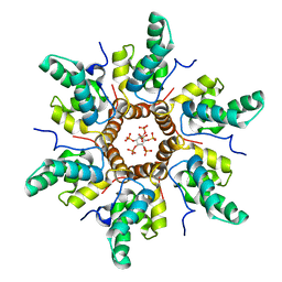
Immature HIV-1 CACTD-SP1 lattice with Inositol hexakisphosphate (IP6)
Descriptor: Gag polyprotein, INOSITOL HEXAKISPHOSPHATE
Authors:Sarkar, S, Zadrozny, K.K, Zadorozhnyi, R, Russell, R.W, Quinn, C.M, Kleinpeter, A, Ablan, S, Meshkin, H, Perilla, J.R, Ganser-Pornillos, B.K, Pornillos, O, Freed, E.O, Gronenborn, A.M, Polenova, T.
Deposit date:2021-06-25
Release date:2023-02-15
Last modified:2023-03-22
Method:SOLID-STATE NMR
Cite:Structural basis of HIV-1 maturation inhibitor binding and activity.
Nat Commun, 14, 2023
7RIK
DownloadVisualize
BU of 7rik by Molmil
Magic-Angle-Spinning NMR Structure of Kinesin-1 Motor Domain Assembled with Microtubules
Descriptor: Kinesin-1 heavy chain
Authors:Zhang, C, Guo, C, Russell, R.W, Quinn, C.M, Li, M, Williams, J.C, Gronenborn, A.M, Polenova, T.
Deposit date:2021-07-20
Release date:2022-11-23
Method:SOLID-STATE NMR
Cite:Magic-angle-spinning NMR structure of the kinesin-1 motor domain assembled with microtubules reveals the elusive neck linker orientation
Nat Commun, 13, 2022
5HIR
DownloadVisualize
BU of 5hir by Molmil
SOLUTION STRUCTURE OF RECOMBINANT HIRUDIN AND THE LYS-47 (RIGHT ARROW) GLU MUTANT. A NUCLEAR MAGNETIC RESONANCE AND HYBRID DISTANCE GEOMETRY-DYNAMICAL SIMULATED ANNEALING STUDY
Descriptor: HIRUDIN VARIANT-1
Authors:Clore, G.M, Gronenborn, A.M.
Deposit date:1990-01-09
Release date:1990-01-15
Last modified:2017-11-29
Method:SOLUTION NMR
Cite:Solution structure of recombinant hirudin and the Lys-47----Glu mutant: a nuclear magnetic resonance and hybrid distance geometry-dynamical simulated annealing study.
Biochemistry, 28, 1989
4Z8L
DownloadVisualize
BU of 4z8l by Molmil
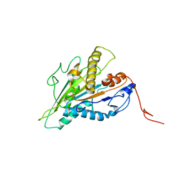
Crystal structure of DCAF1/SIV-MND VPX/MND SAMHD1 NTD ternary complex
Descriptor: Protein VPRBP, SAM domain and HD domain-containing protein, Vpx protein, ...
Authors:Koharudin, L.M, Wu, Y, Calero, G, Ahn, J, Gronenborn, A.M.
Deposit date:2015-04-09
Release date:2015-06-17
Last modified:2023-09-27
Method:X-RAY DIFFRACTION (2.6 Å)
Cite:Structural Basis of Clade-specific Engagement of SAMHD1 (Sterile alpha Motif and Histidine/Aspartate-containing Protein 1) Restriction Factors by Lentiviral Viral Protein X (Vpx) Virulence Factors.
J.Biol.Chem., 290, 2015
4TRX
DownloadVisualize
BU of 4trx by Molmil
HIGH-RESOLUTION THREE-DIMENSIONAL STRUCTURE OF REDUCED RECOMBINANT HUMAN THIOREDOXIN IN SOLUTION
Descriptor: THIOREDOXIN
Authors:Forman-Kay, J.D, Clore, G.M, Gronenborn, A.M.
Deposit date:1990-12-17
Release date:1992-01-15
Last modified:2022-03-16
Method:SOLUTION NMR
Cite:High-resolution three-dimensional structure of reduced recombinant human thioredoxin in solution.
Biochemistry, 30, 1991
6XQJ
DownloadVisualize
BU of 6xqj by Molmil
Structure of HIV-1 Vpr in complex with the human nucleotide excision repair protein hHR23A
Descriptor: Protein Vpr,UV excision repair protein RAD23 homolog A, ZINC ION
Authors:Byeon, I.-J.L, Calero, G, Wu, Y, Byeon, C.H, Gronenborn, A.M.
Deposit date:2020-07-09
Release date:2021-11-17
Last modified:2021-12-08
Method:SOLUTION NMR
Cite:Structure of HIV-1 Vpr in complex with the human nucleotide excision repair protein hHR23A.
Nat Commun, 12, 2021
6X63
DownloadVisualize
BU of 6x63 by Molmil
Atomic-Resolution Structure of HIV-1 Capsid Tubes by Magic Angle Spinning NMR
Descriptor: HIV-1 capsid protein
Authors:Lu, M, Russell, R.W, Bryer, A, Quinn, C.M, Hou, G, Zhang, H, Schwieters, C.D, Perilla, J.R, Gronenborn, A.M, Polenova, T.
Deposit date:2020-05-27
Release date:2020-09-02
Last modified:2023-06-14
Method:SOLID-STATE NMR
Cite:Atomic-resolution structure of HIV-1 capsid tubes by magic-angle spinning NMR.
Nat.Struct.Mol.Biol., 27, 2020
7SD4
DownloadVisualize
BU of 7sd4 by Molmil
SARS-CoV-2 Nucleocapsid N-terminal domain (N-NTD) protein
Descriptor: Nucleoprotein
Authors:Sarkar, S, Runge, B, Russell, R.W, Calero, D, Zeinalilathori, S, Quinn, C.M, Lu, M, Calero, G, Gronenborn, A.M, Polenova, T.
Deposit date:2021-09-29
Release date:2022-06-08
Last modified:2023-06-14
Method:SOLID-STATE NMR
Cite:Atomic-Resolution Structure of SARS-CoV-2 Nucleocapsid Protein N-Terminal Domain.
J.Am.Chem.Soc., 144, 2022
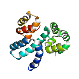
6GAT
DownloadVisualize
BU of 6gat by Molmil
SOLUTION NMR STRUCTURE OF THE L22V MUTANT DNA BINDING DOMAIN OF AREA COMPLEXED TO A 13 BP DNA CONTAINING A TGATA SITE, REGULARIZED MEAN STRUCTURE
Descriptor: DNA (5'-D(*CP*AP*GP*TP*GP*AP*TP*AP*GP*AP*GP*AP*C)-3'), DNA (5'-D(*GP*TP*CP*TP*CP*TP*AP*TP*CP*AP*CP*TP*G)-3'), NITROGEN REGULATORY PROTEIN AREA, ...
Authors:Clore, G.M, Starich, M, Wikstrom, M, Gronenborn, A.M.
Deposit date:1997-11-07
Release date:1998-01-28
Last modified:2021-11-03
Method:SOLUTION NMR
Cite:The solution structure of the Leu22-->Val mutant AREA DNA binding domain complexed with a TGATAG core element defines a role for hydrophobic packing in the determination of specificity.
J.Mol.Biol., 277, 1998
1SAF
DownloadVisualize
BU of 1saf by Molmil
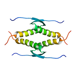
HIGH RESOLUTION SOLUTION NMR STRUCTURE OF THE OLIGOMERIZATION DOMAIN OF P53 BY MULTI-DIMENSIONAL NMR (SAD STRUCTURES)
Descriptor: TUMOR SUPPRESSOR P53
Authors:Clore, G.M, Omichinski, J.G, Gronenborn, A.M.
Deposit date:1995-03-12
Release date:1995-10-15
Last modified:2012-01-11
Method:SOLUTION NMR
Cite:Refined solution structure of the oligomerization domain of the tumour suppressor p53.
Nat.Struct.Biol., 2, 1995
1SAK
DownloadVisualize
BU of 1sak by Molmil
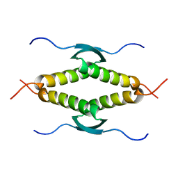
HIGH RESOLUTION SOLUTION NMR STRUCTURE OF THE OLIGOMERIZATION DOMAIN OF P53 BY MULTI-DIMENSIONAL NMR (SAC STRUCTURES)
Descriptor: TUMOR SUPPRESSOR P53
Authors:Clore, G.M, Omichinski, J.G, Gronenborn, A.M.
Deposit date:1995-03-12
Release date:1995-10-15
Last modified:2022-03-02
Method:SOLUTION NMR
Cite:Refined solution structure of the oligomerization domain of the tumour suppressor p53.
Nat.Struct.Biol., 2, 1995
1SAL
DownloadVisualize
BU of 1sal by Molmil
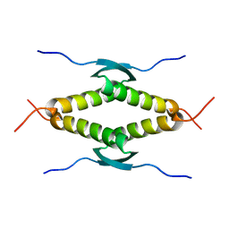
HIGH RESOLUTION SOLUTION NMR STRUCTURE OF THE OLIGOMERIZATION DOMAIN OF P53 BY MULTI-DIMENSIONAL NMR (SAD STRUCTURES)
Descriptor: TUMOR SUPPRESSOR P53
Authors:Clore, G.M, Omichinski, J.G, Gronenborn, A.M.
Deposit date:1995-03-12
Release date:1995-10-15
Last modified:2022-03-02
Method:SOLUTION NMR
Cite:Refined solution structure of the oligomerization domain of the tumour suppressor p53.
Nat.Struct.Biol., 2, 1995

218196

PDB entries from 2024-04-10

PDB statisticsPDBj update infoContact PDBjnumon