Loading
PDBj
MenuPDBj@FacebookPDBj@TwitterPDBj@YouTubewwPDB FoundationwwPDB
RCSB PDBPDBeBMRBAdv. SearchSearch help
Search by PDB author
1PSD
DownloadVisualize
BU of 1psd by Molmil
THE ALLOSTERIC LIGAND SITE IN THE VMAX-TYPE COOPERATIVE ENZYME PHOSPHOGLYCERATE DEHYDROGENASE
Descriptor: D-3-PHOSPHOGLYCERATE DEHYDROGENASE (PHOSPHOGLYCERATE DEHYDROGENASE), NICOTINAMIDE-ADENINE-DINUCLEOTIDE, SERINE
Authors:Schuller, D.J, Grant, G.A, Banaszak, L.J.
Deposit date:1995-05-02
Release date:1995-07-31
Last modified:2024-02-14
Method:X-RAY DIFFRACTION (2.75 Å)
Cite:The allosteric ligand site in the Vmax-type cooperative enzyme phosphoglycerate dehydrogenase.
Nat.Struct.Biol., 2, 1995
4RQO
DownloadVisualize
BU of 4rqo by Molmil
Crystal structure of L-Serine Dehydratase from Legionella pneumophila
Descriptor: CHLORIDE ION, IRON/SULFUR CLUSTER, L-serine dehydratase
Authors:Thoden, J.B, Holden, H.M, Grant, G.A.
Deposit date:2014-11-04
Release date:2014-11-19
Last modified:2014-12-24
Method:X-RAY DIFFRACTION (2.25 Å)
Cite:Structure of l-Serine Dehydratase from Legionella pneumophila: Novel Use of the C-Terminal Cysteine as an Intrinsic Competitive Inhibitor.
Biochemistry, 53, 2014
1YGY
DownloadVisualize
BU of 1ygy by Molmil
Crystal Structure of D-3-Phosphoglycerate dehydrogenase From Mycobacterium tuberculosis
Descriptor: D-3-phosphoglycerate dehydrogenase, L(+)-TARTARIC ACID
Authors:Dey, S, Grant, G.A, Sacchettini, J.C, TB Structural Genomics Consortium (TBSGC)
Deposit date:2005-01-05
Release date:2005-01-25
Last modified:2019-07-10
Method:X-RAY DIFFRACTION (2.3 Å)
Cite:Crystal Structure of Mycobacterium tuberculosis D-3-Phosphoglycerate Dehydrogenase: EXTREME ASYMMETRY IN A TETRAMER OF IDENTICAL SUBUNITS
J.Biol.Chem., 280, 2005
1SC6
DownloadVisualize
BU of 1sc6 by Molmil
Crystal Structure of W139G D-3-Phosphoglycerate dehydrogenase complexed with NAD+
Descriptor: D-3-phosphoglycerate dehydrogenase, NICOTINAMIDE-ADENINE-DINUCLEOTIDE
Authors:Bell, J.K, Grant, G.A, Banaszak, L.J.
Deposit date:2004-02-11
Release date:2005-02-22
Last modified:2021-10-27
Method:X-RAY DIFFRACTION (2.09 Å)
Cite:Multiconformational states in phosphoglycerate dehydrogenase
Biochemistry, 43, 2004
1KBA
DownloadVisualize
BU of 1kba by Molmil
CRYSTAL STRUCTURE OF KAPPA-BUNGAROTOXIN AT 2.3-ANGSTROM RESOLUTION
Descriptor: KAPPA-BUNGAROTOXIN
Authors:Dewan, J.C, Grant, G.A, Sacchettini, J.C.
Deposit date:1994-07-13
Release date:1994-12-20
Last modified:2019-08-14
Method:X-RAY DIFFRACTION (2.3 Å)
Cite:Crystal structure of kappa-bungarotoxin at 2.3-A resolution.
Biochemistry, 33, 1994
1YBA
DownloadVisualize
BU of 1yba by Molmil
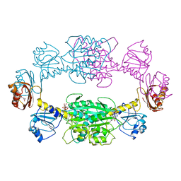
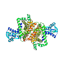
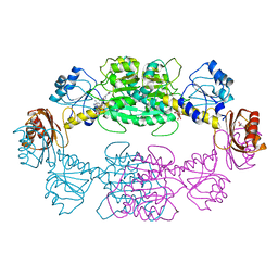
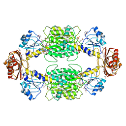
The active form of phosphoglycerate dehydrogenase
Descriptor: 2-OXOGLUTARIC ACID, D-3-phosphoglycerate dehydrogenase, NICOTINAMIDE-ADENINE-DINUCLEOTIDE, ...
Authors:Thompson, J.R, Banaszak, L.J.
Deposit date:2004-12-20
Release date:2005-04-26
Last modified:2024-04-03
Method:X-RAY DIFFRACTION (2.24 Å)
Cite:Vmax Regulation through Domain and Subunit Changes. The Active Form of Phosphoglycerate Dehydrogenase
Biochemistry, 44, 2005
1ZID
DownloadVisualize
BU of 1zid by Molmil
LONG FATTY ACID CHAIN ENOYL-ACP REDUCTASE (INHA) IN COMPLEX WITH AN ISONICOTINIC-ACYL-NADH INHIBITOR
Descriptor: ENOYL-[ACYL-CARRIER-PROTEIN] REDUCTASE, ISONICOTINIC-ACETYL-NICOTINAMIDE-ADENINE DINUCLEOTIDE
Authors:Rozwarski, D.A, Jacobs Jr, W.R, Sacchettini, J.C, TB Structural Genomics Consortium (TBSGC)
Deposit date:1997-03-25
Release date:1998-03-25
Last modified:2023-08-09
Method:X-RAY DIFFRACTION (2.7 Å)
Cite:Modification of the NADH of the isoniazid target (InhA) from Mycobacterium tuberculosis.
Science, 279, 1998
2PA3
DownloadVisualize
BU of 2pa3 by Molmil
crystal structure of serine bound G336V mutant of E.coli phosphoglycerate dehydrogenase
Descriptor: 1,4-DIHYDRONICOTINAMIDE ADENINE DINUCLEOTIDE, D-3-phosphoglycerate dehydrogenase, SERINE
Authors:Dey, S, Sacchettini, J.C.
Deposit date:2007-03-27
Release date:2007-04-24
Last modified:2023-08-30
Method:X-RAY DIFFRACTION (2.74 Å)
Cite:The Effect of Hinge Mutations on Effector Binding and Domain Rotation in Escherichia coli D-3-Phosphoglycerate Dehydrogenase.
J.Biol.Chem., 282, 2007
2P9C
DownloadVisualize
BU of 2p9c by Molmil
Crystal structure of serine bound G336V mutant of E.coli phosphoglycerate dehydrogenase
Descriptor: 1,4-DIHYDRONICOTINAMIDE ADENINE DINUCLEOTIDE, D-3-phosphoglycerate dehydrogenase, SERINE
Authors:Dey, S, Sacchettini, J.C.
Deposit date:2007-03-24
Release date:2007-04-24
Last modified:2023-08-30
Method:X-RAY DIFFRACTION (2.46 Å)
Cite:The Effect of Hinge Mutations on Effector Binding and Domain Rotation in Escherichia coli D-3-Phosphoglycerate Dehydrogenase
J.Biol.Chem., 282, 2007
2P9G
DownloadVisualize
BU of 2p9g by Molmil
Crystal structure of serine bound G336V,G337V double mutant of E.coli phosphoglycerate dehydrogenase
Descriptor: 1,4-DIHYDRONICOTINAMIDE ADENINE DINUCLEOTIDE, D-3-phosphoglycerate dehydrogenase, SERINE
Authors:Dey, S, Sacchettini, J.C.
Deposit date:2007-03-25
Release date:2007-04-24
Last modified:2023-08-30
Method:X-RAY DIFFRACTION (2.8 Å)
Cite:The Effect of Hinge Mutations on Effector Binding and Domain Rotation in Escherichia coli D-3-Phosphoglycerate Dehydrogenase.
J.Biol.Chem., 282, 2007
2P9E
DownloadVisualize
BU of 2p9e by Molmil
Crystal Structure of G336V mutant of E.coli phosphoglycerate dehydrogenase
Descriptor: 1,4-DIHYDRONICOTINAMIDE ADENINE DINUCLEOTIDE, CITRIC ACID, D-3-phosphoglycerate dehydrogenase, ...
Authors:Dey, S, Sacchettini, J.C.
Deposit date:2007-03-25
Release date:2007-04-24
Last modified:2023-08-30
Method:X-RAY DIFFRACTION (2.6 Å)
Cite:The Effect of Hinge Mutations on Effector Binding and Domain Rotation in Escherichia coli D-3-Phosphoglycerate Dehydrogenase
J.Biol.Chem., 282, 2007
3DC2
DownloadVisualize
BU of 3dc2 by Molmil
Crystal structure of serine bound D-3-phosphoglycerate dehydrogenase from Mycobacterium tuberculosis
Descriptor: D-3-phosphoglycerate dehydrogenase, L(+)-TARTARIC ACID, SERINE
Authors:Dey, S, Sacchettini, J.C.
Deposit date:2008-06-03
Release date:2008-07-22
Last modified:2023-11-01
Method:X-RAY DIFFRACTION (2.7 Å)
Cite:Structural analysis of substrate and effector binding in Mycobacterium tuberculosis D-3-phosphoglycerate dehydrogenase
Biochemistry, 47, 2008
3DDN
DownloadVisualize
BU of 3ddn by Molmil
Crystal structure of hydroxypyruvic acid phosphate bound D-3-phosphoglycerate dehydrogenase in mycobacterium tuberculosis
Descriptor: 2-oxo-3-(phosphonooxy)propanoic acid, D-3-phosphoglycerate dehydrogenase, L(+)-TARTARIC ACID
Authors:Dey, S, Sacchettini, J.C.
Deposit date:2008-06-05
Release date:2008-07-22
Last modified:2023-11-01
Method:X-RAY DIFFRACTION (2.4 Å)
Cite:Structural analysis of substrate and effector binding in Mycobacterium tuberculosis D-3-phosphoglycerate dehydrogenase
Biochemistry, 47, 2008

218500

PDB entries from 2024-04-17

PDB statisticsPDBj update infoContact PDBjnumon