Loading
PDBj
MenuPDBj@FacebookPDBj@TwitterPDBj@YouTubewwPDB FoundationwwPDB
RCSB PDBPDBeBMRBAdv. SearchSearch help
Search by PDB author
4RSK
DownloadVisualize
BU of 4rsk by Molmil
STRUCTURE OF THE K7A/R10A/K66A VARIANT OF RIBONUCLEASE A COMPLEXED WITH 3'-UMP
Descriptor: 3'-URIDINEMONOPHOSPHATE, RIBONUCLEASE A
Authors:Schultz, L.W, Fisher, B.M, Raines, R.T.
Deposit date:1998-04-09
Release date:1998-12-09
Last modified:2023-08-09
Method:X-RAY DIFFRACTION (2.1 Å)
Cite:Coulombic Effects of Remote Subsites on the Active Site of Ribonuclease A
Biochemistry, 37, 1998
3RSK
DownloadVisualize
BU of 3rsk by Molmil
STRUCTURE OF THE K7A/R10A/K66A VARIANT OF RIBONUCLEASE A
Descriptor: ACETATE ION, RIBONUCLEASE A
Authors:Schultz, L.W, Fisher, B.M, Raines, R.T.
Deposit date:1998-04-09
Release date:1998-07-15
Last modified:2023-08-09
Method:X-RAY DIFFRACTION (2 Å)
Cite:Coulombic Effects of Remote Subsites on the Active Site of Ribonuclease A
Biochemistry, 37, 1998
1N3Z
DownloadVisualize
BU of 1n3z by Molmil
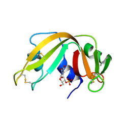
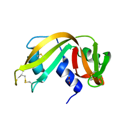
Crystal structure of the [S-carboxyamidomethyl-Cys31, S-carboxyamidomethyl-Cys32] monomeric derivative of the bovine seminal ribonuclease in the liganded state
Descriptor: 3'-URIDINEMONOPHOSPHATE, ADENOSINE, Ribonuclease, ...
Authors:Sica, F, Di Fiore, A, Zagari, A, Mazzarella, L.
Deposit date:2002-10-30
Release date:2003-08-26
Last modified:2011-07-13
Method:X-RAY DIFFRACTION (1.65 Å)
Cite:The unswapped chain of bovine seminal ribonuclease: Crystal structure of the free and liganded monomeric derivative
Proteins, 52, 2003

218500

PDB entries from 2024-04-17

PDB statisticsPDBj update infoContact PDBjnumon