Loading
PDBj
MenuPDBj@FacebookPDBj@TwitterPDBj@YouTubewwPDB FoundationwwPDB
RCSB PDBPDBeBMRBAdv. SearchSearch help
Search by PDB author
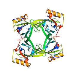
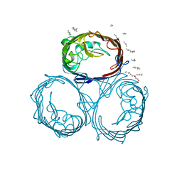
2OMF
DownloadVisualize
BU of 2omf by Molmil
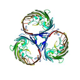
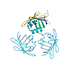
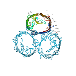
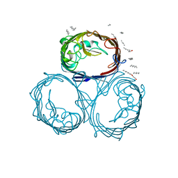
OMPF PORIN
Descriptor: (HYDROXYETHYLOXY)TRI(ETHYLOXY)OCTANE, MATRIX PORIN OUTER MEMBRANE PROTEIN F
Authors:Cowan, S.W.
Deposit date:1995-02-28
Release date:1995-12-07
Last modified:2024-02-21
Method:X-RAY DIFFRACTION (2.4 Å)
Cite:The Refined Structure of Ompf Porin from E.Coli at 2.4 Angstroms Resolution
To be Published
1CRB
DownloadVisualize
BU of 1crb by Molmil
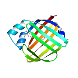
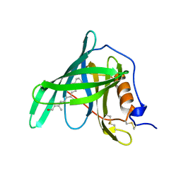
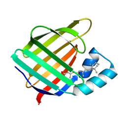
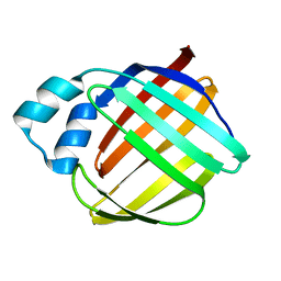
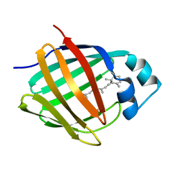
CRYSTALLOGRAPHIC STUDIES ON A FAMILY OF CELLULAR LIPOPHILIC TRANSPORT PROTEINS. REFINEMENT OF P2 MYELIN PROTEIN AND THE STRUCTURE DETERMINATION AND REFINEMENT OF CELLULAR RETINOL-BINDING PROTEIN IN COMPLEX WITH ALL-TRANS-RETINOL
Descriptor: CADMIUM ION, CELLULAR RETINOL BINDING PROTEIN, RETINOL
Authors:Cowan, S.W, Jones, T.A.
Deposit date:1993-02-10
Release date:1994-12-20
Last modified:2024-02-07
Method:X-RAY DIFFRACTION (2.1 Å)
Cite:Crystallographic studies on a family of cellular lipophilic transport proteins. Refinement of P2 myelin protein and the structure determination and refinement of cellular retinol-binding protein in complex with all-trans-retinol.
J.Mol.Biol., 230, 1993
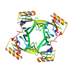
1OPF
DownloadVisualize
BU of 1opf by Molmil
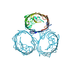
THE STRUCTURE OF OMPF PORIN IN A TETRAGONAL CRYSTAL FORM
Descriptor: MATRIX PORIN OUTER MEMBRANE PROTEIN F
Authors:Cowan, S.W, Schirmer, T, Pauptit, R.A, Jansonius, J.N.
Deposit date:1994-11-21
Release date:1995-02-07
Last modified:2024-02-14
Method:X-RAY DIFFRACTION (3.2 Å)
Cite:The structure of OmpF porin in a tetragonal crystal form.
Structure, 3, 1995
1PMP
DownloadVisualize
BU of 1pmp by Molmil
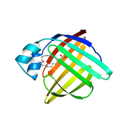
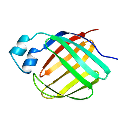
CRYSTALLOGRAPHIC STUDIES ON A FAMILY OF CELLULAR LIPOPHILIC TRANSPORT PROTEINS. REFINEMENT OF P2 MYELIN PROTEIN AND THE STRUCTURE DETERMINATION AND REFINEMENT OF CELLULAR RETINOL-BINDING PROTEIN IN COMPLEX WITH ALL-TRANS-RETINOL
Descriptor: OLEIC ACID, P2 MYELIN PROTEIN
Authors:Cowan, S.W, Newcomer, M.E, Jones, T.A.
Deposit date:1993-02-10
Release date:1995-01-26
Last modified:2024-02-14
Method:X-RAY DIFFRACTION (2.7 Å)
Cite:Crystallographic studies on a family of cellular lipophilic transport proteins. Refinement of P2 myelin protein and the structure determination and refinement of cellular retinol-binding protein in complex with all-trans-retinol.
J.Mol.Biol., 230, 1993
1PHO
DownloadVisualize
BU of 1pho by Molmil
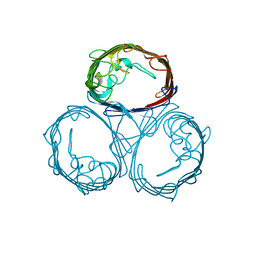
CRYSTAL STRUCTURES EXPLAIN FUNCTIONAL PROPERTIES OF TWO E. COLI PORINS
Descriptor: PHOSPHOPORIN
Authors:Schirmer, T, Cowan, S.W, Jansonius, J.N.
Deposit date:1993-01-15
Release date:1993-10-31
Last modified:2024-02-14
Method:X-RAY DIFFRACTION (3 Å)
Cite:Crystal structures explain functional properties of two E. coli porins.
Nature, 358, 1992
1RBP
DownloadVisualize
BU of 1rbp by Molmil
CRYSTALLOGRAPHIC REFINEMENT OF HUMAN SERUM RETINOL BINDING PROTEIN AT 2 ANGSTROMS RESOLUTION
Descriptor: PLASMA RETINOL-BINDING PROTEIN PRECURSOR, RETINOL
Authors:Jones, T.A, Newcomer, M.E, Cowan, S.W.
Deposit date:1990-04-02
Release date:1991-07-15
Last modified:2017-11-29
Method:X-RAY DIFFRACTION (2 Å)
Cite:Crystallographic refinement of human serum retinol binding protein at 2A resolution.
Proteins, 8, 1990
2DKB
DownloadVisualize
BU of 2dkb by Molmil
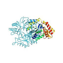
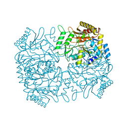
DIALKYLGLYCINE DECARBOXYLASE STRUCTURE: BIFUNCTIONAL ACTIVE SITE AND ALKALI METAL BINDING SITES
Descriptor: 2,2-DIALKYLGLYCINE DECARBOXYLASE (PYRUVATE), 2-(N-MORPHOLINO)-ETHANESULFONIC ACID, PYRIDOXAL-5'-PHOSPHATE, ...
Authors:Toney, M.D, Hohenester, E, Jansonius, J.N.
Deposit date:1994-07-12
Release date:1994-10-15
Last modified:2017-11-29
Method:X-RAY DIFFRACTION (2.1 Å)
Cite:Dialkylglycine decarboxylase structure: bifunctional active site and alkali metal sites.
Science, 261, 1993
1GUH
DownloadVisualize
BU of 1guh by Molmil
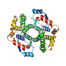
Structure determination and refinement of human alpha class glutathione transferase A1-1, and a comparison with the MU and PI class enzymes
Descriptor: GLUTATHIONE S-TRANSFERASE A1-1, S-BENZYL-GLUTATHIONE
Authors:Sinning, I, Kleywegt, G.J, Jones, T.A.
Deposit date:1993-02-24
Release date:1993-10-31
Last modified:2024-02-07
Method:X-RAY DIFFRACTION (2.6 Å)
Cite:Structure determination and refinement of human alpha class glutathione transferase A1-1, and a comparison with the Mu and Pi class enzymes.
J.Mol.Biol., 232, 1993
1CBR
DownloadVisualize
BU of 1cbr by Molmil
CRYSTAL STRUCTURE OF CELLULAR RETINOIC-ACID-BINDING PROTEINS I AND II IN COMPLEX WITH ALL-TRANS-RETINOIC ACID AND A SYNTHETIC RETINOID
Descriptor: CELLULAR RETINOIC ACID BINDING PROTEIN TYPE I, RETINOIC ACID
Authors:Kleywegt, G.J, Bergfors, T, Jones, T.A.
Deposit date:1994-09-28
Release date:1995-01-26
Last modified:2024-02-07
Method:X-RAY DIFFRACTION (2.9 Å)
Cite:Crystal structures of cellular retinoic acid binding proteins I and II in complex with all-trans-retinoic acid and a synthetic retinoid.
Structure, 2, 1994
1CBS
DownloadVisualize
BU of 1cbs by Molmil
CRYSTAL STRUCTURE OF CELLULAR RETINOIC-ACID-BINDING PROTEINS I AND II IN COMPLEX WITH ALL-TRANS-RETINOIC ACID AND A SYNTHETIC RETINOID
Descriptor: CELLULAR RETINOIC ACID BINDING PROTEIN TYPE II, RETINOIC ACID
Authors:Kleywegt, G.J, Bergfors, T, Jones, T.A.
Deposit date:1994-09-28
Release date:1995-01-26
Last modified:2024-02-07
Method:X-RAY DIFFRACTION (1.8 Å)
Cite:Crystal structures of cellular retinoic acid binding proteins I and II in complex with all-trans-retinoic acid and a synthetic retinoid.
Structure, 2, 1994
1CBQ
DownloadVisualize
BU of 1cbq by Molmil
CRYSTAL STRUCTURE OF CELLULAR RETINOIC-ACID-BINDING PROTEINS I AND II IN COMPLEX WITH ALL-TRANS-RETINOIC ACID AND A SYNTHETIC RETINOID
Descriptor: 6-(2,3,4,5,6,7-HEXAHYDRO-2,4,4-TRIMETHYL-1-METYLENEINDEN-2-YL)-3-METHYLHEXA-2,4-DIENOIC ACID, CELLULAR RETINOIC ACID BINDING PROTEIN TYPE II, PHOSPHATE ION
Authors:Kleywegt, G.J, Bergfors, T, Jones, T.A.
Deposit date:1994-09-28
Release date:1995-01-26
Last modified:2024-02-07
Method:X-RAY DIFFRACTION (2.2 Å)
Cite:Crystal structures of cellular retinoic acid binding proteins I and II in complex with all-trans-retinoic acid and a synthetic retinoid.
Structure, 2, 1994
5JCU
DownloadVisualize
BU of 5jcu by Molmil
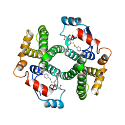
Crystal Structure of hGSTA1-1 with Glutathione Adduct of Phenethyl Isothiocyanate and Cystein Adduct of Phenethyl Isothiocyanate
Descriptor: 1,2-ETHANEDIOL, Glutathione S-transferase A1, L-gamma-glutamyl-S-[(2-phenylethyl)carbamothioyl]-L-cysteinylglycine
Authors:Kumari, V, Ji, X.
Deposit date:2016-04-15
Release date:2016-10-12
Last modified:2023-09-27
Method:X-RAY DIFFRACTION (1.93 Å)
Cite:Irreversible Inhibition of Glutathione S-Transferase by Phenethyl Isothiocyanate (PEITC), a Dietary Cancer Chemopreventive Phytochemical.
Plos One, 11, 2016
1DKA
DownloadVisualize
BU of 1dka by Molmil
DIALKYLGLYCINE DECARBOXYLASE STRUCTURE: BIFUNCTIONAL ACTIVE SITE AND ALKALI METAL BINDING SITES
Descriptor: 2,2-DIALKYLGLYCINE DECARBOXYLASE (PYRUVATE), 2-(N-MORPHOLINO)-ETHANESULFONIC ACID, POTASSIUM ION, ...
Authors:Toney, M.D, Hohenester, E, Jansonius, J.N.
Deposit date:1993-06-18
Release date:1994-10-15
Last modified:2017-11-29
Method:X-RAY DIFFRACTION (2.6 Å)
Cite:Dialkylglycine decarboxylase structure: bifunctional active site and alkali metal sites.
Science, 261, 1993
2A2G
DownloadVisualize
BU of 2a2g by Molmil
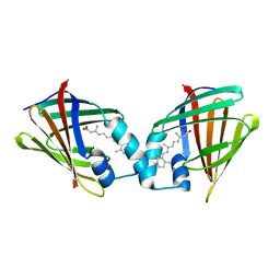
THE CRYSTAL STRUCTURES OF A2U-GLOBULIN AND ITS COMPLEX WITH A HYALINE DROPLET INDUCER.
Descriptor: D-LIMONENE 1,2-EPOXIDE, PROTEIN (ALPHA-2U-GLOBULIN)
Authors:Chaudhuri, B.N, Kleywegt, G.J, Jones, T.A.
Deposit date:1998-11-19
Release date:1999-08-13
Last modified:2024-04-03
Method:X-RAY DIFFRACTION (2.9 Å)
Cite:The structures of alpha 2u-globulin and its complex with a hyaline droplet inducer.
Acta Crystallogr.,Sect.D, 55, 1999
2A2U
DownloadVisualize
BU of 2a2u by Molmil
THE CRYSTAL STRUCTURES OF A2U-GLOBULIN AND ITS COMPLEX WITH A HYALINE DROPLET INDUCER.
Descriptor: PROTEIN (ALPHA-2U-GLOBULIN)
Authors:Chaudhuri, B.N, Kleywegt, G.J, Jones, T.A.
Deposit date:1998-11-19
Release date:1999-08-26
Last modified:2024-04-03
Method:X-RAY DIFFRACTION (2.5 Å)
Cite:The structures of alpha 2u-globulin and its complex with a hyaline droplet inducer.
Acta Crystallogr.,Sect.D, 55, 1999
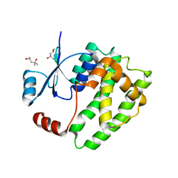
6ATP
DownloadVisualize
BU of 6atp by Molmil
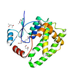
Crystal structure of apo-hGSTA1-1 exhibiting a new conformation of C-terminal helix
Descriptor: (4S)-2-METHYL-2,4-PENTANEDIOL, Glutathione S-transferase A1
Authors:Kumari, V, Ji, X.
Deposit date:2017-08-29
Release date:2018-09-12
Last modified:2023-10-04
Method:X-RAY DIFFRACTION (1.7 Å)
Cite:The dynamic nature of hGSTA1-1 C-terminal helix
To be published
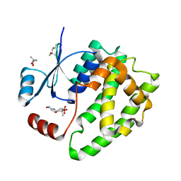
6ATQ
DownloadVisualize
BU of 6atq by Molmil
Crystal structure of apo-hGSTA1-1 exhibiting a new conformation of C-terminal helix
Descriptor: (4S)-2-METHYL-2,4-PENTANEDIOL, 2-(N-MORPHOLINO)-ETHANESULFONIC ACID, Glutathione S-transferase A1
Authors:Kumari, V, Ji, X.
Deposit date:2017-08-29
Release date:2018-09-12
Last modified:2023-10-04
Method:X-RAY DIFFRACTION (2 Å)
Cite:The dynamic nature of hGSTA1-1 C-terminal helix
To be published
6ATO
DownloadVisualize
BU of 6ato by Molmil
Crystal structure of hGSTA1-1 complexed with GSH and MPD in each subunit
Descriptor: (4S)-2-METHYL-2,4-PENTANEDIOL, GLUTATHIONE, Glutathione S-transferase A1
Authors:Kumari, V, Ji, X.
Deposit date:2017-08-29
Release date:2018-09-12
Last modified:2023-10-04
Method:X-RAY DIFFRACTION (1.55 Å)
Cite:The dynamic nature of hGSTA1-1 C-terminal helix
To be published
6ATR
DownloadVisualize
BU of 6atr by Molmil
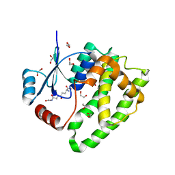
Crystal structure of hGSTA1-1 complexed with two GSH analogues in each subunit
Descriptor: 1,2-ETHANEDIOL, 2-AMINO-5-[1-(CARBOXYLATOMETHYLCARBAMOYL)-2-NITROSOSULFANYL-ETHYL]AMINO-5-OXO-PENTANOATE, Glutathione S-transferase A1, ...
Authors:Kumari, V, Ji, X.
Deposit date:2017-08-29
Release date:2018-09-12
Last modified:2023-10-04
Method:X-RAY DIFFRACTION (1.29 Å)
Cite:hGSTA1 apo structure and dynamic c-terminus helix
To be published
1LPJ
DownloadVisualize
BU of 1lpj by Molmil
Human cRBP IV
Descriptor: Retinol-binding protein IV, cellular
Authors:Calderone, V, Zanotti, G, Berni, R, Folli, C.
Deposit date:2002-05-08
Release date:2003-01-14
Last modified:2023-08-16
Method:X-RAY DIFFRACTION (2 Å)
Cite:Ligand binding and structural analysis of a human putative cellular retinol-binding protein
J.Biol.Chem., 277, 2002
1MPF
DownloadVisualize
BU of 1mpf by Molmil
STRUCTURAL AND FUNCTIONAL ALTERATIONS OF A COLICIN RESISTANT MUTANT OF OMPF-PORIN FROM ESCHERICHIA COLI
Descriptor: (HYDROXYETHYLOXY)TRI(ETHYLOXY)OCTANE, MATRIX PORIN OUTER MEMBRANE PROTEIN F
Authors:Schirmer, T.
Deposit date:1994-08-10
Release date:1995-02-07
Last modified:2024-02-14
Method:X-RAY DIFFRACTION (3 Å)
Cite:Structural and functional alterations of a colicin-resistant mutant of OmpF porin from Escherichia coli.
Proc.Natl.Acad.Sci.USA, 91, 1994
1MX8
DownloadVisualize
BU of 1mx8 by Molmil
Two homologous rat cellular retinol-binding proteins differ in local structure and flexibility
Descriptor: CELLULAR RETINOL-BINDING PROTEIN I, HOLO, RETINOL
Authors:Lu, J, Cistola, D.P, Li, E.
Deposit date:2002-10-01
Release date:2003-07-29
Last modified:2022-02-23
Method:SOLUTION NMR
Cite:Two homologous rat cellular retinol-binding proteins differ in local conformational flexibility.
J.Mol.Biol., 330, 2003
1MX7
DownloadVisualize
BU of 1mx7 by Molmil
Two homologous rat cellular retinol-binding proteins differ in local structure and flexibility
Descriptor: CELLULAR RETINOL-BINDING PROTEIN I, APO
Authors:Lu, J, Cistola, D.P, Li, E.
Deposit date:2002-10-01
Release date:2003-07-29
Last modified:2022-02-23
Method:SOLUTION NMR
Cite:Two Homologous Rat Cellular Retinol-binding Proteins Differ in Local Conformational Flexibility.
J.Mol.Biol., 330, 2003
1BT9
DownloadVisualize
BU of 1bt9 by Molmil
OMPF PORIN MUTANT D74A
Descriptor: PROTEIN (MATRIX PORIN OUTER MEMBRANE PROTEIN F)
Authors:Philippsen, A, Schirmer, T.
Deposit date:1998-09-01
Release date:1999-01-13
Last modified:2024-02-07
Method:X-RAY DIFFRACTION (3 Å)
Cite:Stability of trimeric OmpF porin: the contributions of the latching loop L2.
Biochemistry, 37, 1998
1GFM
DownloadVisualize
BU of 1gfm by Molmil
OMPF PORIN (MUTANT D113G)
Descriptor: (HYDROXYETHYLOXY)TRI(ETHYLOXY)OCTANE, MATRIX PORIN OUTER MEMBRANE PROTEIN F
Authors:Lou, K.-L, Schirmer, T.
Deposit date:1996-05-08
Release date:1996-12-07
Last modified:2024-02-07
Method:X-RAY DIFFRACTION (3.5 Å)
Cite:Structural and functional characterization of OmpF porin mutants selected for larger pore size. I. Crystallographic analysis.
J.Biol.Chem., 271, 1996


12>

218196

PDB entries from 2024-04-10

PDB statisticsPDBj update infoContact PDBjnumon