Loading
PDBj
MenuPDBj@FacebookPDBj@TwitterPDBj@YouTubewwPDB FoundationwwPDB
RCSB PDBPDBeBMRBAdv. SearchSearch help
Search by PDB author
7XR8
DownloadVisualize
BU of 7xr8 by Molmil
Structure of the S8 family protease A4095
Descriptor: Major intracellular serine protease
Authors:Cheng, J.H, Cao, H.Y, Wang, P.
Deposit date:2022-05-09
Release date:2023-05-17
Last modified:2023-11-29
Method:X-RAY DIFFRACTION (1.94 Å)
Cite:Structure of the S8 family protease A4095
To Be Published
5YNZ
DownloadVisualize
BU of 5ynz by Molmil
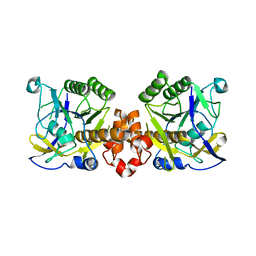
Crystal structure of the dihydroorotase domain (K1556A) of human CAD
Descriptor: CAD protein, ZINC ION
Authors:Huang, Y.H, Chen, K.L, Cheng, J.H, Huang, C.Y.
Deposit date:2017-10-26
Release date:2018-10-24
Last modified:2023-11-22
Method:X-RAY DIFFRACTION (2.774 Å)
Cite:Crystal structures of monometallic dihydropyrimidinase and the human dihydroorotase domain K1556A mutant reveal no lysine carbamylation within the active site
Biochem. Biophys. Res. Commun., 505, 2018
5YKD
DownloadVisualize
BU of 5ykd by Molmil
Crystal structure of dihydropyrimidinase from Pseudomonas aeruginosa PAO1 at 2.17 angstrom resolution
Descriptor: D-hydantoinase/dihydropyrimidinase, ZINC ION
Authors:Huang, Y.H, Huang, C.Y.
Deposit date:2017-10-14
Release date:2018-02-21
Last modified:2023-11-22
Method:X-RAY DIFFRACTION (2.17 Å)
Cite:Structural Basis for pH-Dependent Oligomerization of Dihydropyrimidinase fromPseudomonas aeruginosaPAO1.
Bioinorg Chem Appl, 2018, 2018
6AJD
DownloadVisualize
BU of 6ajd by Molmil
Crystal structure of a monometallic dihydropyrimidinase from Pseudomonas aeruginosa PAO1 reveals no lysine carbamylation within the active site
Descriptor: D-hydantoinase/dihydropyrimidinase, ZINC ION
Authors:Huang, Y.H, Huang, C.Y.
Deposit date:2018-08-27
Release date:2018-10-24
Last modified:2023-11-22
Method:X-RAY DIFFRACTION (2.223 Å)
Cite:Crystal structures of monometallic dihydropyrimidinase and the human dihydroorotase domain K1556A mutant reveal no lysine carbamylation within the active site
Biochem. Biophys. Res. Commun., 505, 2018
6NYH
DownloadVisualize
BU of 6nyh by Molmil
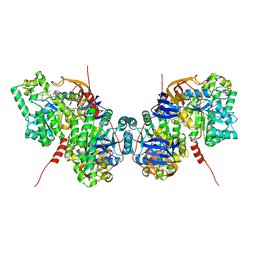
Structure of human RIPK1 kinase domain in complex with GNE684
Descriptor: (5S)-N-[(3S)-7-methoxy-1-methyl-2-oxo-2,3,4,5-tetrahydro-1H-pyrido[3,4-b]azepin-3-yl]-5-phenyl-6,7-dihydro-5H-pyrrolo[1,2-b][1,2,4]triazole-2-carboxamide, IODIDE ION, Receptor-interacting serine/threonine-protein kinase 1
Authors:Fong, R, Lupardus, P.J.
Deposit date:2019-02-11
Release date:2019-05-29
Last modified:2023-10-11
Method:X-RAY DIFFRACTION (2.1 Å)
Cite:RIP1 inhibition blocks inflammatory diseases but not tumor growth or metastases.
Cell Death Differ., 27, 2020
5YYU
DownloadVisualize
BU of 5yyu by Molmil
Crystal structure of Staphylococcus aureus single-stranded DNA-binding protein SsbB
Descriptor: Single-stranded DNA-binding protein
Authors:Huang, Y.H, Huang, C.Y.
Deposit date:2017-12-11
Release date:2018-08-15
Last modified:2023-11-22
Method:X-RAY DIFFRACTION (2.984 Å)
Cite:Characterization of single-stranded DNA-binding protein SsbB from Staphylococcus aureus: SsbB cannot stimulate PriA helicase.
Rsc Adv, 8, 2018

218853

PDB entries from 2024-04-24

PDB statisticsPDBj update infoContact PDBjnumon