Loading
PDBj
MenuPDBj@FacebookPDBj@TwitterPDBj@YouTubewwPDB FoundationwwPDB
RCSB PDBPDBeBMRBAdv. SearchSearch help
Search by PDB author
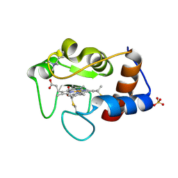
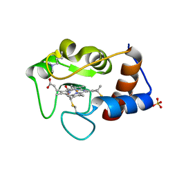
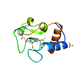
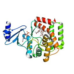
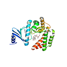
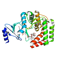
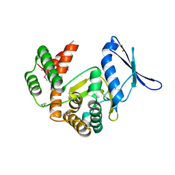
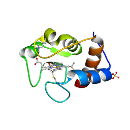
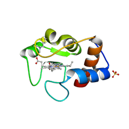
1CRH
DownloadVisualize
BU of 1crh by Molmil
THE ROLE OF A CONSERVED INTERNAL WATER MOLECULE AND ITS ASSOCIATED HYDROGEN BOND NETWORK IN CYTOCHROME C
Descriptor: CYTOCHROME C, HEME C, SULFATE ION
Authors:Berghuis, A.M, Brayer, G.D.
Deposit date:1993-08-06
Release date:1994-01-31
Last modified:2021-03-03
Method:X-RAY DIFFRACTION (1.9 Å)
Cite:The role of a conserved internal water molecule and its associated hydrogen bond network in cytochrome c.
J.Mol.Biol., 236, 1994
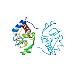
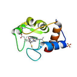
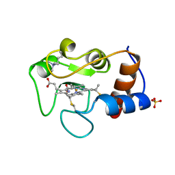
1CRI
DownloadVisualize
BU of 1cri by Molmil
THE ROLE OF A CONSERVED INTERNAL WATER MOLECULE AND ITS ASSOCIATED HYDROGEN BOND NETWORK IN CYTOCHROME C
Descriptor: CYTOCHROME C, PROTOPORPHYRIN IX CONTAINING FE, SULFATE ION
Authors:Berghuis, A.M, Brayer, G.D.
Deposit date:1993-08-06
Release date:1994-01-31
Last modified:2017-11-29
Method:X-RAY DIFFRACTION (2 Å)
Cite:The role of a conserved internal water molecule and its associated hydrogen bond network in cytochrome c.
J.Mol.Biol., 236, 1994
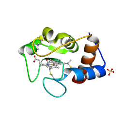
1CRG
DownloadVisualize
BU of 1crg by Molmil
THE ROLE OF A CONSERVED INTERNAL WATER MOLECULE AND ITS ASSOCIATED HYDROGEN BOND NETWORK IN CYTOCHROME C
Descriptor: CYTOCHROME C, HEME C, SULFATE ION
Authors:Berghuis, A.M, Brayer, G.D.
Deposit date:1993-08-06
Release date:1994-01-31
Last modified:2021-03-03
Method:X-RAY DIFFRACTION (2 Å)
Cite:The role of a conserved internal water molecule and its associated hydrogen bond network in cytochrome c.
J.Mol.Biol., 236, 1994
1CRJ
DownloadVisualize
BU of 1crj by Molmil
THE ROLE OF A CONSERVED INTERNAL WATER MOLECULE AND ITS ASSOCIATED HYDROGEN BOND NETWORK IN CYTOCHROME C
Descriptor: CYTOCHROME C, HEME C, SULFATE ION
Authors:Berghuis, A.M, Brayer, G.D.
Deposit date:1993-08-06
Release date:1994-01-31
Last modified:2021-03-03
Method:X-RAY DIFFRACTION (2.05 Å)
Cite:The role of a conserved internal water molecule and its associated hydrogen bond network in cytochrome c.
J.Mol.Biol., 236, 1994
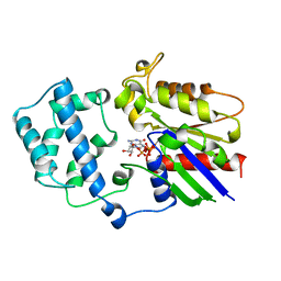
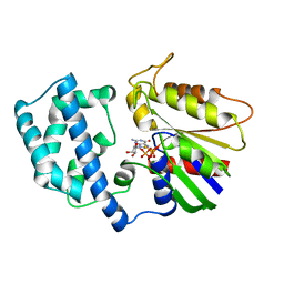
1GIT
DownloadVisualize
BU of 1git by Molmil
STRUCTURE OF GTP-BINDING PROTEIN
Descriptor: G PROTEIN GI ALPHA 1, GUANOSINE-5'-DIPHOSPHATE, PHOSPHATE ION
Authors:Berghuis, A.M, Lee, E, Sprang, S.R.
Deposit date:1996-10-16
Release date:1997-02-12
Last modified:2024-02-07
Method:X-RAY DIFFRACTION (2.6 Å)
Cite:Structure of the GDP-Pi complex of Gly203-->Ala gialpha1: a mimic of the ternary product complex of galpha-catalyzed GTP hydrolysis.
Structure, 4, 1996
1CTY
DownloadVisualize
BU of 1cty by Molmil
MUTATION OF TYROSINE-67 IN CYTOCHROME C SIGNIFICANTLY ALTERS THE LOCAL HEME ENVIRONMENT
Descriptor: CYTOCHROME C, HEME C, SULFATE ION
Authors:Berghuis, A.M, Brayer, G.D.
Deposit date:1993-02-15
Release date:1993-07-15
Last modified:2021-03-03
Method:X-RAY DIFFRACTION (2.2 Å)
Cite:Mutation of tyrosine-67 to phenylalanine in cytochrome c significantly alters the local heme environment.
J.Mol.Biol., 235, 1994
1CTZ
DownloadVisualize
BU of 1ctz by Molmil
MUTATION OF TYROSINE-67 IN CYTOCHROME C SIGNIFICANTLY ALTERS THE LOCAL HEME ENVIRONMENT
Descriptor: CYTOCHROME C, HEME C, SULFATE ION
Authors:Berghuis, A.M, Brayer, G.D.
Deposit date:1993-02-15
Release date:1993-07-15
Last modified:2021-03-03
Method:X-RAY DIFFRACTION (1.9 Å)
Cite:Mutation of tyrosine-67 to phenylalanine in cytochrome c significantly alters the local heme environment.
J.Mol.Biol., 235, 1994
2YCC
DownloadVisualize
BU of 2ycc by Molmil
OXIDATION STATE-DEPENDENT CONFORMATIONAL CHANGES IN CYTOCHROME C
Descriptor: CYTOCHROME C, HEME C, SULFATE ION
Authors:Berghuis, A.M, Brayer, G.D.
Deposit date:1991-01-29
Release date:1992-07-15
Last modified:2021-03-03
Method:X-RAY DIFFRACTION (1.9 Å)
Cite:Oxidation state-dependent conformational changes in cytochrome c.
J.Mol.Biol., 223, 1992
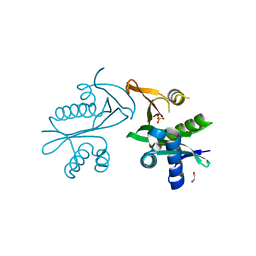
5E96
DownloadVisualize
BU of 5e96 by Molmil
Crystal structure of aminoglycoside 6'-acetyltransferase type Ii
Descriptor: 1,2-ETHANEDIOL, Aminoglycoside 6'-acetyltransferase, PHOSPHATE ION
Authors:Berghuis, A.M, Burk, D.L, Baettig, O.M, Shi, K.
Deposit date:2015-10-14
Release date:2016-07-06
Last modified:2023-09-27
Method:X-RAY DIFFRACTION (2.1 Å)
Cite:Comprehensive characterization of ligand-induced plasticity changes in a dimeric enzyme.
Febs J., 283, 2016
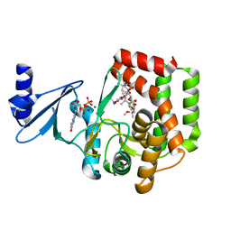
3TM0
DownloadVisualize
BU of 3tm0 by Molmil
Crystal Structure of 3',5"-Aminoglycoside Phosphotransferase Type IIIa AMPPNP Butirosin A Complex
Descriptor: (2S)-4-amino-N-[(1R,2S,3R,4R,5S)-5-amino-4-[(2,6-diamino-2,6-dideoxy-alpha-D-glucopyranosyl)oxy]-2-hydroxy-3-(beta-D-xylofuranosyloxy)cyclohexyl]-2-hydroxybutanamide, Aminoglycoside 3'-phosphotransferase, MAGNESIUM ION, ...
Authors:Berghuis, A.M, Fong, D.H.
Deposit date:2011-08-30
Release date:2011-10-19
Last modified:2023-09-13
Method:X-RAY DIFFRACTION (2.1 Å)
Cite:Structural basis of APH(3')-IIIa-mediated resistance to N1-substituted aminoglycoside antibiotics.
Antimicrob.Agents Chemother., 53, 2009
1IRV
DownloadVisualize
BU of 1irv by Molmil
CYTOCHROME C ISOZYME 1, REDUCED, MUTANT WITH ILE 75 REPLACED BY MET AND CYS 102 REPLACED BY THR
Descriptor: CYTOCHROME C, PROTOPORPHYRIN IX CONTAINING FE, SULFATE ION
Authors:Berghuis, A.M, Brayer, G.D.
Deposit date:1996-06-27
Release date:1997-01-11
Last modified:2021-11-03
Method:X-RAY DIFFRACTION (1.9 Å)
Cite:Mechanistic and structural contributions of critical surface and internal residues to cytochrome c electron transfer reactivity.
Biochemistry, 35, 1996
1IRW
DownloadVisualize
BU of 1irw by Molmil
CYTOCHROME C ISOZYME 1, REDUCED, MUTANT WITH ASN 52 REPLACED BY ALA AND CYS 102 REPLACED BY THR
Descriptor: CYTOCHROME C, PROTOPORPHYRIN IX CONTAINING FE, SULFATE ION
Authors:Berghuis, A.M, Brayer, G.D.
Deposit date:1996-06-27
Release date:1997-01-11
Last modified:2021-11-03
Method:X-RAY DIFFRACTION (2 Å)
Cite:Mechanistic and structural contributions of critical surface and internal residues to cytochrome c electron transfer reactivity.
Biochemistry, 35, 1996
1GIA
DownloadVisualize
BU of 1gia by Molmil
STRUCTURE OF ACTIVE CONFORMATIONS OF GIA1 AND THE MECHANISM OF GTP HYDROLYSIS
Descriptor: 5'-GUANOSINE-DIPHOSPHATE-MONOTHIOPHOSPHATE, G PROTEIN GI ALPHA 1, MAGNESIUM ION
Authors:Berghuis, A.M, Coleman, D.E, Sprang, S.R.
Deposit date:1994-07-20
Release date:1994-09-30
Last modified:2024-02-07
Method:X-RAY DIFFRACTION (2 Å)
Cite:Structures of active conformations of Gi alpha 1 and the mechanism of GTP hydrolysis.
Science, 265, 1994
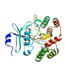
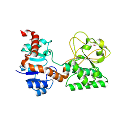
2Q99
DownloadVisualize
BU of 2q99 by Molmil
Crystal Structure of Saccharopine Dehydrogenase from Saccharomyces cerevisiae
Descriptor: Saccharopine dehydrogenase [NAD+, L-lysine-forming
Authors:Berghuis, A.M, Burk, D.L.
Deposit date:2007-06-12
Release date:2007-11-06
Last modified:2017-10-18
Method:X-RAY DIFFRACTION (1.64 Å)
Cite:Structural Studies of the Final Enzyme in the alpha-Aminoadipate Pathway-Saccharopine Dehydrogenase from Saccharomyces cerevisiae
J.Mol.Biol., 373, 2007
3Q2M
DownloadVisualize
BU of 3q2m by Molmil
Crystal Structure of Spectinomycin Phosphotransferase, APH(9)-Ia, Protein Kinase Inhibitor CKI-7 Complex
Descriptor: N-(2-AMINOETHYL)-5-CHLOROISOQUINOLINE-8-SULFONAMIDE, NICKEL (II) ION, Spectinomycin phosphotransferase
Authors:Berghuis, A.M, Fong, D.H, Xiong, B, Hwang, J.
Deposit date:2010-12-20
Release date:2011-05-18
Last modified:2023-09-13
Method:X-RAY DIFFRACTION (2.9 Å)
Cite:Crystal structures of two aminoglycoside kinases bound with a eukaryotic protein kinase inhibitor.
Plos One, 6, 2011
3I1A
DownloadVisualize
BU of 3i1a by Molmil
Crystal Structure of apo Spectinomycin Phosphotransferase, APH(9)-Ia
Descriptor: 2-(N-MORPHOLINO)-ETHANESULFONIC ACID, DIMETHYL SULFOXIDE, Spectinomycin phosphotransferase, ...
Authors:Berghuis, A.M, Fong, D.H, Lemke, C.T, Hwang, J, Xiong, B.
Deposit date:2009-06-25
Release date:2010-01-19
Last modified:2024-02-21
Method:X-RAY DIFFRACTION (1.7 Å)
Cite:Structure of the antibiotic resistance factor spectinomycin phosphotransferase from Legionella pneumophila.
J.Biol.Chem., 285, 2010
3I0Q
DownloadVisualize
BU of 3i0q by Molmil
Crystal Structure of the AMP-bound complex of Spectinomycin Phosphotransferase, APH(9)-Ia
Descriptor: ADENOSINE MONOPHOSPHATE, NICKEL (II) ION, Spectinomycin phosphotransferase
Authors:Berghuis, A.M, Fong, D.H, Lemke, C.T, Hwang, J.-Y, Xiong, B.
Deposit date:2009-06-25
Release date:2010-01-19
Last modified:2023-09-06
Method:X-RAY DIFFRACTION (2.8 Å)
Cite:Structure of the antibiotic resistance factor spectinomycin phosphotransferase from Legionella pneumophila.
J.Biol.Chem., 285, 2010
5IGH
DownloadVisualize
BU of 5igh by Molmil
Macrolide 2'-phosphotransferase type I
Descriptor: Macrolide 2'-phosphotransferase, SULFATE ION
Authors:Berghuis, A.M, Fong, D.H.
Deposit date:2016-02-28
Release date:2017-04-26
Last modified:2020-01-08
Method:X-RAY DIFFRACTION (1.55 Å)
Cite:Structural Basis for Kinase-Mediated Macrolide Antibiotic Resistance.
Structure, 25, 2017
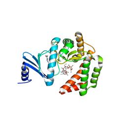
5IGV
DownloadVisualize
BU of 5igv by Molmil
Macrolide 2'-phosphotransferase type II - complex with GDP and azithromycin
Descriptor: AZITHROMYCIN, CALCIUM ION, GUANOSINE-5'-DIPHOSPHATE, ...
Authors:Berghuis, A.M, Fong, D.H.
Deposit date:2016-02-28
Release date:2017-04-26
Last modified:2023-09-27
Method:X-RAY DIFFRACTION (1.55 Å)
Cite:Structural Basis for Kinase-Mediated Macrolide Antibiotic Resistance.
Structure, 25, 2017
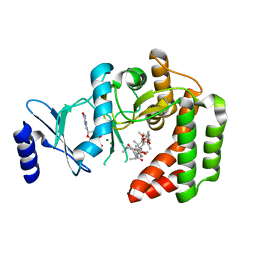
5IGR
DownloadVisualize
BU of 5igr by Molmil
Macrolide 2'-phosphotransferase type I - complex with GDP and oleandomycin
Descriptor: (3S,5R,6S,7R,8R,11R,12S,13R,14S,15S)-6-HYDROXY-5,7,8,11,13,15-HEXAMETHYL-4,10-DIOXO-14-{[3,4,6-TRIDEOXY-3-(DIMETHYLAMINO)-BETA-D-XYLO-HEXOPYRANOSYL]OXY}-1,9-DIOXASPIRO[2.13]HEXADEC-12-YL 2,6-DIDEOXY-3-O-METHYL-ALPHA-L-ARABINO-HEXOPYRANOSIDE, AMMONIUM ION, GUANOSINE-5'-DIPHOSPHATE, ...
Authors:Berghuis, A.M, Fong, D.H.
Deposit date:2016-02-28
Release date:2017-04-26
Last modified:2024-03-06
Method:X-RAY DIFFRACTION (1.6 Å)
Cite:Structural Basis for Kinase-Mediated Macrolide Antibiotic Resistance.
Structure, 25, 2017
5IH1
DownloadVisualize
BU of 5ih1 by Molmil
Macrolide 2'-phosphotransferase type II - complex with GDP and phosphorylated josamycin
Descriptor: CALCIUM ION, GUANOSINE-5'-DIPHOSPHATE, MAGNESIUM ION, ...
Authors:Berghuis, A.M, Fong, D.H.
Deposit date:2016-02-28
Release date:2017-04-26
Last modified:2024-03-06
Method:X-RAY DIFFRACTION (1.31 Å)
Cite:Structural Basis for Kinase-Mediated Macrolide Antibiotic Resistance.
Structure, 25, 2017
5IGU
DownloadVisualize
BU of 5igu by Molmil
Macrolide 2'-phosphotransferase type II
Descriptor: Macrolide 2'-phosphotransferase II
Authors:Berghuis, A.M, Fong, D.H.
Deposit date:2016-02-28
Release date:2017-04-26
Last modified:2020-01-08
Method:X-RAY DIFFRACTION (2.1 Å)
Cite:Structural Basis for Kinase-Mediated Macrolide Antibiotic Resistance.
Structure, 25, 2017
5IH0
DownloadVisualize
BU of 5ih0 by Molmil
Macrolide 2'-phosphotransferase type II Y92M mutant - complex with GDP and erythromycin
Descriptor: ACETATE ION, ERYTHROMYCIN A, GUANOSINE-5'-DIPHOSPHATE, ...
Authors:Berghuis, A.M, Fong, D.H.
Deposit date:2016-02-28
Release date:2017-04-26
Last modified:2023-09-27
Method:X-RAY DIFFRACTION (1.65 Å)
Cite:Structural Basis for Kinase-Mediated Macrolide Antibiotic Resistance.
Structure, 25, 2017
5IGJ
DownloadVisualize
BU of 5igj by Molmil
Macrolide 2'-phosphotransferase type I - complex with guanosine and clarithromycin
Descriptor: CLARITHROMYCIN, GUANOSINE, ISOPROPYL ALCOHOL, ...
Authors:Berghuis, A.M, Fong, D.H.
Deposit date:2016-02-28
Release date:2017-04-26
Last modified:2023-09-27
Method:X-RAY DIFFRACTION (1.4 Å)
Cite:Structural Basis for Kinase-Mediated Macrolide Antibiotic Resistance.
Structure, 25, 2017
5IGW
DownloadVisualize
BU of 5igw by Molmil
Macrolide 2'-phosphotransferase type II - complex with GDP and clarithromycin
Descriptor: CLARITHROMYCIN, GUANOSINE-5'-DIPHOSPHATE, MAGNESIUM ION, ...
Authors:Berghuis, A.M, Fong, D.H.
Deposit date:2016-02-28
Release date:2017-04-26
Last modified:2023-09-27
Method:X-RAY DIFFRACTION (2.096 Å)
Cite:Structural Basis for Kinase-Mediated Macrolide Antibiotic Resistance.
Structure, 25, 2017

218500

PDB entries from 2024-04-17

PDB statisticsPDBj update infoContact PDBjnumon