Loading
PDBj
MenuPDBj@FacebookPDBj@TwitterPDBj@YouTubewwPDB FoundationwwPDB
RCSB PDBPDBeBMRBAdv. SearchSearch help
Search by PDB author
6ID6
DownloadVisualize
BU of 6id6 by Molmil
Crystal structure of Rv0731c from Mycobacterium tuberculosis at 1.63 Angstroms.
Descriptor: MAGNESIUM ION, Putative S-adenosyl-L-methionine-dependent methyltransferase Rv0731c, TRIETHYLENE GLYCOL
Authors:Bansia, H, Kalladi, S.M, Nagaraja, V, Ramakumar, S.
Deposit date:2018-09-08
Release date:2019-09-11
Last modified:2024-03-27
Method:X-RAY DIFFRACTION (1.63 Å)
Cite:Crystal structure of Rv0731c from Mycobacterium tuberculosis.
To Be Published
5XC0
DownloadVisualize
BU of 5xc0 by Molmil
Crystal structure of an aromatic mutant (W6A) of an alkali thermostable GH10 Xylanase from Bacillus sp. NG-27
Descriptor: Beta-xylanase, MAGNESIUM ION, SODIUM ION
Authors:Bansia, H, Mahanta, P, Ramakumar, S.
Deposit date:2017-03-21
Release date:2018-03-28
Last modified:2023-11-22
Method:X-RAY DIFFRACTION (2.32 Å)
Cite:Small Glycols Discover Cryptic Pockets on Proteins for Fragment-Based Approaches.
J.Chem.Inf.Model., 2021
5XC1
DownloadVisualize
BU of 5xc1 by Molmil
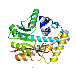
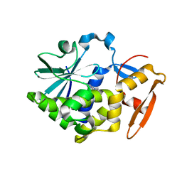
Crystal structure of the complex of an aromatic mutant (W6A) of an alkali thermostable GH10 Xylanase from Bacillus sp. NG-27 with S-1,2-Propanediol
Descriptor: Beta-xylanase, MAGNESIUM ION, S-1,2-PROPANEDIOL, ...
Authors:Bansia, H, Mahanta, P, Ramakumar, S.
Deposit date:2017-03-21
Release date:2018-03-28
Last modified:2023-11-22
Method:X-RAY DIFFRACTION (2.26 Å)
Cite:Small Glycols Discover Cryptic Pockets on Proteins for Fragment-Based Approaches.
J.Chem.Inf.Model., 2021
5Z3J
DownloadVisualize
BU of 5z3j by Molmil
Crystal Structure of Abrin A chain (Recombinant) in complex with Nicotinamide at 1.7 Angstroms
Descriptor: Abrin A-chain, IMIDAZOLE, NICOTINAMIDE
Authors:Bansia, H, Karande, A.A, Ramakumar, S.
Deposit date:2018-01-08
Release date:2019-01-02
Last modified:2023-11-22
Method:X-RAY DIFFRACTION (1.7 Å)
Cite:Structural basis for neutralization of cytotoxic abrin by monoclonal antibody D6F10.
FEBS J., 286, 2019
5Z3I
DownloadVisualize
BU of 5z3i by Molmil
Crystal Structure of Abrin A chain (Recombinant) in complex with Adenine at 1.65 Angstroms
Descriptor: ADENINE, Abrin A-chain
Authors:Bansia, H, Karande, A.A, Ramakumar, S.
Deposit date:2018-01-08
Release date:2019-01-02
Last modified:2023-11-22
Method:X-RAY DIFFRACTION (1.65 Å)
Cite:Structural basis for neutralization of cytotoxic abrin by monoclonal antibody D6F10.
FEBS J., 286, 2019
5Z37
DownloadVisualize
BU of 5z37 by Molmil
Crystal Structure of Abrin A chain (Recombinant) at 1.3 Angstroms
Descriptor: Abrin A-chain, IMIDAZOLE
Authors:Bansia, H, Karande, A.A, Ramakumar, S.
Deposit date:2018-01-05
Release date:2019-01-02
Last modified:2023-11-22
Method:X-RAY DIFFRACTION (1.3 Å)
Cite:Structural basis for neutralization of cytotoxic abrin by monoclonal antibody D6F10.
FEBS J., 286, 2019
7ZQS
DownloadVisualize
BU of 7zqs by Molmil
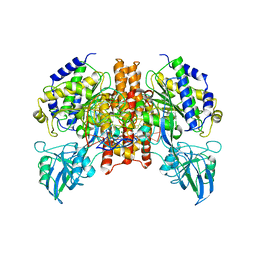
Cryo-EM Structure of Human Transferrin Receptor 1 bound to DNA Aptamer
Descriptor: 2-acetamido-2-deoxy-beta-D-glucopyranose, 2-acetamido-2-deoxy-beta-D-glucopyranose-(1-4)-2-acetamido-2-deoxy-beta-D-glucopyranose, DNA (30-MER), ...
Authors:Bansia, H, Wang, T, Gutierrez, D, des Georges, A.
Deposit date:2022-05-02
Release date:2022-08-17
Method:ELECTRON MICROSCOPY (2.54 Å)
Cite:Discovery of a Transferrin Receptor 1-Binding Aptamer and Its Application in Cancer Cell Depletion for Adoptive T-Cell Therapy Manufacturing.
J.Am.Chem.Soc., 144, 2022
5EFD
DownloadVisualize
BU of 5efd by Molmil
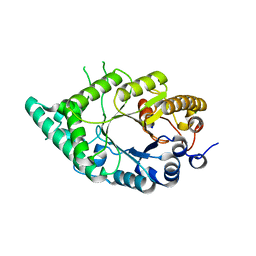
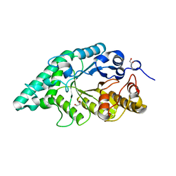
Crystal structure of a surface pocket creating mutant (W6A) of an alkali thermostable GH10 xylanase from Bacillus sp. NG-27
Descriptor: 1,2-ETHANEDIOL, Beta-xylanase, CHLORIDE ION, ...
Authors:Mahanta, P, Bhardwaj, A, Reddy, V.S, Ramakumar, S.
Deposit date:2015-10-23
Release date:2016-10-26
Last modified:2024-03-20
Method:X-RAY DIFFRACTION (1.674 Å)
Cite:Small Glycols Discover Cryptic Pockets on Proteins for Fragment-Based Approaches.
J.Chem.Inf.Model., 2021

218500

PDB entries from 2024-04-17

PDB statisticsPDBj update infoContact PDBjnumon