Loading
PDBj
MenuPDBj@FacebookPDBj@TwitterPDBj@YouTubewwPDB FoundationwwPDB
RCSB PDBPDBeBMRBAdv. SearchSearch help
Search by PDB author
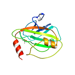
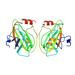
1QOU
DownloadVisualize
BU of 1qou by Molmil
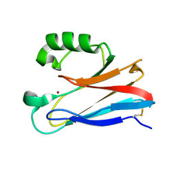
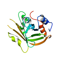
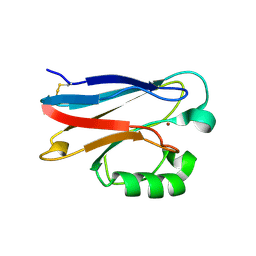
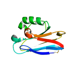
CEN (Centroradialis) protein from Antirrhinum
Descriptor: CEN
Authors:Banfield, M.J, Brady, R.L.
Deposit date:1999-11-17
Release date:2000-03-31
Last modified:2023-12-13
Method:X-RAY DIFFRACTION (1.9 Å)
Cite:The Structure of Antirrhinum Centroradialis Protein (Cen) Suggests a Role as a Kinase Regulator
J.Mol.Biol., 297, 2000
1BD9
DownloadVisualize
BU of 1bd9 by Molmil
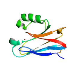
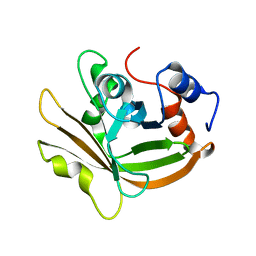
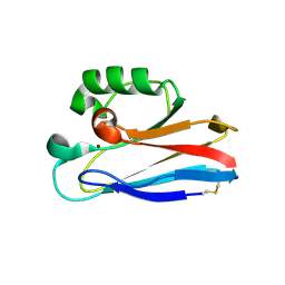
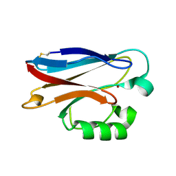
HUMAN PHOSPHATIDYLETHANOLAMINE BINDING PROTEIN
Descriptor: PHOSPHATIDYLETHANOLAMINE BINDING PROTEIN
Authors:Banfield, M.J, Barker, J.J, Perry, A.C.F, Brady, R.L.
Deposit date:1998-05-12
Release date:1998-09-16
Last modified:2024-02-07
Method:X-RAY DIFFRACTION (2.05 Å)
Cite:Function from structure? The crystal structure of human phosphatidylethanolamine-binding protein suggests a role in membrane signal transduction.
Structure, 6, 1998
1BEH
DownloadVisualize
BU of 1beh by Molmil
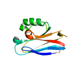
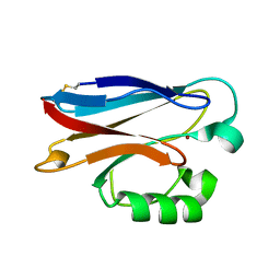
HUMAN PHOSPHATIDYLETHANOLAMINE BINDING PROTEIN IN COMPLEX WITH CACODYLATE
Descriptor: CACODYLATE ION, PHOSPHATIDYLETHANOLAMINE BINDING PROTEIN
Authors:Banfield, M.J, Barker, J.J, Perry, A, Brady, R.L.
Deposit date:1998-05-14
Release date:1998-09-16
Last modified:2023-08-02
Method:X-RAY DIFFRACTION (1.75 Å)
Cite:Function from structure? The crystal structure of human phosphatidylethanolamine-binding protein suggests a role in membrane signal transduction.
Structure, 6, 1998
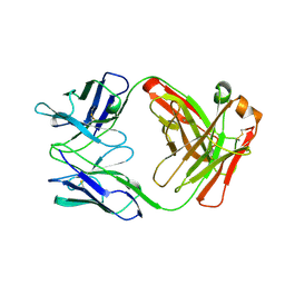
1AD0
DownloadVisualize
BU of 1ad0 by Molmil
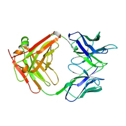
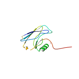
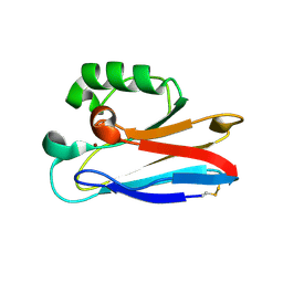
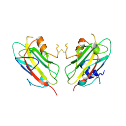
FAB FRAGMENT OF ENGINEERED HUMAN MONOCLONAL ANTIBODY A5B7
Descriptor: ANTIBODY A5B7 (HEAVY CHAIN), ANTIBODY A5B7 (LIGHT CHAIN)
Authors:Banfield, M.J, Brady, R.L.
Deposit date:1997-02-19
Release date:1998-02-25
Last modified:2023-08-02
Method:X-RAY DIFFRACTION (2.5 Å)
Cite:VL:VH domain rotations in engineered antibodies: crystal structures of the Fab fragments from two murine antitumor antibodies and their engineered human constructs.
Proteins, 29, 1997
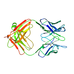
1AE6
DownloadVisualize
BU of 1ae6 by Molmil
IGG-FAB FRAGMENT OF MOUSE MONOCLONAL ANTIBODY CTM01
Descriptor: IGG CTM01 FAB (HEAVY CHAIN), IGG CTM01 FAB (LIGHT CHAIN)
Authors:Banfield, M.J, Brady, R.L.
Deposit date:1997-03-06
Release date:1998-03-18
Last modified:2023-08-02
Method:X-RAY DIFFRACTION (3 Å)
Cite:VL:VH domain rotations in engineered antibodies: crystal structures of the Fab fragments from two murine antitumor antibodies and their engineered human constructs.
Proteins, 29, 1997
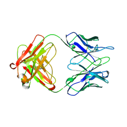
1AD9
DownloadVisualize
BU of 1ad9 by Molmil
IGG-FAB FRAGMENT OF ENGINEERED HUMAN MONOCLONAL ANTIBODY CTM01
Descriptor: IGG CTM01 FAB (HEAVY CHAIN), IGG CTM01 FAB (LIGHT CHAIN), SULFATE ION
Authors:Banfield, M.J, Brady, R.L.
Deposit date:1997-02-24
Release date:1998-02-25
Last modified:2023-08-02
Method:X-RAY DIFFRACTION (2.8 Å)
Cite:VL:VH domain rotations in engineered antibodies: crystal structures of the Fab fragments from two murine antitumor antibodies and their engineered human constructs.
Proteins, 29, 1997
1CLO
DownloadVisualize
BU of 1clo by Molmil
ANTI-CARCINOEMBRYONIC ANTIGEN MONOCLONAL ANTIBODY A5B7
Descriptor: A5B7 MONOCLONAL ANTIBODY
Authors:Banfield, M.J, King, D.J, Mountain, A, Brady, R.L.
Deposit date:1996-05-01
Release date:1997-05-15
Last modified:2011-07-13
Method:X-RAY DIFFRACTION (2.1 Å)
Cite:VL:VH domain rotations in engineered antibodies: crystal structures of the Fab fragments from two murine antitumor antibodies and their engineered human constructs.
Proteins, 29, 1997
1HCF
DownloadVisualize
BU of 1hcf by Molmil
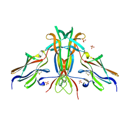
Crystal structure of TrkB-d5 bound to neurotrophin-4/5
Descriptor: BDNF/NT-3 GROWTH FACTORS RECEPTOR, NEUROTROPHIN-4, SULFATE ION
Authors:Banfield, M.J, Naylor, R.L, Robertson, A.G.S, Allen, S.J, Dawbarn, D, Brady, R.L.
Deposit date:2001-05-03
Release date:2001-12-06
Last modified:2023-12-13
Method:X-RAY DIFFRACTION (2.7 Å)
Cite:Specificity in Trk-Receptor:Neurotrophin Interaction: The Crystal Structure of Trkb-D5 in Complex with Neurotrophin-4/5
Structure, 9, 2001
1H5Y
DownloadVisualize
BU of 1h5y by Molmil
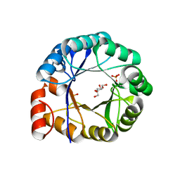
HisF protein from Pyrobaculum aerophilum
Descriptor: GLYCEROL, HISF, PHOSPHATE ION
Authors:Banfield, M.J, Lott, J.S, McCarthy, A.A, Baker, E.N.
Deposit date:2001-05-31
Release date:2001-06-01
Last modified:2023-12-13
Method:X-RAY DIFFRACTION (2 Å)
Cite:Structure of Hisf, a Histidine Biosynthetic Protein from Pyrobaculum Aerophilum
Acta Crystallogr.,Sect.D, 57, 2001
1E3J
DownloadVisualize
BU of 1e3j by Molmil
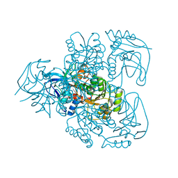
Ketose reductase (sorbitol dehydrogenase) from silverleaf whitefly
Descriptor: BORIC ACID, NADP(H)-DEPENDENT KETOSE REDUCTASE, PHOSPHATE ION, ...
Authors:Banfield, M.J, Salvucci, M.E, Baker, E.N, Smith, C.A.
Deposit date:2000-06-19
Release date:2001-02-04
Last modified:2011-07-13
Method:X-RAY DIFFRACTION (2.3 Å)
Cite:Crystal Structure of Nadp(H)-Dependent Ketose Reductase from Besimia Argentifolii at 2.3 Angstrom Resolution
J.Mol.Biol., 306, 2001
3FSA
DownloadVisualize
BU of 3fsa by Molmil
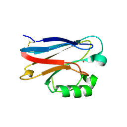
Pseudomonas aeruginosa Azurin with mutated metal-binding loop sequence (CAAHAAM); chemically reduced.
Descriptor: Azurin, COPPER (I) ION
Authors:Banfield, M.J.
Deposit date:2009-01-09
Release date:2009-03-10
Last modified:2017-11-01
Method:X-RAY DIFFRACTION (0.98 Å)
Cite:Metal-binding loop length and not sequence dictates structure.
Proc.Natl.Acad.Sci.USA, 106, 2009
3FS9
DownloadVisualize
BU of 3fs9 by Molmil
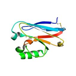
Pseudomonas aeruginosa Azurin with mutated metal-binding loop sequence (CAAHAAM)
Descriptor: Azurin, COPPER (II) ION
Authors:Banfield, M.J.
Deposit date:2009-01-09
Release date:2009-03-10
Last modified:2023-11-01
Method:X-RAY DIFFRACTION (1.05 Å)
Cite:Metal-binding loop length and not sequence dictates structure.
Proc.Natl.Acad.Sci.USA, 106, 2009
3FSV
DownloadVisualize
BU of 3fsv by Molmil
Pseudomonas aeruginosa Azurin with mutated metal-binding loop sequence (CAAAHAAAM)
Descriptor: Azurin, COPPER (II) ION
Authors:Banfield, M.J.
Deposit date:2009-01-12
Release date:2009-03-10
Last modified:2023-11-01
Method:X-RAY DIFFRACTION (2.3 Å)
Cite:Metal-binding loop length and not sequence dictates structure.
Proc.Natl.Acad.Sci.USA, 106, 2009
3FN5
DownloadVisualize
BU of 3fn5 by Molmil
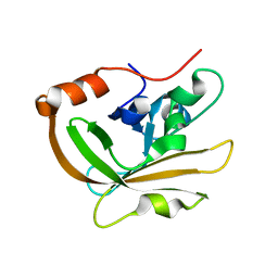
Crystal structure of sortase A (Spy1154) from Streptococcus pyogenes serotype M1 strain SF370
Descriptor: 4-(2-HYDROXYETHYL)-1-PIPERAZINE ETHANESULFONIC ACID, Sortase A
Authors:Banfield, M.J.
Deposit date:2008-12-23
Release date:2009-01-06
Last modified:2023-09-06
Method:X-RAY DIFFRACTION (1.5 Å)
Cite:Crystal Structure of Streptococcus pyogenes Sortase A: Implications for Sortase mechanism
J.Biol.Chem., 284, 2009
3FN6
DownloadVisualize
BU of 3fn6 by Molmil
Crystal structure of sortase A from Streptococcus pyogenes serotype M1 strain SF370 with the active site Cys in its sulphenic acid form
Descriptor: Sortase A
Authors:Banfield, M.J.
Deposit date:2008-12-23
Release date:2009-01-06
Last modified:2023-09-06
Method:X-RAY DIFFRACTION (1.9 Å)
Cite:Crystal Structure of Streptococcus pyogenes Sortase A: Implications for Sortase mechanism
J.Biol.Chem., 284, 2009
3FN7
DownloadVisualize
BU of 3fn7 by Molmil
Crystal structure of sortase A (Spy1154) from Streptococcus pyogenes serotype M1 strain SF370
Descriptor: Sortase A
Authors:Banfield, M.J.
Deposit date:2008-12-23
Release date:2009-01-06
Last modified:2023-09-06
Method:X-RAY DIFFRACTION (1.75 Å)
Cite:Crystal Structure of Streptococcus pyogenes Sortase A: Implications for Sortase mechanism
J.Biol.Chem., 284, 2009
3FT0
DownloadVisualize
BU of 3ft0 by Molmil
Pseudomonas aeruginosa Azurin with mutated metal-binding loop sequence (CAAAAHAAAAM), chemically reduced
Descriptor: Azurin, COPPER (I) ION
Authors:Banfield, M.J.
Deposit date:2009-01-12
Release date:2009-03-10
Last modified:2023-11-01
Method:X-RAY DIFFRACTION (1.8 Å)
Cite:Metal-binding loop length and not sequence dictates structure.
Proc.Natl.Acad.Sci.USA, 106, 2009
3FSW
DownloadVisualize
BU of 3fsw by Molmil
Pseudomonas aeruginosa Azurin with mutated metal-binding loop sequence (CAAAAHAAAM)
Descriptor: Azurin, COPPER (II) ION
Authors:Banfield, M.J.
Deposit date:2009-01-12
Release date:2009-03-10
Last modified:2023-11-01
Method:X-RAY DIFFRACTION (2 Å)
Cite:Metal-binding loop length and not sequence dictates structure.
Proc.Natl.Acad.Sci.USA, 106, 2009
3FSZ
DownloadVisualize
BU of 3fsz by Molmil
Pseudomonas aeruginosa Azurin with mutated metal-binding loop sequence (CAAAAHAAAAM)
Descriptor: Azurin, COPPER (II) ION
Authors:Banfield, M.J.
Deposit date:2009-01-12
Release date:2009-03-10
Last modified:2014-04-02
Method:X-RAY DIFFRACTION (2 Å)
Cite:Metal-binding loop length and not sequence dictates structure.
Proc.Natl.Acad.Sci.USA, 106, 2009
2HXA
DownloadVisualize
BU of 2hxa by Molmil
Crystal structure of Cu(I) Azurin with the metal-binding loop sequence "CTFPGHSALM" replaced with "CSPHQGAGM", at pH3.5
Descriptor: Azurin, COPPER (I) ION
Authors:Banfield, M.J.
Deposit date:2006-08-03
Release date:2007-01-30
Last modified:2011-07-13
Method:X-RAY DIFFRACTION (2.21 Å)
Cite:Engineering Copper Sites in Proteins: Loops Confer Native Structures and Properties to Chimeric Cupredoxins.
J.Am.Chem.Soc., 129, 2007
2HX9
DownloadVisualize
BU of 2hx9 by Molmil
Crystal structure of Cu(I) Azurin with the metal-binding loop sequence "CTFPGHSALM" replaced with "CSPHQGAGM", at pH4
Descriptor: Azurin, COPPER (I) ION
Authors:Banfield, M.J.
Deposit date:2006-08-03
Release date:2007-01-30
Last modified:2011-07-13
Method:X-RAY DIFFRACTION (1.7 Å)
Cite:Engineering Copper Sites in Proteins: Loops Confer Native Structures and Properties to Chimeric Cupredoxins.
J.Am.Chem.Soc., 129, 2007
2HX7
DownloadVisualize
BU of 2hx7 by Molmil
Crystal structure of Cu(II) Azurin with the metal-binding loop sequence "CTFPGHSALM" replaced with "CSPHQGAGM"
Descriptor: Azurin, COPPER (II) ION
Authors:Banfield, M.J.
Deposit date:2006-08-03
Release date:2007-01-30
Last modified:2023-08-30
Method:X-RAY DIFFRACTION (1.55 Å)
Cite:Engineering Copper Sites in Proteins: Loops Confer Native Structures and Properties to Chimeric Cupredoxins.
J.Am.Chem.Soc., 129, 2007
2HX8
DownloadVisualize
BU of 2hx8 by Molmil
Crystal structure of Cu(I) Azurin with the metal-binding loop sequence "CTFPGHSALM" replaced with "CSPHQGAGM", at pH5
Descriptor: Azurin, COPPER (I) ION
Authors:Banfield, M.J.
Deposit date:2006-08-03
Release date:2007-01-30
Last modified:2024-04-03
Method:X-RAY DIFFRACTION (1.6 Å)
Cite:Engineering Copper Sites in Proteins: Loops Confer Native Structures and Properties to Chimeric Cupredoxins.
J.Am.Chem.Soc., 129, 2007
2FT6
DownloadVisualize
BU of 2ft6 by Molmil
Structure of Cu(II)azurin with the metal-binding loop sequence "CTFPGHSALM" replaced with "CTPHPM"
Descriptor: Azurin, COPPER (II) ION
Authors:Banfield, M.J.
Deposit date:2006-01-24
Release date:2006-04-11
Last modified:2023-08-30
Method:X-RAY DIFFRACTION (1.25 Å)
Cite:Basic requirements for a metal-binding site in a protein: The influence of loop shortening on the cupredoxin azurin.
Proc.Natl.Acad.Sci.Usa, 103, 2006
2FT8
DownloadVisualize
BU of 2ft8 by Molmil
Structure of Cu(I)azurin, pH8, with the metal-binding loop sequence "CTFPGHSALM" replaced with "CTPHPM"
Descriptor: Azurin, COPPER (I) ION
Authors:Banfield, M.J.
Deposit date:2006-01-24
Release date:2006-04-11
Last modified:2011-07-13
Method:X-RAY DIFFRACTION (1.55 Å)
Cite:Basic requirements for a metal-binding site in a protein: The influence of loop shortening on the cupredoxin azurin.
Proc.Natl.Acad.Sci.Usa, 103, 2006

218853

PDB entries from 2024-04-24

PDB statisticsPDBj update infoContact PDBjnumon